Chinese Journal of Tissue Engineering Research ›› 2026, Vol. 30 ›› Issue (31): 8181-8189.doi: 10.12307/2026.438
Previous Articles Next Articles
Single-cell transcriptomic analysis of somatostatin receptor-mediated regulation of adrenal corticosteroid synthesis
Deng Siyu1, 2, Chen Zhongyuan3, Jiang Jinghang4, Guo Yi2, Zheng Jie2, Wang Honghong2, Wang Yifu1, Mo Zengnan1, Jiang Yonghua1, 2
- 1Institute of Life Sciences, Guangxi Medical University, Nanning 530021, Guangxi Zhuang Autonomous Region, China; 2Center for Genomic and Personalized Medicine, Guangxi Key Laboratory for Genomic and Personalized Medicine, Guangxi Collaborative Innovation Center for Genomic and Personalized Medicine, Guangxi Medical University, Nanning 530021, Guangxi Zhuang Autonomous Region, China; 3Chongming Hospital Affiliated to Shanghai University of Medicine and Health Sciences, Shanghai 202150, China; 4Jingmen People's Hospital Affiliated to Jingchu University of Technology, Jingmen 448000, Hubei Province, China
-
Received:2025-09-17Accepted:2026-01-24Online:2026-11-08Published:2026-05-25 -
Contact:Jiang Yonghua, PhD, Professor, Institute of Life Sciences, Guangxi Medical University, Nanning 530021, Guangxi Zhuang Autonomous Region, China; Center for Genomic and Personalized Medicine, Guangxi Key Laboratory for Genomic and Personalized Medicine, Guangxi Collaborative Innovation Center for Genomic and Personalized Medicine, Guangxi Medical University, Nanning 530021, Guangxi Zhuang Autonomous Region, China -
About author:Deng Siyu, MS candidate, Institute of Life Sciences, Guangxi Medical University, Nanning 530021, Guangxi Zhuang Autonomous Region, China; Center for Genomic and Personalized Medicine, Guangxi Key Laboratory for Genomic and Personalized Medicine, Guangxi Collaborative Innovation Center for Genomic and Personalized Medicine, Guangxi Medical University, Nanning 530021, Guangxi Zhuang Autonomous Region, China Chen Zhongyuan, MS, Junior laboratory technician, Chongming Hospital Affiliated to Shanghai University of Medicine and Health Sciences, Shanghai 202150, China Deng Siyu and Chen Zhongyuan contributed equally to this article. -
Supported by:Guangxi Natural Science Foundation, No. 2021JJA140912 (to JYH); Guangxi Key Research and Development Program, No. GuikeAB21196022, GuikeAA22412, GuikeAA22398 (to MZN); Guangxi Innovation-Driven Development Program, No. AA18118016 (to MZN); Nanning Municipal Science and Technology Development Program Major Project, No. 20221023 (to JYH); National Natural Science Foundation of China, No. 82460160 (to JYH); National Natural Science Foundation of China, No. 82270806 (to MZN); Hubei Provincial Natural Science Foundation, No. 2024AFB776 (to JJH)

CLC Number:
Cite this article
Deng Siyu, Chen Zhongyuan, Jiang Jinghang, Guo Yi, Zheng Jie, Wang Honghong, Wang Yifu, Mo Zengnan, Jiang Yonghua. Single-cell transcriptomic analysis of somatostatin receptor-mediated regulation of adrenal corticosteroid synthesis[J]. Chinese Journal of Tissue Engineering Research, 2026, 30(31): 8181-8189.
share this article
Add to citation manager EndNote|Reference Manager|ProCite|BibTeX|RefWorks
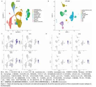
2.1 生长抑素-生长抑素受体配受体对在胚胎和成人肾上腺的单细胞转录组图谱分析 对胎儿和成人肾上腺单细胞测序数据进行分析。首先通过降维和聚类分析,结果提示胚胎肾上腺的细胞类型主要为胚胎免疫细胞、成纤维细胞、类固醇生成细胞、髓质细胞和胚胎上皮细胞,成人肾上腺的细胞类型主要为成人免疫细胞、束状带细胞、成人上皮细胞、网状带细胞、球状带细胞、平滑肌细胞和胚胎髓质细胞(图2A,B)。数据分析发现,无论胚胎还是成人的肾上腺中主要的生长抑素受体为生长抑素受体2(图2C,D),在胚胎肾上腺中表达生长抑素受体2的细胞主要是胚胎上皮细胞和类固醇生成细胞,成人肾上腺中表达生长抑素受体2的细胞主要是球状带细胞、成人上皮细胞和网状带细胞(图2C,D)。以上数据表明生长抑素可能会通过生长抑素受体2作用于肾上腺。因而此次研究进一步在细胞实验上进行验证。 "
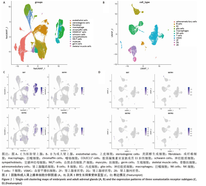
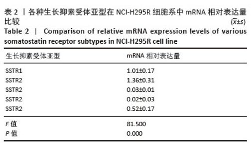
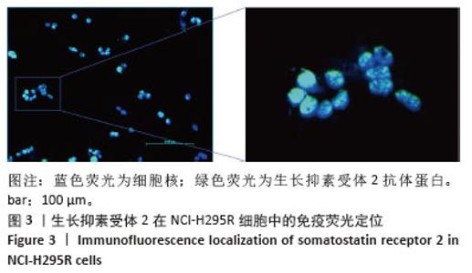
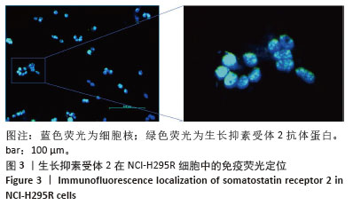
2.2 细胞实验探索生长抑素对肾上腺皮质激素合成的调控机制 2.2.1 qRT-PCR和免疫荧光定位生长抑素受体 在此次研究中采用人肾上腺皮质癌细胞H295R作为体外干预对象,通过qRT-PCR检测NCI-H295R细胞中生长抑素受体1-5不同亚型的基因表达水平(表2)。结果表明在NCI-H295R细胞中主要表达的是生长抑素受体2,随后通过细胞免疫荧光确认了生长抑素受体2在NCI-H295R细胞中存在蛋白表达(图3)。 "
"
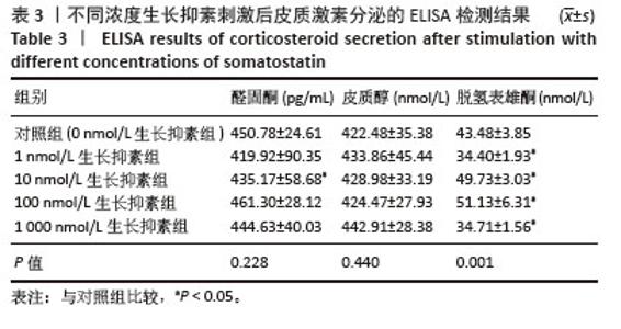
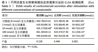
2.2.2 ELISA法检测生长抑素对人肾上腺皮质癌细胞主要类固醇激素合成的影响 在确认人肾上腺皮质癌细胞存在生长抑素受体存在后,使用梯度浓度(0,1,10,100,1 000 nmol/L)的生长抑素处理NCI-H295R细胞。24 h后收集细胞上清,通过ELISA实验测定培养液中人肾上腺皮质癌细胞主要产生3种类固醇激素的含量(表3)。与对照组相比,10 nmol/L生长抑素组细胞醛固酮分泌有减少趋势(P < 0.05),皮质醇的分泌没有明显改变(P > 0.05)。在1 nmol/L生长抑素刺激下,可以有效抑制人肾上腺皮质癌细胞脱氢表雄酮的分泌;10 nmol/L和100 nmol/L生长抑素组细胞的脱氢表雄酮分泌明显增加,分别为(49.73±3.03) nmol/L,(51.13±6.31) nmol/L(P均 < 0.05),这种现象可能是激活其他G蛋白亚型(如Gq)或受体二聚化(如与生长抑素受体5异源二聚)触发非经典信号[30],导致CYB5A上调并促进脱氢表雄酮合成;而1 000 nmol/L生长抑素组细胞脱氢表雄酮的分泌又受到抑制(34.71±1.56) nmol/L (P < 0.05)。以上结果表明,生长抑素干预可以影响人肾上腺皮质癌细胞的脱氢表雄酮和醛固酮分泌,但不影响皮质醇的分泌。考虑到10 nmol/L和100 nmol/L生长抑素刺激后脱氢表雄酮的产量相差不大,在后续实验中选取1 nmol/L和10 nmol/L生长抑素用作基因和蛋白水平的验证。"
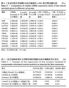
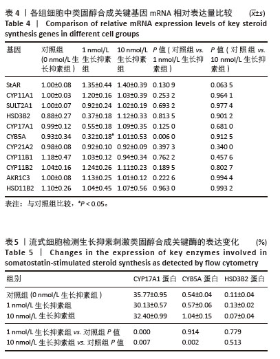
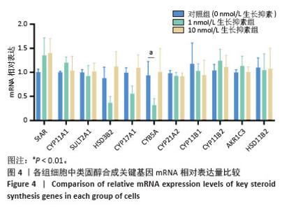
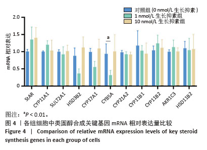
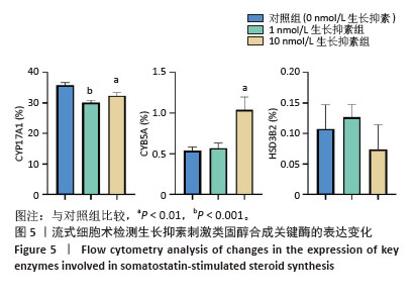
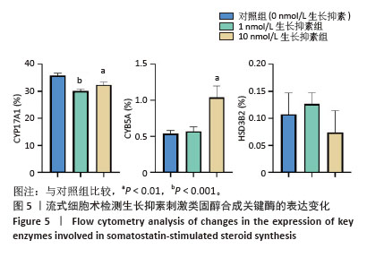
2.2.3 生长抑素对人肾上腺皮质癌细胞类固醇激素关键基因表达量的影响 首先通过qRT-PCR检测类固醇合成通路关键代谢酶基因StAR、CYP11A1、SULT2A1、HSD3B2、CYP17A1、CYB5A、CYP21A2、CYP11B1、CYP11B2、AKR1C3、HSD11B2的表达变化(图4,表4)。检测结果显示,1 nmol/L生长抑素组与对照组相比,CYB5A降低为(0.32±0.18) nmol/L(P < 0.05),说明1 nmol/L生长抑素可以抑制人肾上腺皮质癌细胞脱氢表雄酮合成通路关键基因CYB5A的表达水平。之后用流式细胞术检测CYP17A1、HSD3B2、CYP5A蛋白水平的表达变化,与对照组相比,1 nmol/L生长抑素组细胞CYP17A1的比例[(30.13±0.57)%]明显降低(P < 0.05),CYB5A和HSD3B2的比例无明显变化(P > 0.05)。10 nmol/L组细胞的CYP17A1比例[(32.40±0.99)%]也出现明显降低(P < 0.05),CYB5A的比例[(1.04±0.15)%]明显增加(P < 0.05),而HSD3B2的比例无明显改变(P > 0.05)(图5,表5)。以上结果提示,1 nmol/L生长抑素对脱氢表雄酮合成的抑制作用可能是通过下调CYP17A1产生的,而10 nmol/L生长抑素对脱氢表雄酮合成的促进作用可能是通过上调CYB5A促进了脱氢表雄酮的分泌。"
"
"
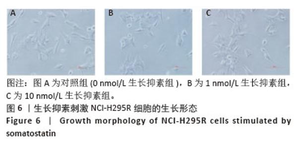
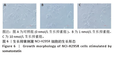
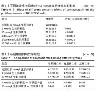
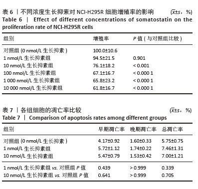
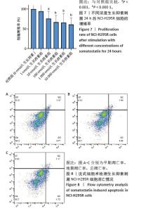
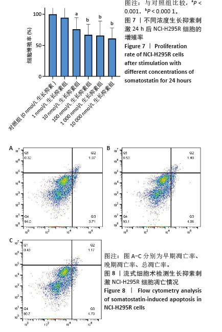
2.2.4 生长抑素对人肾上腺皮质癌细胞增殖和凋亡的影响 生长抑素具有抑制细胞增殖和促进细胞凋亡的作用。该实验为了验证生长抑素是否对人肾上腺皮质有类似的调控作用,观察了人肾上腺皮质癌细胞在1 nmol/L和10 nmol/L浓度下的生长情况。光镜下观察细胞形态发现,与对照组相比,1 nmol/L和10 nmol/L浓度下人肾上腺皮质癌细胞的细胞形态并无明显改变(图6)。随后进行了CCK8细胞增殖率和流式细胞凋亡检测,在不同浓度(0,1,10,100,1 000,10 000 nmol/L)的生长抑素刺激下,CCK8结果显示随着生长抑素浓度的增加,细胞的增殖率逐渐下降,与对照组相比,生长抑素在100,1 000,10 000 nmol/L浓度下,细胞增殖率下降显著(均P < 0.05)(图7,表6)。流式细胞凋亡实验结果显示,与对照组相比,1 nmol/L和10 nmol/L生长抑素组细胞的早、晚期凋亡率和总凋亡率与对照组相比变化不大(图8,表7)。以上结果提示高浓度生长抑素能抑制肾上腺皮质细胞的增殖,但对凋亡未有显著调控作用。 此次研究发现,不同浓度的生长抑素对皮质醇的合成分泌没有明显影响(P > 0.05),而对醛固酮和肾上腺雄激素脱氢表雄酮的合成产生影响。 "
"
"
| [1] ALTIERI B, SECENER AK, SAI S, et al. Single-nucleus and spatial transcriptome reveal adrenal homeostasis in normal and tumoural adrenal glands. Clin Transl Med. 2024;14(8):e1798. [2] HANEMAAIJER ES, MARGARITIS T, SANDERS K, et al. Single-cell atlas of developing murine adrenal gland reveals relation of Schwann cell precursor signature to neuroblastoma phenotype. Proc Natl Acad Sci U S A. 2021;118(5):e2022350118. [3] DEL VALLE I, YOUNG MD, KILDISIUTE G, et al. An integrated single-cell analysis of human adrenal cortex development. JCI Insight. 2023;8(14):e168177. [4] SUCHESTON ME, CANNON MS. Development of zonular patterns in the human adrenal gland. J Morphol. 1968;126(4):477-491. [5] LUCHETTI S, LIERE P, PIANOS A, et al. Disease stage-dependent changes in brain levels and neuroprotective effects of neuroactive steroids in Parkinson’s disease. Neurobiol Dis. 2023;183:106169. [6] SCHWARTZ AG. Dehydroepiandrosterone, Cancer, and Aging. Aging Dis. 2022; 13(2):423-432. [7] LI L, WANG H, YAO Y, et al. The sex steroid precursor dehydroepiandrosterone prevents nonalcoholic steatohepatitis by activating the AMPK pathway mediated by GPR30. Redox Biol. 2021;48:102187. [8] ZHANG S, ZHOU J, LI L, et al. Effect of dehydroepiandrosterone on atherosclerosis in postmenopausal women. Biosci Trends. 2022;15(6):353-364. [9] WANG Y, GUO B, GUO Y, et al. A spatiotemporal steroidogenic regulatory network in human fetal adrenal glands and gonads. Front Endocrinol. 2022;13: 1036517. [10] DAY R, DONG W, PANETTA R, et al. Expression of mRNA for somatostatin receptor (sstr) types 2 and 5 in individual rat pituitary cells. A double labeling in situ hybridization analysis. Endocrinology. 1995;136(11):5232-5235. [11] AMPOFO E, NALBACH L, MENGER MD, et al. Regulatory mechanisms of somatostatin expression. Int J Mol Sci. 2020;21(11): 4170. [12] 孙烁烁,韦晓,张少红,等.生长抑素治疗肥胖的分子机制研究进展[J].中国医药导报,2023,20(15):53-56. [13] STENGEL A, TACHÉ YF. Activation of Brain Somatostatin Signaling Suppresses CRF Receptor-Mediated Stress Response. Front Neurosci. 2017;11:231. [14] PATEL YC. Somatostatin and its receptor family. Front Neuroendocrinol. 1999; 20(3):157-198. [15] AGUILERA G, HARWOOD JP, CATT KJ. Somatostatin modulates effects of angiotensin II in adrenal glomerulosa zone. Nature. 1981;292(5820):262-263. [16] GÜNTHER T, TULIPANO G, DOURNAUD P, et al. International Union of Basic and Clinical Pharmacology. CV. Somatostatin receptors: structure, function, ligands, and new nomenclature. Pharmacol Rev. 2018;70(4):763-835. [17] 李卓凡,黄佳琪,孔炜.生长抑素受体2的功能与研究进展[J].生理科学进展,2022,53(5):385-389. [18] ULANER GA, VANDERMOLEN LA, LI G, et al. Dotatate PET/CT and 225Ac-Dotatate Therapy for Somatostatin Receptor-expressing Metastatic Breast Cancer. Radiology. 2024;312(1):e233408. [19] PELLEGRINO C, FAVALLI N, VOLTA L, et al. Peptide-guided adaptor-CAR T-Cell therapy for the treatment of SSTR2-expressing neuroendocrine tumors. Oncoimmunology. 2024;13(1):2412371. [20] THAKUR S, DALEY B, MILLO C, et al. 177Lu-DOTA-EB-TATE, a Radiolabeled Analogue of Somatostatin Receptor Type 2, for the Imaging and Treatment of Thyroid Cancer. Clin Cancer Res. 2021;27(5):1399-1409. [21] HAN G, HWANG E, LIN F, et al. RYZ101 (Ac-225 DOTATATE) Opportunity beyond Gastroenteropancreatic Neuroendocrine Tumors: Preclinical Efficacy in Small-Cell Lung Cancer. Mol Cancer Ther. 2023;22(12):1434-1443. [22] HEO Y, YOON E, JEON YE, et al. Cryo-EM structure of the human somatostatin receptor 2 complex with its agonist somatostatin delineates the ligand-binding specificity. Elife. 2022;11:e76823. [23] CAMPANA C, AMARÙ J, MILIOTO A, et al. Digital quantification of somatostatin receptor subtypes 2 and 5 in growth hormone-secreting pituitary tumors. Eur J Endocrinol. 2025;192(1):K6-K14. [24] WU J, DING Y, WANG J, et al. Single-cell RNA sequencing in oral science: Current awareness and perspectives. Cell Prolif. 2022;55(10):e13287. [25] LIANG DM, DU PF. scMUG: deep clustering analysis of single-cell RNA-seq data on multiple gene functional modules. Brief Bioinform. 2025;26(2):bbaf138. [26] JOVIC D, LIANG X, ZENG H, et al. Single-cell RNA sequencing technologies and applications: A brief overview. Clin Transl Med. 2022;12(3):e694. [27] CHEN Y, SONG J, RUAN Q, et al. Single-Cell Sequencing Methodologies: From Transcriptome to Multi-Dimensional Measurement. Small Methods. 2021;5(6): e2100111. [28] FIGUEIREDO ML. Applications of single-cell RNA sequencing in rheumatoid arthritis. Front Immunol. 2024;15:1491318. [29] CHENG C, CHEN W, JIN H, et al. A Review of Single-Cell RNA-Seq Annotation, Integration, and Cell-Cell Communication. Cells. 2023;12(15):1970. [30] YANG M, MANDAL K, SÖDERGREN M, et al. Real-time detection of somatostatin release from single islets reveals hypersecretion in type 2 diabetes. Acta Physiol (Oxf). 2025;241(2):e14268. [31] MILEWSKA-KRANC A, ĆWIKŁA JB, KOLASINSKA-ĆWIKŁA A. The Role of Receptor-Ligand Interaction in Somatostatin Signaling Pathways: Implications for Neuroendocrine Tumors. Cancers (Basel). 2023;16(1):116. [32] GERMANO A, RAPA I, DUREGON E, et al. Tissue Expression and Pharmacological In Vitro Analyses of mTOR and SSTR Pathways in Adrenocortical Carcinoma. Endocr Pathol. 2017;28(2):95-102. [33] SPIGA F, ZHAO Z, LIGHTMAN SL. Prolonged treatment with the synthetic glucocorticoid methylprednisolone affects adrenal steroidogenic function and response to inflammatory stress in the rat. Brain Behav Immun. 2020;87:703-714. [34] ZHANG X, SAARINEN AM, CAMPBELL LE, et al. Regulation of Lipolytic Response and Energy Balance by Melanocortin 2 Receptor Accessory Protein (MRAP) in Adipocytes. Diabetes. 2018;67(2):222-234. [35] MILLER WL. MECHANISMS IN ENDOCRINOLOGY: Rare defects in adrenal steroidogenesis. Eur J Endocrinol. 2018;179(3):R125-R141. [36] CAROCCIA B, VANDERRIELE PE, SECCIA TM, et al. Aldosterone and cortisol synthesis regulation by angiotensin-(1-7) and angiotensin-converting enzyme 2 in the human adrenal cortex. J Hypertens. 2021;39(8):1577-1585. [37] ELLIOTT AD, USTIONE A, PISTON DW. Somatostatin and insulin mediate glucose-inhibited glucagon secretion in the pancreatic α-cell by lowering cAMP. Am J Physiol Endocrinol Metab. 2015;308(2):E130-E143. [38] CHEN S, TENG X, ZHENG S. Molecular basis for the selective G protein signaling of somatostatin receptors. Nat Chem Biol. 2023;19(2):133-140. [39] ROBERTSON MJ, MEYEROWITZ JG, PANOVA O, et al. Plasticity in ligand recognition at somatostatin receptors. Nat Struct Mol Biol. 2022;29(3):210-217. [40] BEN-SHLOMO A, DENG N, DING E, et al. DNA damage and growth hormone hypersecretion in pituitary somatotroph adenomas. J Clin Invest. 2020;130(11): 5738-5755. [41] EVANS JS, BEAUMONT J, BRAGA M, et al. Epigenetic potentiation of somatostatin-2 by guadecitabine in neuroendocrine neoplasias as a novel method to allow delivery of peptide receptor radiotherapy. Eur J Cancer. 2022;176: 110-120. [42] JEPSEN SL, ALBRECHTSEN NJW, WINDELØV JA, et al. Antagonizing somatostatin receptor subtype 2 and 5 reduces blood glucose in a gut- and GLP-1R-dependent manner. JCI Insight. 2021;6(4):e143228. [43] HIRSCH A, HAHN D, KEMPNÁ P, et al. Role of AMP-activated protein kinase on steroid hormone biosynthesis in adrenal NCI-H295R cells. PLoS One. 2012;7(1): e30956. [44] LEE JHZ, COLEMAN T, MCLEAN MA, et al. Selective α-Hydroxyketone Formation and Subsequent C-C Bond Cleavage by Cytochrome P450 Monooxygenase Enzymes. ACS Catal. 2024;14(11):8958-8971. [45] XIAO F, SONG X, TIAN P, et al. Comparative Dynamics and Functional Mechanisms of the CYP17A1 Tunnels Regulated by Ligand Binding. J Chem Inf Model. 2020; 60(7):3632-3647. [46] LIU Y, DENISOV IG, SLIGAR SG, et al. Substrate-Specific Allosteric Effects on the Enhancement of CYP17A1 Lyase Efficiency by Cytochrome b5. J Am Chem Soc. 2021;143(10):3729-3733. [47] MAK PJ, DUGGAL R, DENISOV IG, et al. Human Cytochrome CYP17A1: The Structural Basis for Compromised Lyase Activity with 17-Hydroxyprogesterone. J Am Chem Soc. 2018;140(23):7324-7331. [48] WRÓBEL TM, JØRGENSEN FS, PANDEY AV, et al. Non-steroidal CYP17A1 Inhibitors: Discovery and Assessment. J Med Chem. 2023;66(10):6542-6566. [49] WRÓBEL TM, BARTUZI D, KACZOR AA. Secondary Binding Site of CYP17A1 in Enhanced Sampling Simulations. J Chem Inf Model. 2024;64(19):7679-7686. [50] SINGH H, KUMAR R, MAZUMDER A, et al. Insights into Interactions of Human Cytochrome P450 17A1: A Review. Curr Drug Metab. 2022;23(3):172-187. [51] PENHALE SH, PICCI G, OTT LR, et al. Impacts of adrenarcheal DHEA levels on spontaneous cortical activity during development. Dev Cogn Neurosci. 2022; 57:101153. [52] VASANKARI T, KUJALA U, TAIMELA S, et al. Effects of a long acting somatostatin analog on pituitary, adrenal, and testicular function during rest and acute exercise: unexpected stimulation of testosterone secretion. J Clin Endocrinol Metab. 1995;80(11):3298-3303. [53] WELLMAN K, FU R, BALDWIN A, et al. Transcriptomic Response Dynamics of Human Primary and Immortalized Adrenocortical Cells to Steroidogenic Stimuli. Cells. 2021;10(9):2376. [54] MIERKE CT. Physical and biological advances in endothelial cell-based engineered co-culture model systems. Semin Cell Dev Biol. 2023;147:58-69. [55] QI Y, YU L, TIAN F, et al. In vitro models to study human gut-microbiota interactions: Applications, advances, and limitations. Microbiol Res. 2023;270: 127336. [56] DABKEVIČIŪTĖ G, PETRIKAITĖ V. Insights into 2D and 3D cell culture models for nanoparticle-based drug delivery to glioblastoma. Biochem Pharmacol. 2025;237:116931. [57] PARENTE IA, CHIARA L, BERTONI S. Exploring the potential of human intestinal organoids: Applications, challenges, and future directions. Life Sci. 2024;352: 122875. [58] JIN H, XUE Z, LIU J, et al. Advancing Organoid Engineering for Tissue Regeneration and Biofunctional Reconstruction. Biomater Res. 2024;28:0016. [59] UPTON TJ, ZAVALA E, METHLIE P, et al. High-resolution daily profiles of tissue adrenal steroids by portable automated collection. Sci Transl Med. 2023;15(701): eadg8464. [60] DEVASIA TP, DEWARAJA YK, FREY KA, et al. A Novel Time-Activity Information-Sharing Approach Using Nonlinear Mixed Models for Patient-Specific Dosimetry with Reduced Imaging Time Points: Application in SPECT/CT After 177Lu-DOTATATE. J Nucl Med. 2021;62(8):1118-1125. [61] WU D, PLYKU D, KULKARNI K, et al. Optimal Time for 124I PET/CT Imaging in Metastatic Differentiated Thyroid Cancer. Clin Nucl Med. 2021;46(4):283-288. [62] BROSCH-LENZ J, DELKER A, VÖLTER F, et al. Toward Single-Time-Point Image-Based Dosimetry of 177Lu-PSMA-617 Therapy. J Nucl Med. 2023;64(5):767-774. |
| [1] | Yang Wenji, Dai Zhu. Applications and research advances of single-cell RNA sequencing in diseases of knee joint tissues [J]. Chinese Journal of Tissue Engineering Research, 2026, 30(31): 8237-8246. |
| [2] | Qiu Xuedi, Guo Chao, He Jiayue, Zhou Zheng. Single-cell sequencing data identifies differentially expressed genes and immune cell subtypes in periodontitis patients [J]. Chinese Journal of Tissue Engineering Research, 2026, 30(19): 4949-4964. |
| [3] | Cao Lifang, Chen Tao, Shou Jiayin, Fan Fangfang. Hot spot and current status of single-cell RNA sequencing in stroke field [J]. Chinese Journal of Tissue Engineering Research, 2026, 30(13): 3446-3457. |
| [4] | Yu Manya, Cui Xing. Contribution and interaction of various cells in bone marrow microenvironment to exosomal circular RNA associated with multiple myeloma bone disease [J]. Chinese Journal of Tissue Engineering Research, 2026, 30(1): 101-110. |
| [5] | Liu Zhaofeng, Yang Xuejun. Targeted biomarkers of single-cell RNA sequencing in intervertebral disc degeneration [J]. Chinese Journal of Tissue Engineering Research, 2025, 29(20): 4351-4360. |
| [6] | Wang Peigeng, Ye Dongping. Application of single-cell RNA sequencing in spinal cord injury [J]. Chinese Journal of Tissue Engineering Research, 2025, 29(13): 2822-2831. |
| [7] | Yan Ru, Wang Kairu, Zhang Feiyan, Jia Shaobin, Cong Guangzhi. Endothelial cell-specific bone morphogenetic protein 2 affects angiogenesis: bioinformatics analysis and experimental validation [J]. Chinese Journal of Tissue Engineering Research, 2025, 29(1): 103-110. |
| [8] | Cheng Haotian, Zhao Xiaofeng, Lu Xiangdong, Zhao Yibo, Fan Zhifeng, Qi Detai, Wang Xiaonan, Zhou Runtian, Jin Xinjie, Zhao Bin. Single-cell RNA sequencing and the pathogenesis of intervertebral disc degeneration [J]. Chinese Journal of Tissue Engineering Research, 2024, 28(1): 93-99. |
| [9] | Long Qingxi, Zhang Pingshu, Liu Qing, Ou Ya, Zhang Lili, Yuan Xiaodong. Single-cell RNA sequencing reveals the heterogeneity of astrocytes [J]. Chinese Journal of Tissue Engineering Research, 2024, 28(1): 139-146. |
| [10] | Zhang Shijing, Huang Zhuojian, Tao Quyuan, Hou Jiangtao, Peng Bin, Luo Yongxin, Li Yanwu, Li Huibiao, Zhong Jiamin, Chen Xinlin. Molecular characteristic of immune cells in ulcerative colitis patients with spleen deficiency and dampness syndrome based on single-cell RNA sequencing [J]. Chinese Journal of Tissue Engineering Research, 2023, 27(33): 5357-5362. |
| [11] | Wu Hao, Ye Dongping. Application of single-cell RNA sequencing in intervertebral disc degeneration [J]. Chinese Journal of Tissue Engineering Research, 2023, 27(33): 5370-5371. |
| [12] | Huang Chang, Ying Peixi, Yi Guoguo, Fu Sheng, Wang Yan, Guo Xi, Zhang Yuxi, Fu Min. Application of single-cell RNA sequencing in diagnosis and treatment of corneal and retinal diseases [J]. Chinese Journal of Tissue Engineering Research, 2022, 26(13): 2106-2113. |
| [13] | Liu Zhengpeng, Wang Yahui, Zhang Yilong, Ming Ying, Sun Zhijie, Sun He. Application of 3D printed interbody fusion cage for cervical spondylosis of spinal cord type: half-year follow-up of recovery of cervical curvature and intervertebral height [J]. Chinese Journal of Tissue Engineering Research, 2021, 25(6): 849-853. |
| [14] | Zhu Bin, Yan Yiguo. Application of single-cell RNA sequencing technology in the study of skeletal system biology [J]. Chinese Journal of Tissue Engineering Research, 2021, 25(26): 4204-4210. |
| [15] | Liu Yongpan, Gong Xiaofang. Effects of dexmedetomidine on perioperative brain protection in patients undergoing craniocerebral surgery under inhalation anesthesia with sevoflurane: a randomized controlled study [J]. Chinese Journal of Tissue Engineering Research, 2020, 24(35): 5688-5694. |
| Viewed | ||||||
|
Full text |
|
|||||
|
Abstract |
|
|||||