Chinese Journal of Tissue Engineering Research ›› 2025, Vol. 29 ›› Issue (24): 5102-5108.doi: 10.12307/2025.730
Previous Articles Next Articles
Accuracy of orthodontic micro-implant placement guided by a 3D-printed guide plate
Fan Jiabing1, Fu Xuefei1, Zhang Junmei1, Zhou Suodi2, Mo Chaolun1
- 1Stomatology Hospital of Guizhou Medical University, Guiyang 550004, Guizhou Province, China; 2Affiliated Wudang Hospital of Guizhou Medical University, Guiyang 550018, Guizhou Province, China
-
Received:2024-06-25Accepted:2024-09-06Online:2025-08-28Published:2025-01-23 -
Contact:Mo Chaolun, MS, Attending physician, Stomatology Hospital of Guizhou Medical University, Guiyang 550004, Guizhou Province, China -
About author:Fan Jiabing, MS, Attending physician, Stomatology Hospital of Guizhou Medical University, Guiyang 550004, Guizhou Province, China -
Supported by:1Stomatology Hospital of Guizhou Medical University, Guiyang 550004, Guizhou Province, China; 2Affiliated Wudang Hospital of Guizhou Medical University, Guiyang 550018, Guizhou Province, China
CLC Number:
Cite this article
Fan Jiabing, Fu Xuefei, Zhang Junmei, Zhou Suodi, Mo Chaolun. Accuracy of orthodontic micro-implant placement guided by a 3D-printed guide plate[J]. Chinese Journal of Tissue Engineering Research, 2025, 29(24): 5102-5108.
share this article
Add to citation manager EndNote|Reference Manager|ProCite|BibTeX|RefWorks
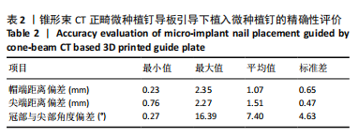
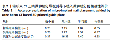
2.1 参与者数量分析 16例患者全部进入结果分析。 2.2 微种植体植入精确性评价 测量结果显示,基于锥形束CT正畸微种植钉导板引导下植入微种植钉与设计植入微种植钉的帽端偏差为(1.07±0.65) mm,支抗钉尖端偏差为(1.51±0.47) mm,冠部与尖部角度偏差为(7.40±4.63)°,见表2。"
| [1] CHEN YJ, CHANG HH, LIN HY, et al. Stability of miniplates and miniscrews used for orthodontic anchorage: experience with 492 temporary anchorage devices. Clin Oral Implants Res. 2008;19(11):1188-1196. [2] BADIALI G, BEVINI M, GULOTTA C, et al. Three-dimensional cephalometric outcome predictability of virtual orthodontic-surgical planning in surgery-first approach. Prog Orthod. 2022;23(1):51. [3] BOURASSA C, HOSEIN YK, POLLMANN SI, et al. In-vitro comparison of different palatal sites for orthodontic miniscrew insertion: Effect of bone quality and quantity on primary stability. Am J Orthod Dentofacial Orthop. 2018;154(6):809-819. [4] 李巍然.种植体支抗在正畸的应用现状与思考[J].中华口腔正畸学杂志,2020,27(1):2-3. [5] MOTOYOSHI M, HIRABAYASHI M, UEMURA M, et al. Recommended placement torque when tightening an orthodontic mini-implant. Clin Oral Implants Res. 2006;17(1):109-114. [6] JING Z, WU Y, JIANG W, et al. Factors Affecting the Clinical Success Rate of Miniscrew Implants for Orthodontic Treatment. Int J Oral Maxillofac Implants. 2016;31(4):835-841. [7] JEDLIŃSKI M, JANISZEWSKA-OLSZOWSKA J, MAZUR M, et al. How Does Orthodontic Mini-Implant Thread Minidesign Influence the Stability?-Systematic Review with Meta-Analysis. J Clin Med. 2022;11(18):5304. [8] HUJA SS, RAO J, STRUCKHOFF JA, et al. Biomechanical and histomorphometric analyses of monocortical screws at placement and 6 weeks postinsertion. J Oral Implantol. 2006;32(3):110-116. [9] 田青鹭,赵志河.微型种植体在口腔正畸中稳定性的研究进展[J].国际口腔医学杂志,2020,47(2):212-218. [10] LEE Y, CHOI SH, YU HS, et al. Stability and success rate of dual-thread miniscrews. Angle Orthod. 2021;91(4):509-514. [11] POUYAFAR V, MESHKABADI R, SADR HAGHIGHI AH, et al. Finite element simulation and statistical investigation of an orthodontic mini-implant’s stability in a novel screw design. Proc Inst Mech Eng H. 2021;235(9):1046-1057. [12] 金海茹.正畸微种植钉成功率及相关风险因素的系统评价[D].济南:山东大学,2020. [13] JARAMILLO-BEDOYA D, VILLEGAS-GIRALDO G, AGUDELO-SUÁREZ AA, et al. A Scoping Review about the Characteristics and Success-Failure Rates of Temporary Anchorage Devices in Orthodontics. Dent J (Basel). 2022;10(5):78. [14] FRANCISCO I, RIBEIRO MP, MARQUES F, et al. Application of Three-Dimensional Digital Technology in Orthodontics: The State of the Art. Biomimetics (Basel). 2022;7(1):23. [15] KNIHA K, BRANDT M, BOCK A, et al. Accuracy of fully guided orthodontic mini-implant placement evaluated by cone-beam computed tomography: a study involving human cadaver heads. Clin Oral Investig. 2021;25(3):1299-1306. [16] MÖHLHENRICH SC, BRANDT M, KNIHA K, et al. Accuracy of orthodontic mini-implants placed at the anterior palate by tooth-borne or gingiva-borne guide support: a cadaveric study. Clin Oral Investig. 2019;23(12):4425-4431. [17] POZZAN L, MIGLIORATI M, DINELLI L, et al. Accuracy of the digital workflow for guided insertion of orthodontic palatal TADs: a step-by-step 3D analysis. Prog Orthod. 2022;23(1):27. [18] LEE JA, AHN HW, OH SH, et al. Evaluation of interradicular space, soft tissue, and hard tissue of the posterior palatal alveolar process for orthodontic mini-implant, using cone-beam computed tomography. Am J Orthod Dentofacial Orthop. 2021;159(4):460-469. [19] MALLICK S, MURALI PS, KUTTAPPA MN, et al. Optimal sites for mini-implant insertion in the lingual or palatal alveolar cortical bone as assessed by cone beam computed tomography in South Indian population. Orthod Craniofac Res. 2021;24(1):121-129. [20] DU B, ZHU J, LI L, et al. Bone depth and thickness of different infrazygomatic crest miniscrew insertion paths between the first and second maxillary molars for distal tooth movement: A 3-dimensional assessment. Am J Orthod Dentofacial Orthop. 2021;160(1):113-123. [21] 徐静,胡敏.正畸微种植体定位方法研究进展[J].口腔医学研究, 2023,39(2):101-104. [22] LANDIN M, JADHAV A, YADAV S, et al. A comparative study between currently used methods and Small Volume-Cone Beam Tomography for surgical placement of mini implants. Angle Orthod. 2015;85(3):446-453. [23] SHARMA K, SANGWAN A. K.s. Micro-implant placement guide. Ann Med Health Sci Res. 2014;4(Suppl 3):S326-S328. [24] MIYAZAWA K, KAWAGUCHI M, TABUCHI M, et al. Accurate pre-surgical determination for self-drilling miniscrew implant placement using surgical guides and cone-beam computed tomography. Eur J Orthod. 2010;32:735-740. [25] MIYAZAWA K, KAWAGUCHI M, TABUCHI M, et al. Accurate pre-surgical determination for self-drilling miniscrew implant placement using surgical guides and cone-beam computed tomography. Eur J Orthod. 2010;32(6):735-740. [26] 赵岩,吴平.应用锥形束CT探究上颌前牙区颌骨微种植支抗钉植入的安全区[J].实用口腔医学杂志,2012,28(4):486-489. [27] AL-GAZZAWI AMQ, KNODE V, LUDWIG B, et al. Midpalatal miniscrew insertion: The accuracy of digital planning and surgical placement. Am J Orthod Dentofacial Orthop. 2024;166(1):69-75. [28] ELKOLALY MA, HASAN HS. MH cortical screws, a revolutionary orthodontic TADs design. J Orthod Sci. 2022;11:53. [29] PETTE GA, NORKIN FJ, GANELES J, et al. Incidental findings from a retrospective study of 318 cone beam computed tomography consultation reports. Int J Oral Maxillofac Implants. 2012;27(3):595-603. [30] 樊佳兵,张军梅.成年女性不同垂直骨面型下颌骨形态的测量分析[J].中国组织工程研究,2021,25(8):1177-1183. [31] SAHOTA J, BHATIA A, GUPTA M, et al. Reliability of Orthopantomography and Cone-beam Computed Tomography in Presurgical Implant Planning: A Clinical Study. J Contemp Dent Pract. 2017;18(8):665-669. [32] CHAWSHLI OF, HASAN HS, YALDA FA, et al. The success rate of mini-screws for incisors intrusion and patient age, gender, and insertion angle in the maxilla using CBCT and implant-guided surgery. A split-mouth, randomized control trail. Orthod Craniofac Res. 2024;27(1): 118-125. [33] 唐敏,陈妍曲,邹道星,等.高精度三维整合牙颌模型在计算机辅助设计与制作个体化微种植体手术导板中的应用研究[J].中华口腔正畸学杂志,2018,25(2):88-91. [34] 仇玲玲,厉松,白玉兴.基于锥形束CT的正畸种植体导板设计及其引导下种植体植入安全性和稳定性的初步评价[J].中华口腔医学杂志,2016,51(6):336-340. [35] GHOLAMPOUR A, MOLLAEI M, EHSANI H, et al. Evaluation of the accuracy of cone beam computed tomography (CBCT) in the detection of peri-implant fenestration. BMC Oral Health. 2024;24(1):922. [36] SHAN LH, GUO N, ZHOU GJ, et al. Finite Element Analysis of Bone Stress for Miniscrew Implant Proximal to Root Under Occlusal Force and Implant Loading. J Craniofac Surg. 2015;26(7):2072-2076. [37] MOON YG, LEE KM. Comparison of the accuracy of intraoral scans between complete-arch scan and quadrant scan. Prog Orthod. 2020; 21(1):36. [38] 刘洪良,刘星纲,田月明,等.口内扫描数字化模型检测接触准确率的临床研究[J].中华口腔医学杂志,2020,55(10):737-742. [39] LO RUSSO L, CARADONNA G, TROIANO G, et al. Three-dimensional differences between intraoral scans and conventional impressions of edentulous jaws: A clinical study. J Prosthet Dent. 2020;123(2):264-268. [40] BAE MJ, KIM JY, PARK JT, et al. Accuracy of miniscrew surgical guides assessed from cone-beam computed tomography and digital models. Am J Orthod Dentofacial Orthop. 2013;143(6):893-901. [41] STURSA L, WENDL B, JAKSE N, et al. Accuracy of Palatal Orthodontic Mini-Implants Placed Using Fully Digital Planned Insertion Guides: A Cadaver Study. J Clin Med. 2023;12(21):6782. [42] PAN Y, CHEN S. Contact of the incisive canal and upper central incisors causing root resorption after retraction with orthodontic mini-implants: A CBCT study. Angle Orthod. 2019;89(2):200-205. [43] CASAÑA-RUIZ MD, BELLOT-ARCÍS C, PAREDES-GALLARDO V, et al. Risk factors for orthodontic mini-implants in skeletal anchorage biological stability: a systematic literature review and meta-analysis. Sci Rep. 2020;10(1):5848. [44] BECKER K, UNLAND J, WILMES B, et al. Is there an ideal insertion angle and position for orthodontic mini-implants in the anterior palate? A CBCT study in humans. Am J Orthod Dentofacial Orthop. 2019;156(3):345-354. [45] 陈妍曲.三维整合牙颌模型在计算机辅助设计与制作个体化微种植体手术导板中的应用研究[D].南宁:广西医科大学,2018. [46] 宋毅,朱房勇,徐小红,等.基于椅旁设计和3D打印的数字化栅栏式微种植钉导板的应用研究[J].口腔医学研究,2024,40(6):525-529. [47] AL-GAZZAWI AMQ, KNODE V, LUDWIG B, et al. Midpalatal miniscrew insertion: The accuracy of digital planning and surgical placement. Am J Orthod Dentofacial Orthop. 2024;166(1):69-75. [48] 孙应明,张梦洁,王晓波.三维模板定位引导正畸微种植体植入的成功率[J].中国组织工程研究与临床康复,2011,15(39):7247-7250. [49] QIU L, XU H, FENG P, et al. Clinical effectiveness of orthodontic miniscrew implantation guided by a novel cone beam CT image-based computer aided design and computer aided manufacturing (CAD-CAM) template. Ann Transl Med. 2021;9(12):1025. |
| [1] | Wei Zhiheng, Guan Tianmin, Liu Qing, Gong Jue, Xiang Xianxiang. Application of 3D printing accurate osteotomy guide combined with the revision of anterior cruciate ligament with abnormally increased posterior slope of tibial plateau [J]. Chinese Journal of Tissue Engineering Research, 2025, 29(33): 7130-7136. |
| [2] | Song Yufei, Cheng Huanzhi, Fan Haixia, Hou Meng. Role and advantages of 3D printing technology in stomatology and maxillofacial surgery restoration and reconstruction [J]. Chinese Journal of Tissue Engineering Research, 2025, 29(22): 4823-4831. |
| [3] | Wang Yuning, Zhu Haotian, Liu Kang, Ding Huanwen, Yan Han. Comparison of short-term therapeutic effects between digital precision total knee arthroplasty and traditional methods [J]. Chinese Journal of Tissue Engineering Research, 2025, 29(21): 4521-4528. |
| [4] | Yang Wenjing, Gegentana. Finite element analysis of the mechanical properties of the tooth in endodontic root canal treatment for endodontic periapical disease [J]. Chinese Journal of Tissue Engineering Research, 2025, 29(20): 4295-4304. |
| [5] | Li Zhiyao, Hu Zheng, Li Xuan, Lu Peijun. Effects of enamel adhesives with different components on Porphyromonas gingivalis and Streptococcus mutans [J]. Chinese Journal of Tissue Engineering Research, 2024, 28(3): 329-335. |
| [6] | Cui Jiali, Huang Minhui, Liu Donglin, Jia Ruiming, Li Han. Computer aided design of 3D dental segmentation and its application scenarios [J]. Chinese Journal of Tissue Engineering Research, 2024, 28(2): 252-257. |
| [7] | Zhao Hexiang, Chen Ziyan, Wang Jing, Ge Zhenlin. Biomechanical characteristics of orthodontic tooth movement before and after increasing alveolar bone mass with periodontally accelerated osteogenic orthodontics [J]. Chinese Journal of Tissue Engineering Research, 2024, 28(14): 2133-2139. |
| [8] | Jiang Haifang, Liu Rong, Hu Peng, Chen Wei, Wei Zairong, Yang Chenglan, Nie Kaiyu. Application of 3D printing technology in the precise and personalized treatment of cleft lip and palate [J]. Chinese Journal of Tissue Engineering Research, 2023, 27(3): 413-419. |
| [9] | Liu Wenwen, Cui Zhanqin, Liu Yingqi. Systematic review of the research progress of nickel-titanium arch wires for orthodontics [J]. Chinese Journal of Tissue Engineering Research, 2023, 27(16): 2556-2562. |
| [10] | Sun Xiaotong, Cheng Yi, Bi Lan, Wang Huida. Effect of parathyroid hormone and parathyroid hormone-related peptides on tissue remodeling during orthodontics [J]. Chinese Journal of Tissue Engineering Research, 2023, 27(14): 2234-2241. |
| [11] | Huang Taosheng, Chen Jianquan, Lin Xinyuan, Lyu Zhouming, Chen Maoshui. 3D printing percutaneous puncture guide plate assisted vertebroplasty for single-level osteoporotic vertebral compression fracture with active registration location combined with anatomic marker localization [J]. Chinese Journal of Tissue Engineering Research, 2022, 26(36): 5819-5825. |
| [12] | Sheng Xiaolei, Liu Su, Wang Jin, Zhao Lei, Zhu Yi, Zhang Wei, Gu Qi, Yuan Feng, Tian Shoujin, Ge Jianfei. Femoral neck fractures in middle-aged and young adults using femoral neck system assisted by 3D printed guide plate [J]. Chinese Journal of Tissue Engineering Research, 2022, 26(33): 5290-5296. |
| [13] | Liu Fatai, Yang Jinshun, Zhong Weibin. Application of three-dimensional printed osteotomy guide plate in knee arthroplasty in osteoarthritis patients with concomitant femoral deformity [J]. Chinese Journal of Tissue Engineering Research, 2022, 26(15): 2312-2316. |
| [14] | Gu Yueguang, Shen Jianhuan, Ni Jieli, Guo Shuyu, Yan Zhongyi, Zhang Yang. Effect of osteogenesis in patients with alveolar cleft after bone grafting investigated by volume analysis [J]. Chinese Journal of Tissue Engineering Research, 2022, 26(10): 1501-1504. |
| [15] | Wang Sizhe, Gao Haiyan, Lan Xiaoquan, Wang Pan, Huang Haoran, Wang Jinyu, Ma Jianlin. A short-term evaluation of three-dimensional printed guide plate assisted hollow screw in the treatment of non-displaced femoral neck fracture [J]. Chinese Journal of Tissue Engineering Research, 2021, 25(36): 5753-5758. |
| Viewed | ||||||
|
Full text |
|
|||||
|
Abstract |
|
|||||