Chinese Journal of Tissue Engineering Research ›› 2022, Vol. 26 ›› Issue (29): 4687-4692.doi: 10.12307/2022.898
Previous Articles Next Articles
Displacement and rate changes of orthodontic tooth movement in Sprague-Dawley rats with chronic fluorosis
Ding Xue1, Jia Ying1, Liu Chun1, Yang Shirong1, Lai Lingyan1, Yang Hua1, Ding Qi2
- 1School of Stomatology, Guizhou Medical University, Guiyang 550001, Guizhou Province, China; 2Department of Orthodontics, Affiliated Stomatology Hospital of Guizhou Medical University, Guiyang 550001, Guizhou Province, China
-
Received:2021-10-18Accepted:2021-12-07Online:2022-10-18Published:2022-03-27 -
Contact:Jia Ying, Professor, Master’s supervisor, School of Stomatology, Guizhou Medical University, Guiyang 550001, Guizhou Province, China -
About author:Ding Xue, Master candidate, Physician, School of Stomatology, Guizhou Medical University, Guiyang 550001, Guizhou Province, China -
Supported by:the National Natural Science Foundation of China, No. 81860795 (to JY); Guizhou Provincial Science and Technology Project, No. qkh [2018]2754 (to JY); Guiyang Municipal Science and Technology Project, No. Zhuke contract [2018]1-83 (to YH)
CLC Number:
Cite this article
Ding Xue, Jia Ying, Liu Chun, Yang Shirong, Lai Lingyan, Yang Hua, Ding Qi. Displacement and rate changes of orthodontic tooth movement in Sprague-Dawley rats with chronic fluorosis[J]. Chinese Journal of Tissue Engineering Research, 2022, 26(29): 4687-4692.
share this article
Add to citation manager EndNote|Reference Manager|ProCite|BibTeX|RefWorks

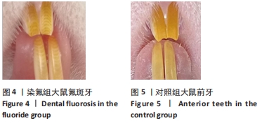
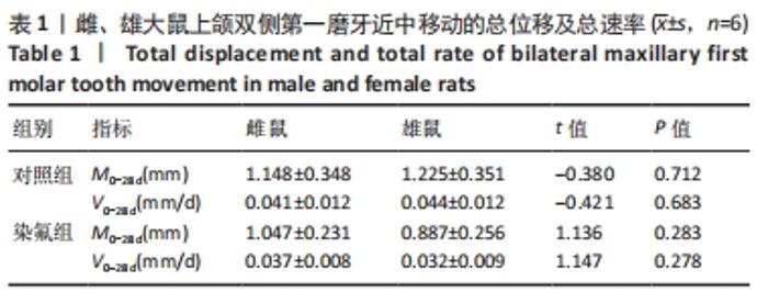
2.1 模型复制鉴定结果 染氟组大鼠饲氟4个月后,前牙牙面出现明显棕、白色相间横纹,即Ⅱ-Ⅲ级氟牙症,见图4,对照组大鼠前牙牙面光滑有光泽,呈半透明状,见图5。尿氟水平:染氟组(45.76±23.02) mg/L>对照组(16.95±9.40) mg/L,差异有显著性意义,P=0.000 < 0.05,提示染氟模型复制成功。"
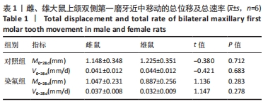
2.2 雌、雄大鼠双侧第一磨牙的总位移及总速率 对照组和染氟组中,雌鼠与雄鼠上颌双侧第一磨牙在一个牙移动周期内(0-28 d)的总位移和总速率无显著差异,见表1。"
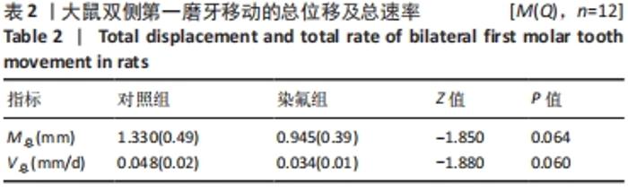
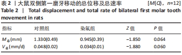
2.3 大鼠双侧第一磨牙的总位移及总速率 染氟组大鼠上颌双侧第一磨牙近中移动的总位移及总速率有低于对照组的趋势,但在观察期内差异无显著性意义,见表2。"
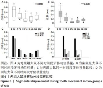
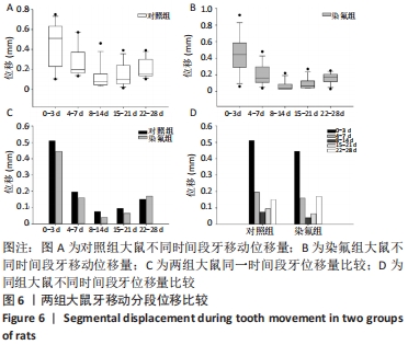
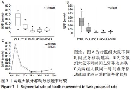
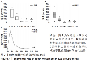
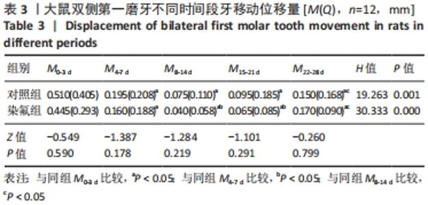
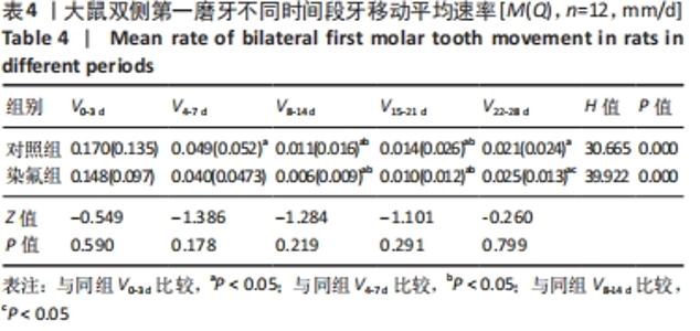
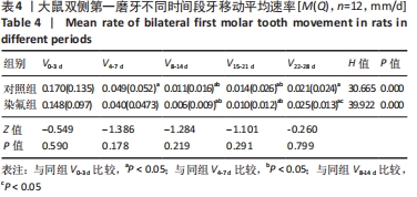
2.4 大鼠双侧第一磨牙的分段位移及分段速率 T0-3 d、T4-7 d、T8-14 d、T15-21 d时段中,染氟组位移及速率有低于对照组的趋势;T22-28 d时段,染氟组位移及速率有高于对照组的趋势,见图6,7,但在观察期内尚未发现统计学差异,见表3,4。随时间延长;两组大鼠牙移动位移及速率呈现先增加后降低然后回升的趋势,对照组M0-3 d>M4-7 d,M8-14 d<M22-28 d,V0-3 d>V4-7 d,V4-7 d>V8-14 d,染氟组M0-3 d>M4-7 d,M4-7 d>M8-14 d,M8-14 d<M22-28 d,V0-3 d>V8-14 d,V8-14 d<V22-28 d,差异均有显著性意义,见表3,4。"
"
"
"
| [1] ASIRY MA. Biological aspects of orthodontic tooth movement: A review of literature. Saudi J Biol Sci. 2018;25(6):1027-1032. [2] JEON HH, TEIXEIRA H, TSAI A. Mechanistic Insight into Orthodontic Tooth Movement Based on Animal Studies: A Critical Review. J Clin Med. 2021;10(8):1733. [3] LI Y, ZHAN Q, BAO M, et al. Biomechanical and biological responses of periodontium in orthodontic tooth movement: up-date in a new decade. Int J Oral Sci. 2021;13(1):20. [4] WEI Y, ZENG B, ZHANG H, et al. iTRAQ-Based Proteomics Analysis of Serum Proteins in Wistar Rats Treated with Sodium Fluoride: Insight into the Potential Mechanism and Candidate Biomarkers of Fluorosis. Int J Mol Sci. 2016;17(10):1644. [5] ZORLU FY, DARICI H, TURKKAHRAMAN H. Histomorphometric and Histopathologic Evaluation of the Effects of Systemic Fluoride Intake on Orthodontic Tooth Movement. Eur J Dent. 2019;13(3):361-369. [6] 李薛燕,黄文丽.氟中毒致机体损伤及其机制[J] .国外医学(医学地理分册),2015,36(3):186-189. [7] 蒋宁宁.氟对骨细胞调控成骨作用的研究[D].长春:吉林大学,2020. [8] DI GIOVANNI T, ELIADES T, PAPAGEORGIOU SN. Interventions for dental fluorosis: A systematic review. J Esthet Restor Dent. 2018;30(6):502-508. [9] FOSSEY S, VAHLE J, LONG P, et al. Nonproliferative and Proliferative Lesions of the Rat and Mouse Skeletal Tissues (Bones, Joints, and Teeth). J Toxicol Pathol. 2016;29(3 Suppl):49S-103S. [10] DAIWILE AP, SIVANESAN S, IZZOTTI A, et al. Noncoding RNAs: Possible Players in the Development of Fluorosis. Biomed Res Int. 2015;2015: 274852. [11] 张文岚,高申,郑德明,等.慢性氟中毒与成骨细胞激活[J].中国地方病防治杂志,2003,18(4):210-213. [12] 姜国有.氟牙症对正畸的影响研究[J].世界最新医学信息文摘:连续型电子期刊,2015,15(34):54. [13] GONZALES C, HOTOKEZAKA H, KARADENIZ EI, et al. Effects of fluoride intake on orthodontic tooth movement and orthodontically induced root resorption. Am J Orthod Dentofacial Orthop. 2011;139(2):196-205. [14] PERUMAL E, PAUL V, GOVINDARAJAN V, et al. A brief review on experimental fluorosis. Toxicol Lett. 2013;223(2):236-251. [15] NAGENDRA AH, BOSE B, SHENOY PS. Recent advances in cellular effects of fluoride: an update on its signalling pathway and targeted therapeutic approaches. Mol Biol Rep. 2021;48(7):5661-5673. [16] 万良斌,于燕妮,万昌武,等.慢性氟中毒对大鼠骨组织 Dlx5 蛋白及其 mRNA 表达的影响[J].中国预防医学杂志,2012,13(9):641-644. [17] LIU L, ZHANG Y, GU HF, et al. The effect of fluoride on the metabolism of teeth and bone in rats. Shanghai Kou Qiang Yi Xue. 2014;23(2):129-132. [18] ZHAO Q, TAN Z, GUO J, et al. Influences of orthodontic tooth movement on estrous cycle and estrogen in rats. Zhonghua Kou Qiang Yi Xue Za Zhi. 2006;41(2):90-91. [19] DENG L, GUO Y. Estrogen effects on orthodontic tooth movement and orthodontically-induced root resorption. Arch Oral Biol. 2020;118: 104840. [20] ISOLA G, MATARESE G, CORDASCO G, et al. Mechanobiology of the tooth movement during the orthodontic treatment: a literature review. Minerva Stomatol. 2016;65(5):299-327. [21] 贾玉龙,王旭霞,刘舒扬,等.牵引上颌埋伏尖牙移动过程的动态模拟[J].临床口腔医学杂志,2011,27(8):487-489. [22] KOHNO T, MATSUMOTO Y, KANNO Z, et al. Experimental tooth movement under light orthodontic forces: rates of tooth movement and changes of the periodontium. J Orthod. 2002;29(2):129-135. [23] WALDO CM, ROTHBLATT JM. Histologic response to tooth movement in the laboratory rat; procedure and preliminary observations. J Dent Res. 1954;33(4):481-486. [24] KIRSCHNECK C, BAUER M, GUBERNATOR J, et al. Comparative assessment of mouse models for experimental orthodontic tooth movement. Sci Rep. 2020;10(1):12154. [25] REN Y, MALTHA JC, VAN ‘T HOF MA, et al. Age effect on orthodontic tooth movement in rats. J Dent Res. 2003;82(1):38-42. [26] VERNA C, DALSTRA M, MELSEN B. The rate and the type of orthodontic tooth movement is influenced by bone turnover in a rat model. Eur J Orthod. 2000;22(4):343-352. [27] HUANG H, WILLIAMS RC, KYRKANIDES S. Accelerated orthodontic tooth movement: molecular mechanisms. Am J Orthod Dentofacial Orthop. 2014;146(5):620-632. [28] KRISHNAN V, DAVIDOVITCH Z. On a path to unfolding the biological mechanisms of orthodontic tooth movement. J Dent Res. 2009;88(7): 597-608. [29] HENNEMAN S, VON DEN HOFF JW, MALTHA JC. Mechanobiology of tooth movement. Eur J Orthod. 2008;30(3):299-306. [30] MURSHID SA. The role of osteocytes during experimental orthodontic tooth movement: A review. Arch Oral Biol. 2017;73:25-33. [31] HOLLIDAY LS, OSTROV DA, WRONSKI TJ, et al. Osteoclast polarization and orthodontic tooth movement. Orthod Craniofac Res. 2009;12(2): 105-112. [32] 李仲伟.慢性氟中毒对大鼠牙移动过程中张力侧牙周组织VEGF、eNOS和ALP表达的影响[D].贵阳:贵州医科大学,2021. [33] 周星宇.慢性氟中毒对大鼠正畸牙移动压力侧牙周组织改建及相关骨效应变化[D].贵阳:贵州医科大学,2021. [34] IBRAHIM AY, GUDHIMELLA S, PANDRUVADA SN, et al. Resolving differences between animal models for expedited orthodontic tooth movement. Orthod Craniofac Res. 2017;20 Suppl 1:72-76. [35] DAVILA JE, MILLER JR, HODGES JS, et al. Effect of neonatal capsaicin treatment on orthodontic tooth movement in male Sprague-Dawley rats. Am J Orthod Dentofacial Orthop. 2011;139(4):e345-e352. [36] VERNA C, DALSTRA M, MELSEN B. The rate and the type of orthodontic tooth movement is influenced by bone turnover in a rat model. Eur J Orthod. 2000;22(4):343-352. [37] ZHOU J, YANG F, XU X, et al. Dynamic Evaluation of Orthodontically-Induced Tooth Movement, Root Resorption, and Alveolar Bone Remodeling in Rats by in Vivo Micro-Computed Tomography. Med Sci Monit. 2018;24:8306-8314. [38] ZOU M, LI C, ZHENG Z. Remote Corticotomy Accelerates Orthodontic Tooth Movement in a Rat Model. Biomed Res Int. 2019;2019:4934128. [39] 刘红,王毅,张勤.牙周炎大鼠正畸牙移动过程中基质金属蛋白酶7在牙周组织中的表达[J].中国组织工程研究,2019,23(11):1669-1673. [40] 燕滨漪,冯云霞,张亮.不同力值对正畸治疗影响的实验研究[J].山西医科大学学报,2009,40(7):607-611,673. [41] 常悦,王明洁,王月,等.不同正畸力对大鼠牙周组织张力侧BMP-2蛋白表达的影响[J].郑州大学学报(医学版), 2015,50(5): 675-678. [42] THEODOROU CI, KUIJPERS-JAGTMAN AM, BRONKHORST EM, et al. Optimal force magnitude for bodily orthodontic tooth movement with fixed appliances: A systematic review. Am J Orthod Dentofacial Orthop. 2019;156(5):582-592. [43] FLORES-MIR C. Forces Between 50 and 100 cN May be Best for Mesiodistal Orthodontic Tooth Movements by Fixed Appliances. J Evid Based Dent Pract. 2020;20(4):101490. |
| [1] | Liu Ke, Fan Haixia, Wang Hong, Cheng Huanzhi, Geng Haixia. Expression and significance of collagen fiber and matrix metalloproteinase-9 during orthodontic root resorption in rats [J]. Chinese Journal of Tissue Engineering Research, 2022, 26(27): 4288-4292. |
| [2] | Jia Qiyu, Guo Jian, Wei Qin, Guo Xiaobin, Chen Dongsheng, Feng Dongwei, Liu Yanshi, Ma Chuang. Establishment and evaluation of a validation model for the efficacy of external femoral fixator screws in rats [J]. Chinese Journal of Tissue Engineering Research, 2022, 26(18): 2854-2861. |
| [3] | Cheng Yi, Liu Ting, Guo Yujing, Sun Xiaotong, Bi Lan, Zhang Ronghe. Effect of estrogen on bone remodeling and root resorption during orthodontics [J]. Chinese Journal of Tissue Engineering Research, 2022, 26(17): 2782-2788. |
| [4] | Xie Fei, Jia Peng, Liu Jiaxin, Liu Lu, Zhang Chunqiu, Ye Jinduo. Effect of loading mode on stress distribution in head and neck of the femoral stem [J]. Chinese Journal of Tissue Engineering Research, 2022, 26(15): 2302-2306. |
| [5] | Zhang Shuping, Dai Lingli, Wang Yajun, Liu Wenjuan, Huang Zuoyi. Effects of angiotensin converting enzyme inhibitors on oxidative stress related factors in a rat model of beta-amyloid 1-42 dementia [J]. Chinese Journal of Tissue Engineering Research, 2021, 25(35): 5650-5655. |
| [6] | Lü Jie, Wang Yongfeng, Yuan Jie, Xu Zhaojian, Qin Yichuan, Hao Jiaqi. Biomechanical finite element analysis of lateral displacement of the cage after oblique lumbar interbody fusion [J]. Chinese Journal of Tissue Engineering Research, 2021, 25(33): 5301-5306. |
| [7] | Tan Jiachang, Yuan Zhenchao, Wu Zhenjie, Liu Bin, Zhao Jinmin. Biomechanical analysis of elastic nail combined with end caps and wire fixation for long oblique femoral shaft fractures [J]. Chinese Journal of Tissue Engineering Research, 2021, 25(3): 334-338. |
| [8] | Yao Rubin, Wang Shiyong, Yang Kaishun. Kummell’s disease treated by intra-pedicle bone cement perfusion combined with kyphoplasty: To enhance the stability of bone cement mass in the vertebral body [J]. Chinese Journal of Tissue Engineering Research, 2021, 25(28): 4435-4440. |
| [9] | Shu Qihang, Liao Yijia, Xue Jingbo, Yan Yiguo, Wang Cheng. Three-dimensional finite element analysis of a new three-dimensional printed porous fusion cage for cervical vertebra [J]. Chinese Journal of Tissue Engineering Research, 2021, 25(24): 3810-3815. |
| [10] | Han Shichong, Li Chang, Xing Haiyang, Ge Wenlong, Wang Gang . Finite element analysis of two internal fixation methods for treating extra-articular proximal tibial fractures [J]. Chinese Journal of Tissue Engineering Research, 2021, 25(15): 2329-2333. |
| [11] | Yan Jiying. Mechanical simulation analysis of static finite element models of the femur with different material assignments [J]. Chinese Journal of Tissue Engineering Research, 2020, 24(9): 1390-1394. |
| [12] | Li Xiao, Pan Jinbing, Ma Yun, Qian Haoyu, Zhang Quncheng, Wang Zheng. Silicone stent insertion for treating tracheobronchomalacia in adults [J]. Chinese Journal of Tissue Engineering Research, 2020, 24(4): 549-554. |
| [13] | Wang Qiang, Gu Yong, Chen Liang. Advantages and disadvantages of internal fixation with suture anchors and locking plate in the treatment of the greater tuberosity fracture [J]. Chinese Journal of Tissue Engineering Research, 2020, 24(30): 4813-4817. |
| [14] | Ji Haihong, Dong Qiang. Finite element analysis of different fixation methods for mandibular defects reconstructing with fibula flaps [J]. Chinese Journal of Tissue Engineering Research, 2020, 24(24): 3821-3827. |
| [15] | Jiang Tao, Shao Min, Chen Qingzhen, Ling Cuimin, Shen Zhen, Wang Gang, Huo Shaochuan, Lin Yanping, Liu Haiquan, Wang Qinsheng, Zeng Zhenming. Inokosterone effects on proliferation and differentiation of osteoblasts from neonatal Sprague-Dawley rats [J]. Chinese Journal of Tissue Engineering Research, 2020, 24(23): 3636-3642. |
| Viewed | ||||||
|
Full text |
|
|||||
|
Abstract |
|
|||||