Chinese Journal of Tissue Engineering Research ›› 2022, Vol. 26 ›› Issue (18): 2854-2861.doi: 10.12307/2022.694
Previous Articles Next Articles
Establishment and evaluation of a validation model for the efficacy of external femoral fixator screws in rats
Jia Qiyu1, Guo Jian1, Wei Qin2, 3, Guo Xiaobin4, Chen Dongsheng1, Feng Dongwei1, Liu Yanshi1, Ma Chuang1
- 1Department of Microprosthetic Surgery, 4Department of Joint Surgery, Orthopaedic Center, the First Affiliated Hospital of Xinjiang Medical University, Urumqi 830054, Xinjiang Uygur Autonomous Region, China; 2Animal Experiment Center, Xinjiang Medical University, Urumqi 830011, Xinjiang Uygur Autonomous Region, China; 3Key Laboratory for Research on New Medical Animal Models, Urumqi 830054, Xinjiang Uygur Autonomous Region, China
-
Received:2021-08-19Accepted:2021-10-11Online:2022-06-28Published:2022-01-29 -
Contact:Ma Chuang, MD, Chief physician, Master’s supervisor, Department of Microprosthetic Surgery, Orthopaedic Center, the First Affiliated Hospital of Xinjiang Medical University, Urumqi 830054, Xinjiang Uygur Autonomous Region, China -
About author:Jia Qiyu, Master candidate, Department of Microprosthetic Surgery, Orthopaedic Center, the First Affiliated Hospital of Xinjiang Medical University, Urumqi 830054, Xinjiang Uygur Autonomous Region, China -
Supported by:the National Natural Science Foundation of China, No. 81760397 (to MC); the Natural Science Foundation of Xinjiang Uygur Autonomous Region, No. 2020D01C263 (to GXB)
CLC Number:
Cite this article
Jia Qiyu, Guo Jian, Wei Qin, Guo Xiaobin, Chen Dongsheng, Feng Dongwei, Liu Yanshi, Ma Chuang. Establishment and evaluation of a validation model for the efficacy of external femoral fixator screws in rats[J]. Chinese Journal of Tissue Engineering Research, 2022, 26(18): 2854-2861.
share this article
Add to citation manager EndNote|Reference Manager|ProCite|BibTeX|RefWorks
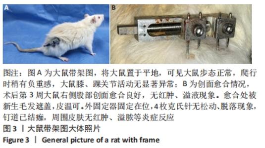
2.1 实验动物数量分析 实验选择大鼠30只,实验过程无脱失,全部进入结果分析。 2.2 大鼠造模时长 大鼠股骨骨折外固定模型造模时长为(20±5) min。 2.3 大鼠活动情况 围术期大鼠精神、饮食、睡眠及活动均正常。术后第3周大鼠右侧股部创面愈合良好,无红肿、溢液现象。愈合处被新生毛发遮盖,皮温可。外固定器固定在位,4枚克氏针无松动、脱落现象,见图3。钉道已结痂,周围皮肤无红肿、溢脓等炎症反应,见图3B。将大鼠置于平地、可见大鼠步态正常、爬行时稍有负重感,见图3A。大鼠膝、踝关节活动无显著异常。"

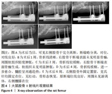
2.4 X射线片观察及标本大体观察结果 于术后当日行X射线摄片,可见右侧股骨干完全离断,断端略分离,对位、对线良好,图4A;术后1周骨折线清晰,右股骨干断端表面未见明显骨痂,周围软组织轻度肿胀,图4B;术后2周骨折线较模糊,右股骨干断端表面骨痂生成,软组织未见明显肿胀,图4C;术后4周骨折线模糊,进一步愈合,髓腔呈再通趋势,图4D;术后6周右侧股骨外固定置留,克氏针均固定在位、无松动,骨形态恢复,原断端位线良好,周围未见游离体,右侧髋膝在位,图4E。"

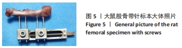
于6周时进行取材,将股骨标本置于无菌单进行大体观察,见图5,共30只股骨带针标本,均实现取材前存活,其中28只(93%)实现骨愈合;1只愈合不良假关节形成,伴螺钉严重松动合并近端钉道周围大面积骨溶解;2只(7%)螺钉轻度松动;1只(3%)出现严重钉道感染。模型总体一致满意率为90%(27/30)。"
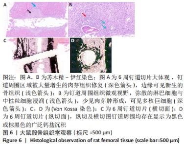
2.5 组织学观察结果 2.5.1 苏木精-伊红染色 显示钉道周围区域被大量增生的肉芽组织修复,伴有弥散的淋巴细胞与中性粒细胞浸润,少见肉芽肿形成,可见多核巨细胞,边缘可见新生的骨组织,见图6A,B。 2.5.2 Von Kossa 染色 纵切及横切图钉道周围均存在显示为黑色或棕黑色的广泛钙盐沉积,见图6C,D。"

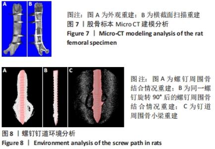
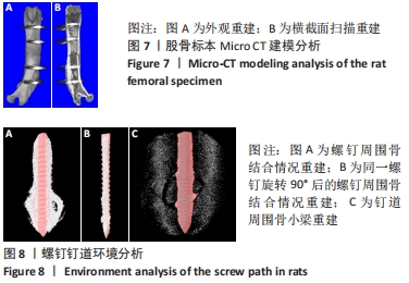
2.6 Micro CT结果分析 通过Micro CT 对股骨标本进行重建,见图7,并通过3D分析对外固定螺钉周围的小梁微结构进行评估,见图8。"
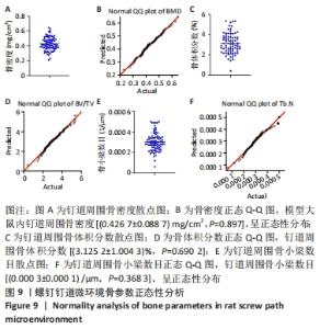
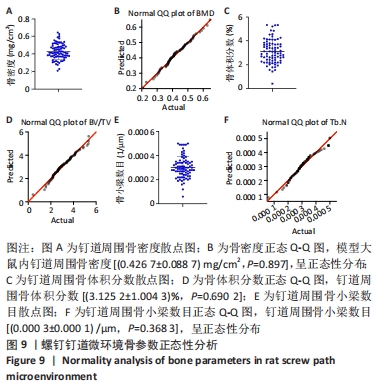
定量分析表明,术后6周,模型大鼠内钉道周围骨密度(0.426 7±0.088 7) mg/cm2,骨体积分数(3.125 2±1.004 3)%,骨小梁数目(0.000 3±0.000 1) /μm。进行模型大鼠骨参数分布情况分析和正态性检验,骨密度(P=0.897),骨体积分数(P=0.690 2),骨小梁数目(P=0.368 3)总体趋于稳定,呈正态性分布,见图9。"
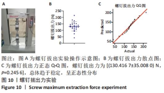
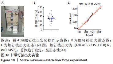
2.7 生物力学测定 对24枚螺钉行螺钉拔出实验,螺钉拔出力为(130.416 7±35.008 0) N,进行模型大鼠分布情况分析和正态性检验,P=0.245 6,总体趋于稳定,呈正态性分布,见图10。"

2.8 材料生物相容性及不良反应 1只大鼠愈合不良假关节形成,伴螺钉严重松动合并近端钉道周围大面积骨溶解;1只大鼠螺钉轻度松动合并严重钉道感染;1只仅出现严重钉道感染。模型总体一致满意率为90%。"

| [1] 阮洪江, 范存义. 改善外固定支架钉-骨界面强度相关技术研究进展[J]. 国际骨科学杂志,2007,28(6):356-358. [2] TUNALI O, SAGLAM Y, BALCI HI, et al. Gustilo type IIIC open tibia fractures with vascular repair: minimum 2-year follow-up. Eur J Trauma Emerg Surg. 2017;43(4):505-512. [3] PALEY D. Problems, Obstacles, and Complications of Limb Lengthening by the Ilizarov Technique. Clin Orthop Relat Res. 1990;(250):81-104. [4] YANAGISAWA Y, ITO A, HARA Y, et al. Initial clinical trial of pins coated with fibroblast growth factor-2–apatite composite layer in external fixation of distal radius fractures. J Orthop. 2018;16(1):69-73. [5] GATHEN M, PLOEGER MM, JAENISCH M, et al. Outcome evaluation of new calcium titanate schanz-screws for external fixators. First clinical results and cadaver studies. J Mater Sci Mater Med. 2019;30(11):124. [6] BAKHSH K, ZIMRI FK, ATIQ UR R, et al. Outcome of complex non-unions of femoral fractures managed with Ilizarov method of distraction osteogenesis. Pak J Med Sci. 2019;35(4):1055-1059. [7] KOSEKI H, ASAHARA T, SHIDA T, et al. Clinical and histomorphometrical study on titanium dioxide-coated external fixation pins. Int J Nanomedicine. 2013;8:593-599. [8] BLUM AL, BONGIOVANNI JC, MORGAN SJ, et al. Complications associated with distraction osteogenesis for infected nonunion of the femoral shaft in the presence of a bone defect: a retrospective series. J Bone Joint Surg Br. 2010;92(4):565-570. [9] BATTLE J, CARMICHAEL KD. Incidence of pin track infections in children’s fractures treated with Kirschner wire fixation. J Pediatr Orthop. 2007;27(2):154-157. [10] MAIMAITI B, ZHANG N, YAN L, et al. Stable ZnO-doped hydroxyapatite nanocoating for anti-infection and osteogenic on titanium. Colloids Surf B Biointerfaces. 2020;186:110731. [11] PATEL A, GHAI A, ANAND A. Clinical Benefit of Hydroxyapatite-Coated Versus Uncoated External Fixation: A Systematic Review. Int J Orthop. 2016;3(3):581-590. [12] PAN G, SUN S, ZHANG W, et al. Biomimetic Design of Mussel-Derived Bioactive Peptides for Dual-Functionalization of Titanium-Based Biomaterials. J Am Chem Soc. 2016;138(45):15078-15086. [13] FU X, LIU P, ZHAO D, et al. Effects of Nanotopography Regulation and Silicon Doping on Angiogenic and Osteogenic Activities of Hydroxyapatite Coating on Titanium Implant. Int J Nanomedicine. 2020;15:4171-4189. [14] KONONOVICH NA, POPKOV AV, GORBACH EN, et al. The Effect of Nanostructured Hydroxyapatite Coating on Distraction Osteogenesis. Key Engineering Materials. 2020;854:216-222. [15] GNEDENKOV SV, SINEBRYUKHOV SL, PUZ AV, et al. In vivo study of osteogenerating properties of calcium-phosphate coating on titanium alloy Ti-6Al-4V. Biomed Mater Eng. 2016;27(6):551-560. [16] JEBAHI S, SAOUDI M, BADRAOUI R, et al. Biologic response to carbonated hydroxyapatite associated with orthopedic device: experimental study in a rabbit modelKorean J Pathol. 2012;46(1):48-54. [17] HARMANKAYA N, IGAWA K, STENLUND P, et al. Healing of complement activating Ti implants compared with non-activating Ti in rat tibia. Acta Biomaterialia. 2012;8(9):3532-3540. [18] SCHELL H, REUTHER T, DUDA GN, et al. The pin-bone interface in external fixator: a standardized analysis in a sheep osteotomy model. J Orthop Trauma. 2011;25(7):438-445. [19] WANG ZL, HE RZ, TU B, et al. Enhanced biocompatibility and osseointegration of calcium titanate coating on titanium screws in rabbit femur. J Huazhong Univ Sci Technolog Med Sci. 2017;37(3):362-370. [20] GLATT V, MATTHYS R. Adjustable stiffness, external fixator for the rat femur osteotomy and segmental bone defect models. J Vis Exp. 2014; (92):e51558. [21] KASPAR K, SCHELL H, TOBEN D, et al. An easily reproducible and biomechanically standardized model to investigate bone healing in rats, using external fixation. Biomed Tech (Berl). 2007;52(6):383-390. [22] MULLER M, HENNIG FF, HOTHORN T, et al. Bone-implant interface shear modulus and ultimate stress in a transcortical rabbit model of open-pore Ti6Al4V implants. J Biomech. 2006;39(11):2123-2132. [23] HARRISON LJ, CUNNINGHAM JL, STROMBERG L, et al. Controlled induction of a pseudarthrosis: a study using a rodent model. J Orthop Trauma. 2003;17(1):11-21. [24] MARK H, BERGHOLM J, NILSSON A, et al. An external fixation method and device to study fracture healing in rats. Acta Orthop Scand. 2003; 74(4):476-482. [25] WILLIE B, ADKINS K, ZHENG X, et al. Mechanical characterization of external fixator stiffness for a rat femoral fracture model. J Orthop Res. 2009;27(5):687-693. [26] 吴硕, 买买艾力·玉山, 魏琴, 等. 大鼠股骨牵张成骨模型的建立[J]. 中华显微外科杂志,2020,43(6):578-582. [27] DROSSE I, VOLKMER E, SEITZ S, et al. Validation of a femoral critical size defect model for orthotopic evaluation of bone healing: a biomechanical, veterinary and trauma surgical perspective. Tissue Eng Part C Methods. 2008;14(1):79-88. [28] ZHAO X, YOU L, WANG T, et al. Enhanced Osseointegration of Titanium Implants by Surface Modification with Silicon-doped Titania Nanotubes.Int J Nanomedicine. 2020;15:8583-8594. [29] CHAI H, GUO L, WANG X, et al. Antibacterial effect of 317L stainless steel contained copper in prevention of implant-related infection in vitro and in vivo. J Mater Sci Mater Med. 2011;22(11) 2525-2535. [30] MORONI A, CADOSSI M, ROMAGNOLI M, et al. A biomechanical and histological analysis of standard versus hydroxyapatite-coated pins for external fixation. J Biomed Mater Res B Appl Biomater. 2008;86(2): 417-421. [31] KALINICHENKO SG, MATVEEVA NY, KOSTIV RY, et al. The topography and proliferative activity of cells immunoreactive to various growth factors in rat femoral bone tissues after experimental fracture and implantation of titanium implants with bioactive biodegradable coatings. Biomed Mater Eng. 2019;30(1):85-95. [32] HERSH-BOYLE RA, KAPATKIN AS, GARCIA TC, et al. Comparison of torsional properties between a Fixateur Externe du Service de Sante des Armees and an acrylic tie-in external skeletal fixator in a red-tailed hawk (Buteo jamaicensis) synthetic tibiotarsal bone model. Am J Vet Res. 2020;81(7):557-564. [33] ZOETIS T, TASSINARI MS, BAGI C, et al. Species comparison of postnatal bone growth and development. Birth Defects Res B Dev Reprod Toxicol. 2003;68(2):86-110. [34] EINHORN TA. The cell and molecular biology of fracture healing. Clin Orthop Relat Res. 1998;(355 Suppl):S7-21. [35] GUO X, LIU Y, BAI J, et al. Efficient Inhibition of Wear-Debris-Induced Osteolysis by Surface Biomimetic Engineering of Titanium Implant with a Mussel-Derived Integrin-Targeting Peptide. Adv Biosyst. 2019; 3(2):e1800253. [36] SMIT TH. The use of a quadruped as an in vivo model for the study of the spine - biomechanical considerations. Eur Spine J. 2002;11(2):137-144. [37] BERGMANN G, GRAICHEN F, ROHLMANN A. Hip joint forces in sheep. J Biomech. 1999;32(8):769-777. [38] IBRAHIMI DISHA S, FURLANI B, DREVENSEK G, et al. The role of endothelin B receptor in bone modelling during orthodontic tooth movement: a study on ETB knockout rats. Sci Rep. 2020;10(1):14226. [39] FUJII T, HIROTA K, YASODA A, et al. Rats deficient C-type natriuretic peptide suffer from impaired skeletal growth without early death. PLoS One. 2018;13(3):e0194812. [40] ANRAKU Y, MIZUTA H, SEI A, et al. The chondrogenic repair response of undifferentiated mesenchymal cells in rat full-thickness articular cartilage defects. Osteoarthritis Cartilage. 2008;16(8):961-964. [41] KERZNER B, MARTIN HL, WEISER M, et al. A Reliable and Reproducible Critical-Sized Segmental Femoral Defect Model in Rats Stabilized with a Custom External Fixator. J Vis Exp. 2019;(145). doi: 10.3791/59206 [42] YANG Y, PAN Q, ZOU K, et al. Administration of allogeneic mesenchymal stem cells in lengthening phase accelerates early bone consolidation in rat distraction osteogenesis model. Stem Cell Res Ther. 2020;11(1): 129. [43] STREET J, BAO M, DEGUZMAN L, et al. Vascular endothelial growth factor stimulates bone repair by promoting angiogenesis and bone turnover. Proc Natl Acad Sci U S A. 2002;99(15):9656-9661. [44] 陈红浩, 康庆林, 贾亚超, 等. 大鼠股骨牵张成骨模型制作技巧[J]. 解剖学杂志,2017,40(1):104-106. |
| [1] | Tan Xinfang, Guo Yanxing, Qin Xiaofei, Zhang Binqing, Zhao Dongliang, Pan Kunkun, Li Yuzhuo, Chen Haoyu. Effect of uniaxial fatigue exercise on patellofemoral cartilage injury in a rabbit [J]. Chinese Journal of Tissue Engineering Research, 2022, 26(在线): 1-6. |
| [2] | Xu Xinzhong, Wu Zhonghan, Yu Shuisheng, Zhao Yao, Xu Chungui, Zhang Xin, Zheng Meige, Jing Juehua. Biomechanical analysis of different ways of inserting Steinmann Pins into the femoral head [J]. Chinese Journal of Tissue Engineering Research, 2022, 26(9): 1313-1317. |
| [3] | Wei Guoqiang, Li Yunfeng, Wang Yi, Niu Xiaofen, Che Lifang, Wang Haiyan, Li Zhijun, Shi Guopeng, Bai Ling, Mo Kai, Zhang Chenchen, Xu Yangyang, Li Xiaohe. Biomechanical analysis of non-uniform material femur under different loads [J]. Chinese Journal of Tissue Engineering Research, 2022, 26(9): 1318-1322. |
| [4] | Pan Baoshun, Fang Zhen, Gao Mingjie, Fang Guiming, Chen Jinshui. Design for posterior atlantoaxial internal fixation system with fusion cage based on imaging data [J]. Chinese Journal of Tissue Engineering Research, 2022, 26(9): 1372-1376. |
| [5] | Li Kun, Gao Erke, Xiong Feng, Wang Xing, Wu Danqi, Li Zhijun, Zhang Shaojie, Liu Yanan, Duo Lan, Li Ziyu. Feasibility of axial transpedicle screw internal fixation in children aged 1 to 6 years [J]. Chinese Journal of Tissue Engineering Research, 2022, 26(9): 1383-1387. |
| [6] | Li Canhui, Wu Zhengjie, Zeng Yanhui, He Yinghao, Situ Xiaopeng, Du Xuelian, Hong Shi, He Jiaxiong. Advantage and disadvantage of robot-assisted sacroiliac screw placement and traditional fluoroscopy in orthopedic surgery [J]. Chinese Journal of Tissue Engineering Research, 2022, 26(9): 1434-1438. |
| [7] | Wang Baojuan, Zheng Shuguang, Zhang Qi, Li Tianyang. Miao medicine fumigation can delay extracellular matrix destruction in a rabbit model of knee osteoarthritis [J]. Chinese Journal of Tissue Engineering Research, 2022, 26(8): 1180-1186. |
| [8] | Lü Yiyan, Li Hanbing, Ma Xiaoqing, Zhang Han, Zhang Yuhang, Li Genlin. Establishment and characteristic analysis of interior heat and diabetes mouse model using compound factors [J]. Chinese Journal of Tissue Engineering Research, 2022, 26(8): 1187-1193. |
| [9] | Zhu Chan, Han Xuke, Yao Chengjiao, Zhang Qiang, Liu Jing, Shao Ming. Acupuncture for Parkinson’s disease: an insight into the action mechanism in animal experiments [J]. Chinese Journal of Tissue Engineering Research, 2022, 26(8): 1272-1277. |
| [10] | Wang Xinmin, Liu Fei, Xu Jie, Bai Yuxi, Lü Jian. Core decompression combined with dental pulp stem cells in the treatment of steroid-associated femoral head necrosis in rabbits [J]. Chinese Journal of Tissue Engineering Research, 2022, 26(7): 1074-1079. |
| [11] | Li Shuo, Su Peng, Zhang Li, Wu Qiulong, Hu Xiangyu, Lai Yuliang. Positive effect of supracondylar femoral osteotomy on the correction of knee varus based on three-dimensional reconstruction and finite element analysis [J]. Chinese Journal of Tissue Engineering Research, 2022, 26(6): 858-863. |
| [12] | Wei Bing, Chang Shan. Finite element analysis of different angles of nail placement in sagittal plane of spinal fracture [J]. Chinese Journal of Tissue Engineering Research, 2022, 26(6): 864-869. |
| [13] | Song Yuxin, Zhang Tongtong, Niu Jianxiong, Wang Zengping, Wen Jie, Zhang Qunli, Xue Wen, Liu Lin. Precise screw placement of 3D printing model and orthopedic robot in spinal deformity [J]. Chinese Journal of Tissue Engineering Research, 2022, 26(6): 904-907. |
| [14] | Yang Jun, Yang Qun, Zhang Rui, Jiang Chang. A novel slidable pedicle screw-rod system for lumbar tuberculosis: promoting bone graft fusion by producing stress stimulation to fused segment [J]. Chinese Journal of Tissue Engineering Research, 2022, 26(6): 914-918. |
| [15] | Guo Xiaohui, Song Xizheng, Xiang Hanrui, Kang Zhaorong, Li Daming, Kang Yu, Hu Jun, Sheng Kai. External spinal fixation elastic stress in the treatment of jumping spinal fracture [J]. Chinese Journal of Tissue Engineering Research, 2022, 26(6): 919-923. |
| Viewed | ||||||
|
Full text |
|
|||||
|
Abstract |
|
|||||