[1] JOSEPH GB, NEVITT MC, MCCULLOCH CE, et al. Associations between molecular biomarkers and MR-based cartilage composition and knee joint morphology: data from the Osteoarthritis Initiative. Osteoarthritis Cartilage. 2018;4(19):1070-1077.
[2] 张子琦,梁佳林,樊立宏,等.开放楔形胫骨高位截骨术治疗膝关节内侧间室骨关节炎疗效观察[J].中国修复重建外科杂志,2018,32(8):997-1000.
[3] SUZANNE W, JILLIAN J, ROBERT L, et al. High Activity Level and Return to Sports Following Knee Osteotomy in Young Athletic Patients. Arthroscop. 2017;33(10): 169-170.
[4] HIROYUKI S, TAKEO N, YASUNORI S, et al. Effect of tibial coronal inclination on hindfoot kinematics: A biomechanical simulation study. Proc Inst Mech Eng Part H-J Eng Med. 2017;231(10):952-958.
[5] JACKSON J, WAUGH W. The technique and complications of upper tibial osteotomy. A review of 226 operations. Bone Joint J. 1974;56(2):236-245.
[6] XIAO S, LIU D, YANG F, et al. Femoral supracondylar osteotomy for the treatment of valgus knee osteoarthritis in young and middle-aged patients. Chin J Orthopa Trauma. 2017;30(8):735-738.
[7] 周绪光,曲卫东,翟连文,等.TKA术前股骨髁精确截骨的影像学分析[J].山东医药,2019,59(28):83-85.
[8] BUVANESVARI V, SUGANTHI M. Three dimensional modelling of MRI knee images using improved edge detection and finite element modelling. Multimed Tools App. 2020;79(3):1-12.
[9] TRAD Z, BARKAOUI A, CHAFRA M, et al. Finite Element Models of the Knee Joint. SpringerBriefs ApplSci Technol. 2018;(2):1-34.
[10] 刘宇,陈胜.医学图像分割方法综述[J].电子科技,2017,30(8):169-172.
[11] WANG Z, WANG Y, JIANG L, et al. An image segmentation method using automatic threshold based on improved genetic selecting algorithm. Autom Control Comp Sci. 2016;50(6):432-440.
[12] 高艳芳,豆贺,韩盛松,等.基于Mimics软件的股骨的提取与三维模型重建[J].中国科技信息,2017(17):99-101.
[13] DONG Y, HU G, ZHANG L, et al. Accurate 3D Reconstruction of Subject-Specific Knee Finite Element Model to Simulate the Articular Cartilage Defects. J Shanghai Jiaotong Univ(Agric Sci). 2011;16(5):620-627.
[14] 高巍. 膝关节组成结构的临床解剖学研究[D].石家庄:河北医科大学,2006.
[15] CAMPBELL J, COOMBS D, RAO M, et al. Automated finite element meshing of the lumbar spine: Verification and validation with 18 specimen-specific models. J Biomech. 2016;49(13):2669-2676.
[16] 姜京伟,程阔.有限元网格划分剖析[J].山西交通科技,2007(5):71-73.
[17] SUN J, YAN S, JIANG Y, et al. Finite element analysis of the valgus knee joint of an obese child. Biomed Eng Online. 2016;15(Suppl 2):158.
[18] PEÑA E, CALVO B, MARTÍNEZ M, et al. A three-dimensional finite element analysis of the combined behavior of ligaments and menisci in the healthy human knee joint. J Biomech. 2006;39(9):1689-1701.
[19] DONG Y, DONG Y, XU Q, et al. The Effect of Varying Degrees of Radial Meniscal Tears on the Knee Contact Stresses: A Finite Element Analysis. Adv Mat Res. 2011; 1362:135-141.
[20] AHMET E. Open Knee: Open Source Modeling and Simulation in Knee Biomechanics. J Knee Surg. 2015;29(2):107-116.
[21] THOMAS G, OLIVER D, DILBAR A, et al. Approaches for process and structural finite element simulations of braided ligament replacements. J Ind Text. 2017; 47(3):408-425.
[22] YOON H, OH S, KYUNGSOO K. Analysis of biomechanical effect of stem-end design in revision TKA using Digital Korean model. Clin Biomech. 2008;23(7):853-858.
[23] ZHU G, GUO W, ZHANG Q, et al. Finite Element Analysis of Mobile-bearing Unicompartmental Knee Arthroplasty: The Influence of Tibial Component Coronal Alignment. Chin Med J. 2015; 128(21):2873-2878.
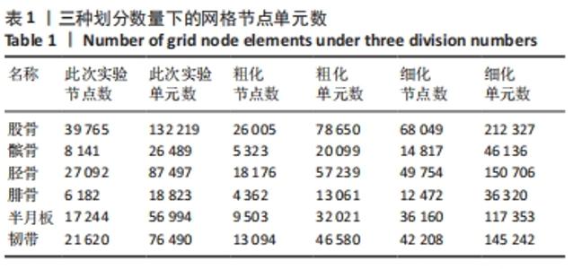
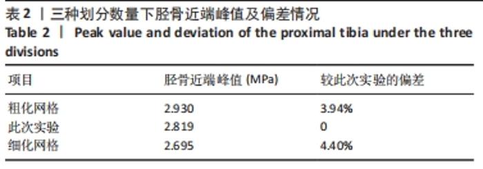
|