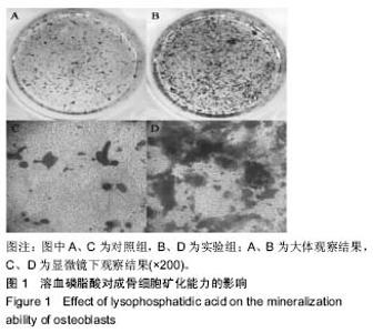
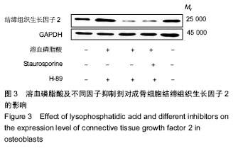
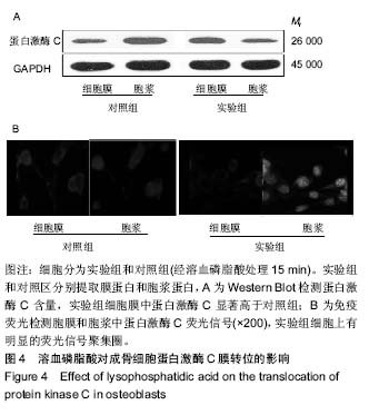
| [1] 杜田田,罗海龙,张忠敏,等.急性进展性脑梗死生物学标记物的研究进展[J].中国现代医生,2017,(25):160-164.[2] 禹萌,任雅芳,张洪涛,等.溶血磷脂酸D-二聚体检测预测进展性脑卒中的临床应用研究[J].中国实用神经疾病杂志, 2017,(8):106, 123.[3] 乌日娜,杨敬平,徐喜媛.溶血磷脂酸及其在肺部疾病中的作用[J].临床肺科杂志,2015,(3):542-543,544.[4] 郑亚,孙红.溶血磷脂酸在卵巢癌进展中的机制研究[J].国际妇产科学杂志,2016,(6):673-676.[5] 赖凡,唐康来.细胞学方法促进腱骨愈合研究进展[J].中国运动医学杂志,2015,34(9):905-909.[6] Mansell JP, Nowghani M, Pabbruwe M, et al. Lysophosphatidic acid and calcitriol co-operate to promote human osteoblastogenesis: requirement of albumin-bound LPA. Prostaglandins Other Lipid Mediat. 2011;95:45-52. [7] 魏福海,陈伟良.骨纤维异常增殖症组织病理学及其发病机制研究进展[J].临床口腔医学杂志,2005,21(8):507-508.[8] 褚伟强,巫津晶,钱文娟.CTGF、HGF在肾病患者血清中的表达及临床意义研究[J].国际检验医学杂志,2017,(24):3393-3395.[9] 刘彩云,郭影.结缔组织生长因子在去卵巢大鼠骨丢失中的作用[J].中国老年学杂志,2018,(2):310-312.[10] Cabello-Verrugio C, Cordova G, Vial C, et al. Connective tissue growth factor induction by lysophosphatidic acid requires transactivation of transforming growth factor type beta receptors and the JNK pathway. Cell Signal. 2011; 23:449-57. [11] Kim KI, Park S, Im GI. Osteogenic differentiation and angiogenesis with cocultured adipose-derived stromal cells and bone marrow stromal cells. Biomaterials. 2014;35: 4792-804. [12] Peyruchaud O, Leblanc R, David M. Pleiotropic activity of lysophosphatidic acid in bone metastasis. Biochim Biophys Acta. 2013;1831:99-104. [13] Wu X, Wang H. The important role of lysophosphatidic acid induced interleukin-6 and -8 syntheses by human osteoblasts in skeletal biology. Bone. 2013;1:268. [14] 赵晓静,徐喜媛,乌日娜,等.溶血磷脂酸在成体干细胞中的作用研究[J].临床肺科杂志,2017,(11):2092-2094.[15] Dziak R. The role of sphingosine-1-phosphate (S1P) and lysophosphatidic acid in regulation of osteoclastic and osteoblastic cells. Immunol Invest. 2013;5(8):510-518. [16] Salles JP. Laurencin-Dalicieux S, Conte-Auriol F, et al. Bone defects in LPA receptor genetically modified mice. Biochim Biophys Acta. 2013;1:93-98. [17] Salles JP, Laurencin-Dalicieux S, Conte-Auriol F, et al. Bone defects in LPA receptor genetically modified mice. Biochim Biophys Acta. 2013;1831:93-98. [18] Gennero I, Laurencin-Dalicieux S, Conte-Auriol F, et al. Absence of the lysophosphatidic acid receptor LPA 1 results in abnormal bone development and decreased bone mass. Bone. 2011;49:395-403. [19] 尹齐,许铭炎,傅玉才,等.蛋白激酶C和JNK在溶血磷脂酸诱导人肺成纤维细胞MCP-1表达中的作用[J].癌变.畸变.突变,2017, (1):31-36.[20] 木其日,阿拉坦高勒.溶血磷脂酸受体3与相关疾病关系的研究进展[J].世界最新医学信息文摘,2015,15(93):35-36+39. [21] 龚晓华,饶勇,马卫列,张志珍.溶血磷脂酸受体2[J].生命的化学, 2013,33(01):24-28.[22] 仲兴,张德志,韩鸿宾,等.地塞米松诱导成骨细胞凋亡的蛋白激酶C途径[J].中国组织工程研究,2013,17(41):7205-7212.[23] 桂雪洋,施鸿飞,陈一心.非经典Wnt信号通路在骨发育和骨代谢中的作用[J].中华实验外科杂志,2017,(8):1437-1440.[24] 张辉,刘窕,李蓓,等.早衰小鼠骨髓间充质干细胞电压门控钙通道的表达检测[J].牙体牙髓牙周病学杂志,2016,(3):140-146.[25] 许莹莹,王敬,韩尚志,等.混旋聚乳酸/纳米羟基磷灰石板对成骨细胞标志性蛋白mRNA表达及细胞培养环境的影响[J].临床和实验医学杂志,2017,(22):2185-2189.[26] Tojima T, Hines JH, Henley JR, et al. Second messengers and membrane trafficking direct and organize growth cone steering. Nat Rev Neurosci. 2011;12:191-203. [27] Panupinthu N, Rogers JT, Zhao L, et al. P2X7 receptors on osteoblasts couple to production of lysophosphatidic acid: a signaling axis promoting osteogenesis. J Cell Biol. 2008; 181:859-871. [28] Aki Y, Kondo A, Nakamura H, et al. Lysophosphatidic acid-stimulated interleukin-6 and -8 synthesis through LPA1 receptors on human osteoblasts. Arch Oral Biol. 2008;53: 207-213. [29] 邹明新,李思颉,邵国,等.电针刺激足三里穴对甲醛溶液所致内脏炎症痛大鼠脊髓蛋白激酶C膜转位水平的影响[J].中国疼痛医学杂志,2016,(3):174-177.[30] 郝松,孟越,李威,等.甲状旁腺素非PLC依赖蛋白激酶C通路激活增强成骨细胞CITED1表达[J].南方医科大学学报, 2015,(4): 486-491. |