Chinese Journal of Tissue Engineering Research ›› 2019, Vol. 23 ›› Issue (18): 2833-2841.doi: 10.3969/j.issn.2095-4344.1671
Previous Articles Next Articles
Preparation and properties of degradable polymethyl methacrylate bone cement incorporated with N-acetyl cysteine
- 1Department of Orthopedics, Zhangjiagang First People’s Hospital, Suzhou 215000, Jiangsu Province, China; 2Department of Orthopedics, the First Affiliated Hospital of Soochow University, Suzhou 215000, Jiangsu Province, China
-
Received:2019-01-14Online:2019-06-28Published:2019-06-28 -
Contact:Wang Liming, Associate chief physician, Department of Orthopedics, Zhangjiagang First People’s Hospital, Suzhou 215000, Jiangsu Province, China -
About author:Zhao Kangquan, Master, Physician, Department of Orthopedics, Zhangjiagang First People’s Hospital, Suzhou 215000, Jiangsu Province, China -
Supported by:the Science and Education Youth Science Foundation of Suzhou, No. KJXW2017059 (to ZKQ)
CLC Number:
Cite this article
Zhao Kangquan, Pi Bin, Sha Weiping, Ge Jianfei, Yang Huilin, Wang Liming. Preparation and properties of degradable polymethyl methacrylate bone cement incorporated with N-acetyl cysteine[J]. Chinese Journal of Tissue Engineering Research, 2019, 23(18): 2833-2841.
share this article

2.1 骨水泥的力学性能 不同配方骨水泥的抗压强度,见图2。"
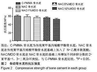
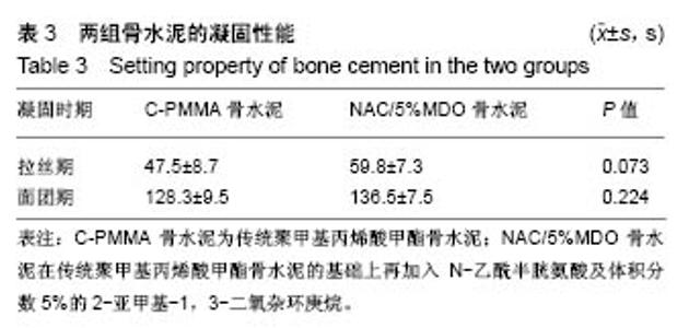
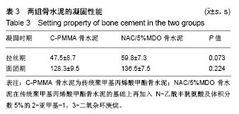
图2可见,随着加入2-亚甲基-1,3-二氧杂环庚烷含量的增加,骨水泥的抗压强度出现了下降趋势,统计学结果提示各组间的抗压强度比较差异有显著性意义(P < 0.05),两两比较后发现,传统骨水泥与NAC/10%MDO骨水泥的抗压强度差异有显著性意义(P < 0.05),而与其他组的比较差异均无显著性意义(P > 0.05)。所以选择第4组NAC/5%MDO骨水泥,即不明显降低抗压强度时含最高2-亚甲基-1,3-二氧杂环庚烷比例的骨水泥,用于后续实验。 2.2 骨水泥凝固时间 骨水泥凝固时间,见表3,两组骨水泥的拉丝期及面团期比较差异均无显著性意义(P > 0.05),提示在骨水泥液体组分中加入25 mmol/L N-乙酰半胱氨酸及体积分数5%2-亚甲基-1,3-二氧杂环庚烷时,不明显影响骨水泥的操作性能。"
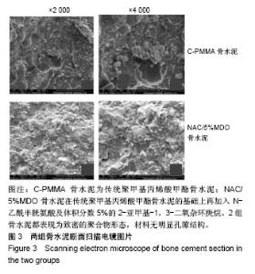
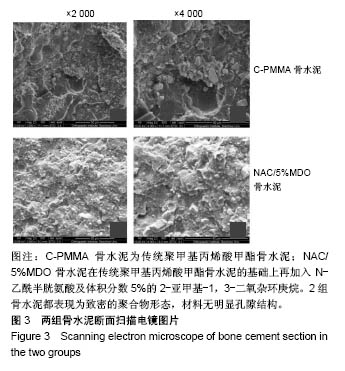
2.3 骨水泥断面扫描电镜 骨水泥断面扫描电镜显示,C-PMMA骨水泥与NAC/5%MDO骨水泥断面都表现为致密的聚合物形态,材料无明显孔隙结构,见图3,说明加入N-乙酰半胱氨酸及2-亚甲基-1,3-二氧杂环庚烷未对材料表观产生影响。"

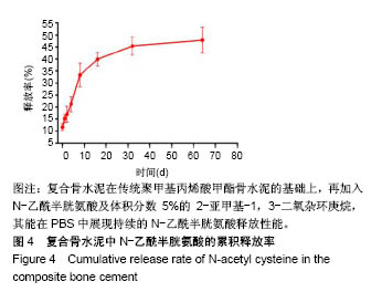
2.4 N-乙酰半胱氨酸体外释放实验 根据图4的结果,NAC/5%MDO骨水泥能在PBS中展现持续的N-乙酰半胱氨酸释放性能,药物突释效应发生在1 h内,1 h内释放率为(11.67±1.38)%,1 d内释放率为(15.16±1.65)%,此后释放率逐渐平稳,能持续释放2个月以上,在64 d时,其药物释放率达到(47.87±5.38)%。"
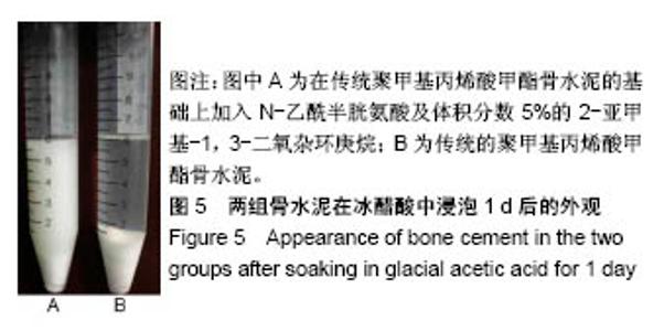
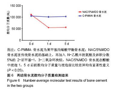
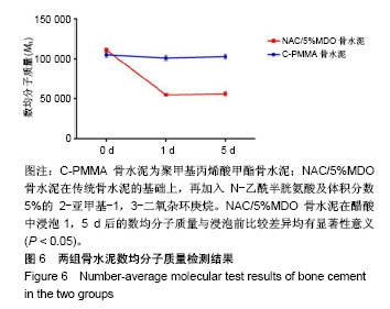
2.5 骨水泥的降解性能 图5显示骨水泥在冰醋酸中浸泡1 d时的外观表现,可见2组骨水泥均发生明显的溶胀坍塌,但C-PMMA骨水泥聚合长链未发生降解,粉体组分仍包埋于聚合体中,液体澄清;NAC/5%MDO骨水泥的聚合长链发生降解断裂,粉体组分无法继续被包埋于聚合体中而扬至周围液体中,使液体明显浑浊。数均分子质量结果证实了这一点,C-PMMA骨水泥浸泡于冰醋酸1,5 d后的数均分子质量较浸泡前未发生统计学意义上的变化(P > 0.05);NAC/5%MDO骨水泥在醋酸中浸泡1 d后的数均分子质量下降50.4%,浸泡1,5 d后的数均分子质量与浸泡前比较差异均有显著性意义(P < 0.05),浸泡1,5 d的数均分子质量比较差异无显著性意义(P > 0.05),见图6。"

"

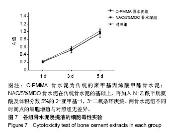
2.6 骨水泥的生物相容性结果 2.6.1 浸提液CCK-8检测结果 图7显示浸提液中的细胞增殖情况,两骨水泥组各时间点上细胞的增殖情况与对照组基本一致,细胞在浸提液中处于对数增长状态,增殖速度快。各检测点上,C-PMMA骨水泥组、NAC/5%MDO骨水泥组与对照组的差异无统计学意义(P > 0.05)。ISO10993-5要求[30],当细胞增殖力下降30%时,提示材料有毒性,实验结果显示两种骨水泥与对照组比不存在明显的细胞毒性。"
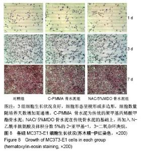
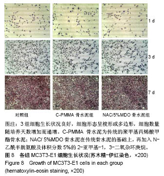
2.6.2 苏木精-伊红染色结果 见图8。 图8显示,对照组、C-PMMA骨水泥组、NAC/5%MDO骨水泥组的MC3T3-E1细胞系生长状况良好,细胞形态呈梭形或多边形,细胞数量随培养天数增加而递增,各组未见明显不同,培养至7 d时,各组细胞几乎完全铺满培养板,胞质胞核染色好,"
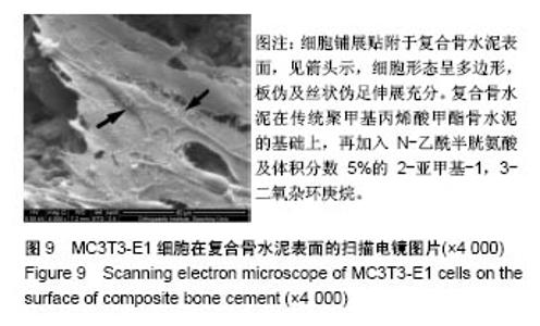
2.6.3 细胞贴附实验结果 电镜下MC3T3-E1细胞生长于NAC/5%MDO骨水泥表面的状态,见图9,图中黑色箭头所指细胞铺展贴附于骨水泥表面,细胞形态呈多边形,板伪及丝状伪足伸展充分,表明骨水泥对直接接触的细胞无毒性反应,不影响细胞形态,不致细胞畸形。"

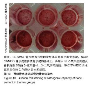
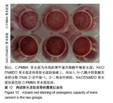
2.7 骨水泥促成骨性能 将MC3T3-E1细胞系于骨水泥表面诱导成骨,茜素红染色后的情况见图10,图片显示NAC/5%MDO骨水泥组染色较C-PMMA骨水泥组深。"
| [1]Rachner TD,Khosla S,Hofbauer LC.Osteoporosis: now and the future. Lancet.2011;377(3):1276-1287.[2]Harrop JS,Prpa B,Reinhardt MK,et al. Primary and secondary osteoporosis' incidence of subsequent vertebral compression fractures after kyphoplasty.Spine (Phila Pa 1976).2004;29(19): 2120-2125..[3]朱汉民.老年人骨折的流行病学及其对生命质量的影响[J].中华老年医学杂志,1993,12(3):168-171.[4]Rousing R,Andersen MO,Jespersen SM,et al.Percutaneous vertebroplasty compared to conservative treatment in patients with painful acute or subacute osteoporotic vertebral fractures: three-months follow-up in a clinical randomized study. Spine (Phila Pa 1976). 2009; 34(13):1349-1354.[5]Kado DM, Browner WS, Palermo L, et al.Vertebral fractures and mortality in olderwomen: a prospective study. Study of Osteoporotic Fractures Research Group. Arch Internal Med.1999;159: 1215-1220.[6]Lewis G. Injectable bone cements for use in vertebroplastyand kyphoplasty: state-of-the-art review. J Biomed MaterRes B Appl Biomater. 2006;76:456-468.[7]Boger A,Heini P,Windolf M,et al.Adjacent vertebralfailure after vertebroplasty: abiomechanical study of lowmodulus PMMA cement.Eur Spine J.2007;16:2118-2125.[8]Berlemann U, Muller CW, Krettek C. Percutaneous cementing techniques of the spine -- chances and limits. Der Orthopade.2004; 33(1):6-12.[9]He Z,Zhai Q,Hu M,et al.Bone cements for percutaneous vertebroplasty and balloon kyphoplasty: Current status and future developments.J Orthop Translat. 2014;3(1):1-11.[10]Granchi D,Cenni E,Savarino L,et al.Bone cement extracts modulatethe osteoprotegerin/osteoprotegerin-ligand expression in MG63 osteoblast-like cells.Biomaterials. 2002;23(11):2359-2365.[11]Delplace V,Tardy A,Harrisson S,et al.Degradable and comb-like PEG-based copolymers by nitroxide-mediate dradicalring-opening polymerization.Biomacromolecules.2013;14(10):3769-3779.[12]Agarwal S,Ren L.Polycaprolactone-Based Novel DegradableIonomers by RadicalRing-Opening Polymerization of 2-Methylene-1,3-dioxepane. Macromolecules.2009;42:1574-1579.[13]Jenny U,Anna F,Ann-Christine A.Adjustable Degradation Properties and Biocompatibility of Amorphous and Functional Poly(ester-acrylate)-Based Materials.Biomacromolecules. 2014; 15(7):2800-2807.[14]Xu Y,Shi Y,Ding S.A chemical approach to stem-cell biology and regenerative medicine.Nature. 2008;453:338-344.[15]Emre N,Coleman R,Ding S.A chemical approach to stem cell biology. Curr Opin Chem Biol. 2007;11:252-258.[16]Takahashi K,Yamanaka S.Induction of pluripotent stem cells from mouse embryonic and adult fibroblast cultures by defined factors. Cell. 2006; 126:663-676.[17]Kennedy JG,O'Grady P,McCarthy DR,et al.An investigation into the role of oxygen free radical scavengers in preventing polymethylmethacrylate- induced necrosis in an osteoblast cell culture.Orthopedics. 2000;23(5): 481-485.[18]Toker H,Ozdemir H,Balc H,et al.N-acetylcysteine decreases alveolar bone loss onexperimental periodontitisin streptozotocin- induceddiabetic rats.J Periodont Res.2012; 47:793-799.[19]Cao JJ, Picklo MJ,et al. N-acetylcysteine supplementation decreases osteoclast differentiation and increases bone mass in mice fed a high-fat diet.J Nutr.2014;144(3):289-296.[20]Tsukeoka T,Suzuki M,Ohtsuki C,et al.Mechanical and histological evaluation of a PMMA-based bone cement modified with gamma- methacryloxypropyltrimethoxysilane and calcium acetate. Biomaterials. 2006;27(21):3897-3903.[21]Sugino A,Ohtsuki C,Miyazaki T.In vivo response of bioactive PMMA-based bone cement modified with alkoxysilane and calcium acetate.J Biomater Appl.2008;23(3):213-228.[22]Tsukimura N,Yamada M,Aita H,et al.N-acetyl cysteine (NAC)-mediated detoxification and functionalization of poly(methyl methacrylate) bone cement.Biomater Appl. 2009;30(3):3378-3389.[23]Yamada M,Tsukimura N,Ikeda T,et al.N-acetyl cysteine as an osteogenesis-enhancing molecule for boneregeneration. Biomater Appl. 2013;34(6):6147-6156.[24]ISO 5833:2002 I.Implants for surgery-Acrylic resin cements.[25]孙强,郑家法,刘德南,等.聚甲基丙烯酸甲酯骨水泥最为抗肿瘤药物缓释载体的实验研究[J].中华肿瘤防治杂志, 2010,17(11):823-826.[26]Feng T,Pi B,Li B,et al.N-Acetyl cysteine (NAC)-mediated reinforcement of alpha-tricalcium phosphate/silk fibroin (α-TCP/SF) cement.Colloids Surf B Biointerfaces. 2015;136:892-899.[27]Ruediger T,Berg A,Guellmar A,et al.Cytocompatibility ofpolymer-based periodontal bone substitutes in gingival fibroblast andMC3T3 osteoblast cell cultures.Dent Mater.2012;28(10): e239-249.[28]ISO 10993 I.Biological evaluation of medical devices.[29]Zhang M,Wu C,Lin K,et al.Biological responses of human bone marrow mesenchymal stem cells to Sr-M-Si (M = Zn, Mg) silicate bioceramics. J Biomed Mater Res. 2012;100(11):2979-2990.[30]Guo JW,Ma Q,Cui Y H,et al.Synthesis and characterization of biodegradable copolymers P( AA-co-MDO) as non-phosphorous detergent builders.Acta Polymerica Sinica. 2012;(9):958-964.[31]Sadowska AM,Manuel-Y-Keenoy B,De Backer WA.Antioxidant and anti-inflammatory efficacy of NAC in the treatment of COPD: discordant in vitro and in vivo dose-effects: a review.Pulm Pharmacol Ther. 2007;20(1):9-22. [32]Zuin R,Palamidese A,Negrin R,et al.High dose N-Acetyicisteine in Patients with Exacerbations of Chronic Obstructive Pulmonary Diseases.Clin Drug Invest. 2005;25(6):401-408.[33]Pela R,Calcagni AM,Subiaco S,et al. N-acetylcysteine reduces the exacerbation rate in patients with moderate to severe COPD. Respiration. 1999;66(6):495-500. [34]Reichenberger F,Tamm M.N-acetylcysteine in the therapy of chronic bronchitis.Pneumologie. 2002;56(12):793-794. [35]Ernst P,Suissa S.N-acetylcysteine is unlikely to reduce hospitalization for chronic obstructive pulmonary diease.Eur Respir J.2003;22(5):865. [36]Kuleci S,Hanta I,Kocabas A,et al.The effect of different treatment modalities on oxidative stress in COPD.Adv Ther.2008;25(7): 710-717.[37]Shinzato S,Nakamura T,Kokubo T,et al.A new bioactive bone cement: effect of glass bead filler content on mechanical and biological properties.J Biomed Mater Res. 2001;54(4):491-500. [38]包利,唐海,王炳强,等.CPC/PMMA双向骨水泥性能及生物相容性的初步研究[J].中国骨质疏松杂志,2012,18(5):420-424.[39]崔维,刘宝戈,王磊,等.经皮椎体后凸成形术中球囊扩张体积与骨水泥注射量的相关性分析[J].中华外科杂志, 2015,53(4):289-293.[40]晏雄伟,张洪燕.椎体后凸成形治疗骨质疏松性脊柱骨折:注入骨水泥的要点[J].中国组织工程研究, 2014,18(9):1471-1476.[41]Kim SY,Jeon SH.Setting properties, mechanical strength and in vivo evaluation of calcium phosphate-based bone cements.J Ind Eng Chem. 2012;18(1):128-136.[42]全仁夫,倪月明,郑宣,等.椎体成形术骨水泥与松质骨的近期与远期改变的实验研究[J].中华骨科杂志, 2013,33(11):1155-1163.[43]Peng W,Bin P,Jin-Ning W.Preparation and properties of calcium sulfate bone cement incorporated with silk fibroin and Sema3A- loaded chitosan microspheres. Front Mater Sci. 2015;9(1):51-65.[44]Fredric, Andrew.Mordem Superabsorbent Polymer Technology, Wiley-VCH, New York,1998.[45]Yuki T,Atsushi S,Toshiki M,et al.Acceleration of calcium phosphate formation on bioactive PMMA-based bone cement by controlling spatial design.Mater Sci Eng. 2010;30(4):624-630. [46]Shinzato S,Nakamura T,Ando K,et al.Mechanical properties and osteoconductivity of new bioactive composites consisting of partially crystallized glass beads and poly(methyl methacrylate).J Biomed MaterRes.2002;60(4):556-563. [47]Charnley J.Anchorage of the femoral head prosthesis to the shaft of the femur. J Bone Joint Surg Br.1960;42:28-30.[48]Yamada M,Ogawa T.Chemodynamics underlying N-acetyl cysteine-mediated bone cement monomer detoxification.Acta Biomater. 2009;5(8):2963-2973.[49]Lin Y,Qu JF.The research progress of polymerization of 1,3-two oxygen heptyl ring and their derivatives.World Sci Tech R&D. 2008;30(1):1-6. |
| [1] | Chen Xinmin, Li Wenbiao, Xiong Kaikai, Xiong Xiaoyan, Zheng Liqin, Li Musheng, Zheng Yongze, Lin Ziling. Type A3.3 femoral intertrochanteric fracture with augmented proximal femoral nail anti-rotation in the elderly: finite element analysis of the optimal amount of bone cement [J]. Chinese Journal of Tissue Engineering Research, 2021, 25(9): 1404-1409. |
| [2] | Yuan Jun, Yang Jiafu. Hemostatic effect of topical tranexamic acid infiltration in cementless total knee arthroplasty [J]. Chinese Journal of Tissue Engineering Research, 2021, 25(6): 873-877. |
| [3] | Hou Guangyuan, Zhang Jixue, Zhang Zhijun, Meng Xianghui, Duan Wen, Gao Weilu. Bone cement pedicle screw fixation and fusion in the treatment of degenerative spinal disease with osteoporosis: one-year follow-up [J]. Chinese Journal of Tissue Engineering Research, 2021, 25(6): 878-883. |
| [4] | Li Li, Ma Li. Immobilization of lactase on magnetic chitosan microspheres and its effect on enzymatic properties [J]. Chinese Journal of Tissue Engineering Research, 2021, 25(4): 576-581. |
| [5] | Liu Fei, Cui Yutao, Liu He. Advantages and problems of local antibiotic delivery system in the treatment of osteomyelitis [J]. Chinese Journal of Tissue Engineering Research, 2021, 25(4): 614-620. |
| [6] | Zhong Yuanming, Wan Tong, Zhong Xifeng, Wu Zhuotan, He Bingkun, Wu Sixian. Meta-analysis of the efficacy and safety of percutaneous curved vertebroplasty and unilateral pedicle approach percutaneous vertebroplasty in the treatment of osteoporotic vertebral compression fracture [J]. Chinese Journal of Tissue Engineering Research, 2021, 25(3): 456-462. |
| [7] | Meng Lingjie, Qian Hui, Sheng Xiaolei, Lu Jianfeng, Huang Jianping, Qi Liangang, Liu Zongbao. Application of three-dimensional printing technology combined with bone cement in minimally invasive treatment of the collapsed Sanders III type of calcaneal fractures [J]. Chinese Journal of Tissue Engineering Research, 2021, 25(24): 3784-3789. |
| [8] | Feng Guancheng, Fang Jianming, Lü Haoran, Zhang Dongsheng, Wei Jiadong, Yu Bingbing. How does bone cement dispersion affect the early outcome of percutaneous vertebroplasty [J]. Chinese Journal of Tissue Engineering Research, 2021, 25(22): 3450-3457. |
| [9] | Liu Jun, Yang Long, Wang Weiyu, Zhou Yuhu, Wu Ying, Lu Tao, Shu Liping, Ma Minxian, Ye Chuan. Preparation and properties of poly3-hydroxybutyrate 4-hydroxybutyrate/polyethylene glycol/graphene oxide tissue-engineered scaffolds [J]. Chinese Journal of Tissue Engineering Research, 2021, 25(22): 3466-3472. |
| [10] | Liu Chang, Li Datong, Liu Yuan, Kong Lingbo, Guo Rui, Yang Lixue, Hao Dingjun, He Baorong. Poor efficacy after vertebral augmentation surgery of acute symptomatic thoracolumbar osteoporotic compression fracture: relationship with bone cement, bone mineral density, and adjacent fractures [J]. Chinese Journal of Tissue Engineering Research, 2021, 25(22): 3510-3516. |
| [11] | He Lin, Wu Xi, He Song, Yang Sen. Hydrophilicity and cell adhesion of hydroxyapatite bioceramics after the coating of polydopamine [J]. Chinese Journal of Tissue Engineering Research, 2021, 25(22): 3540-3544. |
| [12] | Zhou Anqi, Tang Yufei, Wu Bingfeng, Xiang Lin. Designing of periosteum tissue engineering: combination of generality and individuality [J]. Chinese Journal of Tissue Engineering Research, 2021, 25(22): 3551-3557. |
| [13] | Lang Limin, He Sheng, Jiang Zengyu, Hu Yiyi, Zhang Zhixing, Liang Minqian. Application progress of conductive composite materials in the field of tissue engineering treatment of myocardial infarction [J]. Chinese Journal of Tissue Engineering Research, 2021, 25(22): 3584-3590. |
| [14] | Sun Yiqiang, Xing Jianqiang, Li Xuecheng, Wang Xin, Tian Shenglan, Zhao Zihao, Geng Xiaopeng. Kyphoplasty versus vertebroplasty in the treatment of osteoporotic vertebral compression fracture in the elderly: a comparison of vertebral height recovery [J]. Chinese Journal of Tissue Engineering Research, 2021, 25(18): 2851-2855. |
| [15] | Li Qiang, Li Jun, Luan Jian, Jin Canghai, Hao Meng, Lin Yong. Bone cement distribution of percutaneous curved vertebroplasty for the treatment of osteoporotic vertebral compression fractures [J]. Chinese Journal of Tissue Engineering Research, 2021, 25(16): 2466-2471. |
| Viewed | ||||||
|
Full text |
|
|||||
|
Abstract |
|
|||||