Chinese Journal of Tissue Engineering Research ›› 2016, Vol. 20 ›› Issue (47): 7027-7033.doi: 10.3969/j.issn.2095-4344.2016.47.005

Previous Articles     Next Articles

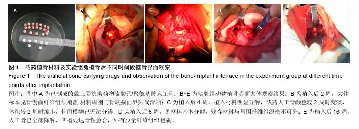
Histological changes of the bone-implant interface after calcium sulfate/ polyaminoacid artificial bone carrying triple-anti-tuberculosis drugs is implanted into the spinal tuberculosis focus

Zhang Zhuo1, Sun Yu-hang2, Geng Guang-qi3, Shi Jian-dang3, Wang Zi-li3, Niu Ning-kui3, Ma Wen-xin3, Liu Hai-tao3, Wang Qian4
  

  1. 1 Department of Orthopaedics, Ningxia People’s Hospital, Yinchuan 750001, Ningxia Hui Autonomous Region, China; 2Ningxia Medical University, Yinchuan 750004, Ningxia Hui Autonomous Region, China; 3Department of Spine Surgery, General Hospital of Ningxia Medical University, Yinchuan 750004, Ningxia Hui Autonomous Region, China; 4College of Pharmacy of University of South Florida in U.S, St. Petersburg 33620, U.S
  • Received:2016-09-10 Online:2016-11-18 Published:2016-11-18
  • Contact: Geng Guang-qi, Master, Associate chief physician, Department of Spine Surgery, General Hospital of Ningxia Medical University, Yinchuan 750004, Ningxia Hui Autonomous Region, China
  • About author:Zhang Zhuo, Master, Physician, Department of Orthopaedics, Ningxia People’s Hospital, Yinchuan 750001, Ningxia Hui Autonomous Region, China Sun Yu-hang, Studying for master’s degree, Ningxia Medical University, Yinchuan 750004, Ningxia Hui Autonomous Region, China Zhang Zhuo and Sun Yu-hang contributed equally to this work.
  • Supported by:

    the National Natural Science Foundation of China, No. 81060149

Abstract:

BACKGROUND: Most researchers have devoted to study the control-release system in spinal tuberculosis focus and implant material for bone defects, but the effective combination to obtain a novel implant material and its treatment outcomes are rarely reported.
OBJECTIVE: To observe the histological change of the implant-bone interface after implantation of the calcium sulfate/polyaminoacid artificial bone carrying triple-anti-tuberculosis drugs including isoniazid, rifampicin and pyrazinamide in a rabbit model of spinal tuberculosis, and to assess the fusion ability of the material.
METHODS: Thirty-six New Zealand rabbits were randomly divided into experiment, control and blank control groups (n=12 per group). Models of spinal tuberculosis were established in the rabbits and randomly assigned into either experiment or control groups, followed by implantation with the calcium sulfate/polyaminoacid artificial bone carrying triple-anti-tuberculosis drugs including isoniazid, rifampicin and pyrazinamide, or the calcium sulfate/polyaminoacid artificial bone, respectively. Healthy controls received the implantation with calcium sulfate/polyaminoacid artificial bone only.
RESULTS AND CONCLUSION: Hematoxylin-eosin staining showed that the artificial bone was covered by fibers and tightly adhered to the wound in each group at 2 weeks after implantation, the graft started to degrade obviously at the 4th week, degraded mostly at the 8th week, and disappeared completely at the 16th week. There was obvious callus formation at 4 weeks after implantation in the experiment group, but the bone healing was later than that of the blank control group. The bone healing ended in the control group. In the blank control group, callus formation and early osteogenesis appeared at the 4th week, and healed completely at the 16th week. Transmission electron microscope and scanning electron microscope revealed that osteoblasts could crawl through the gap of materials to form new bone. These results suggest that the calcium sulfate/polyaminoacid artificial bone carrying triple-anti-tuberculosis drugs can be used as a scaffold, and osteocytes in the bone defect area can crawl through the gap of materials, thereby promoting bone osseointegration at the bone-implant interface.

Key words: Tissue Engineering, Tuberculosis, Spinal, Bony Callus

CLC Number: