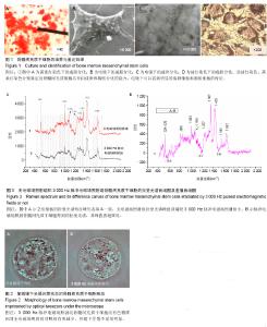
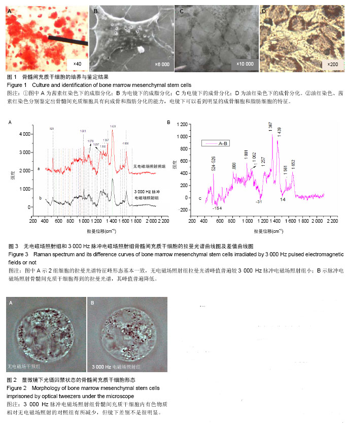
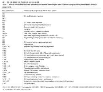
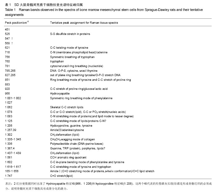
| [1] 荣曦,黄庶识,刘红,等. 拉曼光谱分析技术在细胞生物学研究中的应用进展[J]. 激光生物学报, 2010, 19(1):136-142.
[2] 吴智辉,莫华. 拉曼光镊技术在不同细胞中应用的进展[J]. 中国医学物理学杂志, 2009,26(6):1554-1558.
[3] Xie C, Dinno MA, Li YQ. Near-infrared Raman spectroscopy of single optically trapped biological cells. Opt Lett. 2002; 27(4):249-251.
[4] Chan JW, Winhold H, Corzett MH, et al. Monitoring dynamic protein expression in living E. coli. Bacterial cells by laser tweezers Raman spectroscopy. Cytometry A. 2007;71(7): 468-474.
[5] Liu R, Taylor DS, Matthews DL, et al. Parallel analysis of individual biological cells using multifocal laser tweezers Raman spectroscopy. Appl Spectrosc. 2010;64(11):1308- 1310.
[6] 艾敏,刘军贤,黄庶识,等.拉曼镊子在生物单细胞中的应用与进展[J].分析化学, 2009, 37(5):758-763.
[7] 王桂文,姚辉璐,何碧娟,等.单个血小板的拉曼光谱分析[J].光谱学与光谱分析,2007,27(7):1347-1350.
[8] Huang SS, Chen D, Pelczar PL, et al. Levels of Ca2+-dipicolinic acid in individual bacillus spores determined using microfluidic Raman tweezers. J Bacteriol. 2007;189(13): 4681-4687.
[9] Friedenstein AJ, Petrakova KV, Kurolesova AI, et al. Heterotopic of bone marrow.Analysis of precursor cells for osteogenic and hematopoietic tissues. Transplantation.1968; 6(2):230-247.
[10] Pittenger MF, Mackay AM, Beck SC, et al. Multilineage potential of adult human mesenchymal stem cells. Science. 1999;284(5411):143-147.
[11] Jansen JH, van der Jagt OP, Punt BJ,et al.Stimulation of osteogenic differentiation in human osteoprogenitor cells by pulsed electromagnetic fields: an in vitro study. BMC Musculoskelet Disord. 2010;11:188.
[12] Tsai MT, Li WJ, Tuan RS, et al. Modulation of osteogenesis in human mesenchymal stem cells by specific pulsed electromagnetic field stimulation. J Orthop Res.2009;27(9): 1169-1174.
[13] Canè V, Botti P, Soana S.Pulsed magnetic fields improve osteoblast activity during the repair of an experimental osseous defect. J Orthop Res. 1993;11(5):664-670.
[14] 宋晋刚,许建中,周强,等.不同频率脉冲电磁场促人骨髓间充质干细胞增殖的实验研究[J].中国矫形外科杂志, 2004,12(23): 1867-1869.
[15] 郝海虎,吴华,张海军,等.工频电磁场对骨髓间充质干细胞增殖、分化及其胞浆内钙离子浓度的影响[J].中华物理医学与康复杂志,2007,29(4):217-220.
[16] Dexter TM.Stromal cell associated haemopoiesis.J Cell Physiol Suppl.1982;1:87-94.
[17] Tavassoli M, Friedenstein A. Hemopoietic stromal microenvironment. Am J Hematol.1983;15(2):195-203.
[18] Rosen CJ, Ackert-Bicknell C, Rodriguez JP, et al. Marrow fat and the bone microenvironment: developmental, functional, and pathological implications.Crit Rev Eukaryot Gene Expr. 2009;19(2): 109-124.
[19] 黄超. 基于光镊拉曼光谱的单细胞分析方法研究[D]. 广西师范大学,2007.
[20] 陈秀丽,王桂文,刘军贤,等.基于拉曼光谱的地贫红细胞种类识别方法的研究[J].分析测试学报, 2009,28(4):403-408.
[21] 艾敏,刘军贤,姚辉璐,等. 外周血中单个网织红细胞与小淋巴细胞的拉曼光谱[J]. 光学学报,2009, 29(4):1043-1048.
[22] 邹祖全,刘燕楠,吴瑛,等.臭氧对胎牛血清氧化损伤的表面增强拉曼光谱[J].光谱学与光谱分析, 2007, 27(6):1140-1142.
[23] 段书源,洪元萍,刘拥军,等.羟基磷灰石/胶原矿化机理的研究进展[J].化学通报, 2010,73(4):297-301.
[24] 叶青,谢肇,罗飞,等. 多聚赖氨酸修饰的山羊脱钙骨富集骨髓干细胞的性能评价[J]. 第三军医大学学报, 2009,31(21): 2037-2040.
[25] 韩昕光,毕郑刚,彭伟,等.骨不连接区组织成骨成分改变的研究[A].第二届国际创伤骨科高峰论坛论文集[C]. 2007.
[26] 李志宏,武继民,关静,等.胶原蛋白-壳聚糖/羟基磷灰石复合组织引导再生膜的性能表征[J].中国组织工程研究与临床康复,2009, 13(42): 8388-8392.
[27] 田悦,杜军保.二硫键和巯基在蛋白质结构功能中的作用及分析方法[J].实用儿科临床杂志, 2007, 22 (19):1499-1501.
[28] 张增涛,陈清,罗岚,等.重组人角质细胞生长因子I型缺失突变体二硫键配对与生物活性[J].重庆理工大学学报:自然科学版, 2010, 24(5):44-49.
[29] 朱奇.海南捕鸟蛛毒素-Ⅲ的结构与功能研究及二硫键在蝎毒Leiurotoxin-Ⅰ分子折叠、结构与生理活性中的作用[D].北京大学,2002.
[30] 苏纪勇,刘燕楠,宋昆,等.电流作用后成骨细胞表面增强的拉曼光谱[J].中国组织工程研究与临床康复, 2007,11(44):8839-8843.
[31] 宋昆,苏纪勇,何烁杰,等.低频弱脉冲电磁场作用下成骨细胞的实时表面增强拉曼光谱研究[J].中国组织工程研究与临床康复, 2007, 11(40):8090-8094. |