Chinese Journal of Tissue Engineering Research ›› 2014, Vol. 18 ›› Issue (32): 5203-5208.doi: 10.3969/j.issn.2095-4344.2014.32.020
Previous Articles Next Articles
Chondrogenic differentiation of placenta-derived mesenchymal stem cells in vitro
Li Zhi1, Zhao Wei1, Liu Wei2, Zhou Ye2, Jia Jing-qiao3, Yang Li-feng1
- 1Department of Orthopedics, 2Department of Obstetrics, Central Hospital of Shenyang Medical University, Shenyang 110024, Liaoning Province, China; 3Liaoning Wellcare Stem Cell Biotechnology Co., Ltd., Benxi 117000, Liaoning Province, China
-
Received:2014-06-25Online:2014-08-06Published:2014-09-18 -
Contact:Yang Li-feng, Department of Orthopedics, Central Hospital of Shenyang Medical University, Shenyang 110024, Liaoning Province, China -
About author:Li Zhi, Studying for doctorate, Chief physician, Department of Orthopedics, Central Hospital of Shenyang Medical University, Shenyang 110024, Liaoning Province, China -
Supported by:the Scientific Research Program of Liaoning Science and Technology Bureau, No. 2012225014
CLC Number:
Cite this article
Li Zhi, Zhao Wei, Liu Wei, Zhou Ye, Jia Jing-qiao, Yang Li-feng . Chondrogenic differentiation of placenta-derived mesenchymal stem cells in vitro[J]. Chinese Journal of Tissue Engineering Research, 2014, 18(32): 5203-5208.
share this article
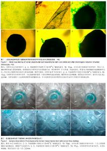
2.1 胎盘来源间充质干细胞形态及细胞表面标志物检测结果 人胎盘间充质干细胞在间充质干细胞培养基中培养,细胞增殖形成大量贴壁细胞,放大40倍倒置显微镜观察,贴壁细胞具有典型的间充质细胞形态,即纺锤体型,折光性强。 用流式细胞仪检测结果显示,阳性表达CD90、CD44、CD105和CD73,不表达CD34、CD14和CD45。流式细胞仪检测结果表明实验培养的细胞具备干细胞的表面标志物,可证实为胎盘间充质干细胞。 2.2 成软骨细胞诱导后阿利新蓝染色结果 倒置显微镜观察:对照组1:形成微团时所用的胎盘来源间充质干细胞的细胞悬液体积为5 μL,细胞悬液浓度为1.6×10¹⁰ L-1,在间充质干细胞培养基中培养,每3 d换液1次。细胞增殖形成大量贴壁细胞,14 d后阿利新蓝染色,放大40倍倒置显微镜观察,贴壁细胞具有典型的间充质细胞形态,即纺锤体型,折光性强,色成淡蓝色(图1A)。 对照组2:形成微团时用细胞浓度为1.6×10¹⁰ L-1的胎盘来源间充质干细胞悬液10 μL,可见细胞增殖形成大量贴壁细胞外,还会形成细胞膜状反折,14 d后阿利新蓝染色,倒置显微镜观察,贴壁细胞具有典型的间充质细胞形态(图1B)。 对照组3:形成微团时用细胞浓度为1.6×10¹⁰ L-1的胎盘来源间充质干细胞悬液15 μL,4 d后行阿利新蓝染色,倒置显微镜观察,微团的基部具有大量贴壁细胞,微团不圆润(图1C)。 实验组1:形成微团时所用的胎盘来源间充质干细胞的细胞悬液体积为5 μL,细胞悬液浓度为1.6×10¹⁰ L-1,在软骨诱导培养基中培养,每3 d换液1次。14 d后行阿利新蓝染色,放大40倍倒置显微镜观察,细胞只会保持微团,不会继续增殖形成贴壁细胞,微团基部无贴壁细胞,微团悬浮培养基中,微团直径约1 mm。倒置显微镜下无法观察到微团内的细胞形态,但可观察到实验组中微团的边缘向外延伸出少数的丝状细胞结构(图1D)。 实验组2,3:形成微团时用细胞浓度为1.6×10¹⁰ L-1的胎盘来源间充质干细胞悬液10,15 μL,倒置显微镜观察悬浮培养基中的微团直径约1.5,2 mm,其形态与实验组1相似(图1E,F)。 大体观察结果:各组胎盘来源间充质干细胞的数码相机照相结果见图2。 对照组1,2中细胞在间充质干细胞培养基中增殖良好,形成大量贴壁细胞长满24孔板(图2A,B);对照组3中有细胞团出现(图2C);实验组细胞在软骨诱导培养基中培养,未见贴壁细胞,微团直径约1.0,1.5,2.0 mm(图2D-F)。 "

| [1] Patel DM, Shah J,Srivastava AS. Therapeutic Potential of Mesenchymal Stem Cells in Regenerative Medicine. Stem Cells International. Stem Cells Int. 2013; 2013: 496218. [2] Syková E, Jendelová P, Urdzíková L, et al.Bone marrow stem cells and polymer hydrogels--two strategies for spinal cord injury repair.Cell Mol Neurobiol. 2006; 26(7-8):1113-1129. [3] Satija NK, Singh VK, Verma YK,et al.Mesenchymal stem cell-based therapy: a new paradigm in regenerative medicine.J Cell Mol Med. 2009;13(11-12):4385-4402. [4] Shin M, Yoshimoto H, Vacanti JP.In vivo bone tissue engineering using mesenchymal stem cells on a novel electrospun nanofibrous scaffold.Tissue Eng. 2004; 10(1-2): 33-41. [5] Shen J, Nair A, Saxena R, et al.Tissue engineering bone using autologous progenitor cells in the peritoneum.PLoS One. 2014; 9(3):e93514. [6] Tuan RS, Boland G, Tuli R.Adult mesenchymal stem cells and cell-based tissue engineering. Arthritis Res Ther. 2003;5(1): 32-45. [7] Schumann D, Kujat R, Nerlich M, et al.Mechanobiological conditioning of stem cells for cartilage tissue engineering.Biomed Mater Eng. 2006; 16(4 Suppl):S37-52. [8] Liu YH, Vaghjiani V, Tee JY, et al.Amniotic epithelial cells from the human placenta potently suppress a mouse model of multiple sclerosis.PLoS One. 2012;7(4):1-8. [9] Hayashi N,Takahashi K,Abe Y, et al. Placental/umbilical cord blood- derived mesenchymal stem cell-like stromal cells support hematopoietic recovery of X-irradiated human CD34~+ cells. Life Sciences. 2009; 84(17-18):598-605. [10] Toubai T, Paczesny S, Shono Y, et al.Mesenchymal stem cells for treatment and prevention of graft-versus-host disease after allogeneic hematopoietic cell transplantation.Curr Stem Cell Res Ther. 2009;4(4):252-259. [11] Banas A. Purification of adipose tissue mesenchymal stem cells and differentiation toward hepatic-like cells.Methods Mol Biol. 2012;826:61-72. [12] Sacchetti B, Funari A, Michienzi S, et al. Self-renewing osteoprogenitors in bone marrow sinusoids can organize a hematopoietic microenvironment. Cell. 2007;131(2):324-336. [13] Aziz Aly LA, Menoufy HE, Ragae A, et al. Adipose stem cells as alternatives for bone marrow mesenchymal stem cells in oral ulcer healing.Int J Stem Cells. 2012;5(2):104-114. [14] Zhu SF, Zhong ZN, Fu XF,et al.Comparison of cell proliferation, apoptosis, cellular morphology and ultrastructure between human umbilical cord and placenta-derived mesenchymal stem cells.Neurosci Lett. 2013;541:77-82. [15] 韩之波,王有为,王涛,等.人胎盘底蜕膜间充质干细胞的分离及其生物学特性研究[J].中国实验血液学杂志,2013,21(3):754-759. [16] Parolini O, Alviano F, Bergwerf I,et al.Toward cell therapy using placenta-derived cells: disease mechanisms, cell biology, preclinical studies, and regulatory aspects at the round table. Stem Cells and Development.2010; 19(2): 143-154. [17] 朱少芳,何援利,卢国辉,等.人胎盘间充质干细胞生物学特性和抗凋亡细胞因子的分泌[J].中国组织工程研究,2011,15(6): 1005-1008. [18] 李芳,苗宗宁,许云云,等. 酶消化和整体灌注分离人胎盘间充质干细胞的比较[J].中国组织工程研究,2010,14(32):5944-5948. [19] 张红艳,杨乃龙.胎盘来源间充质干细胞的生物学特征[J].中国组织工程究,2010,14(40):7535-7538. [20] Miao Z, Jin J, Chen L, et al. Isolation of mesenchymal stem cells from human placenta: Comparison with human bone marrow mesenchymal stem cells. Cell Biol Int. 2006;30(9): 681-687. [21] Wolbank S, Peterbauer A, Fahrner M,et al. Dose-dependent immunomodulatory effect of human stem cells from amniotic membrane: a comparison with human mesenchymal stem cells from adipose tissue. Tissue Eng. 2007;13(6):1173-1183. [22] Alviano F, Fossati V, Marchionni C,et al. Term Amniotic membrane is a high throughput source for multipotent Mesenchymal Stem Cells with the ability to differentiate into endothelial cells in vitro.BMC Dev Biol. 2007;7:11. [23] Portmann-Lanz CB, Schoeberlein A, Portmann R,et al. Turning placenta into brain: placental mesenchymal stem cells differentiate into neurons and oligodendrocytes. Am J Obstet Gynecol. 2010;202(3):294.e1-294.e11. [24] Chang YJ, Hwang SM, Tseng CP,et al. Isolation of mesenchymal stem cells with neurogenic potential from the mesoderm of the amniotic membrane.Cells Tissues Organs. 2010;192(2):93-105. [25] 王珂,徐建敏,华玉明,等.红景天苷诱导人胎盘间充质干细胞分化为肝细胞样细胞[J].江苏医药, 2011,37(15):1765-1767. [26] Ventura C, Cantoni S, Bianchi F,et al. Hyaluronan mixed esters of butyric and retinoic Acid drive cardiac and endothelial fate in term placenta human mesenchymal stem cells and enhance cardiac repair in infarcted rat hearts. J Biol Chem. 2007;282(19):14243-14252. [27] 杜莉莉,金玉楠,李昆,等.胎盘来源的间充质干细胞体外分离培养及向软骨细胞分化的研究[J].解剖科学进展,2008 ,14(1):83-86. [28] 张钰.体外诱导人胎盘间充质干细胞分化为软骨细胞的实验研究[D].中国医科大学,2008. [29] Richardson SM, Hoyland JA, Mobasheri R,et al. Mesenchymal stem cells in regenerative medicine: opportunities and challenges for articular cartilage and intervertebral disc tissue engineering. J Cell Physiol. 2010; 222(1):23-32. [30] Mobasheri A, Csaki C, Clutterbuck AL, et al.Mesenchymal stem cells in connective tissue engineering and regenerative medicine: applications in cartilage repair and osteoarthritis therapy.Histol Histopathol. 2009; 24(3):347-366. [31] Onyekwelu I, Goldring MB, Hidaka C.Chondrogenesis, joint formation, and articular cartilage regeneration.J Cell Biochem. 2009;107(3):383-392. [32] Ramadori G, Mansuroglu T.Mesenchymal origin of hepatic stellate cells, submesothelial cells, and perivascular mesenchymal cells during mouse liver development. Hepatology. 2009;50(1):320; author reply 320. [33] Shimomura K, Ando W, Tateishi K,et al.The influence of skeletal maturity on allogenic synovial mesenchymal stem cell-based repair of cartilage in a large animal model. Biomaterials. 2010;31(31):8004-8011. [34] Dozin B, Malpeli M, Camardella L, et al.Response of young, aged and osteoarthritic human articular chondrocytes to inflammatory cytokines: molecular and cellular aspects.Matrix Biol. 2002;21(5):449-459. [35] Williams JT, Southerland SS, Souza J, et al.Cells isolated from adult human skeletal muscle capable of differentiating into multiple mesodermal phenotypes.Am Surg.1999;65(1): 22-26. [36] Sakaguchi Y, Sekiya I, Yagishita K, et al.Comparison of human stem cells derived from various mesenchymal tissues: superiority of synovium as a cell source.Arthritis Rheum. 2005; 52(8):2521-2529. [37] Dicker A, Le Blanc K, Aström G, et al.Functional studies of mesenchymal stem cells derived from adult human adipose tissue.Exp Cell Res. 2005;308(2):283-290. [38] Aziz Aly LA, Menoufy HE, Ragae A, et al.Adipose stem cells as alternatives for bone marrow mesenchymal stem cells in oral ulcer healing.Int J Stem Cells. 2012; 5(2):104-114. [39] 卢国辉,张世忠,陈强,等.人胎盘底蜕膜间充质干细胞的分离培养及其多向分化潜能的实验研究[J].南方医科大学学报, 2011, 31(2): 262-265. [40] Parolini O, Alviano F, Bagnara GP, et al.Concise review: isolation and characterization of cells from human term placenta: outcome of the first international Workshop on Placenta Derived Stem Cells.Stem Cells. 2008; 26(2):300- 311. [41] Kadam S, Muthyala S, Nair P, et al.Human placenta-derived mesenchymal stem cells and islet-like cell clusters generated from these cells as a novel source for stem cell therapy in diabetes.Rev Diabet Stud. 2010;7(2):168-182. [42] Sabapathy V, Ravi S, Srivastava V,et al.Long-term cultured human term placenta-derived mesenchymal stem cells of maternal origin displays plasticity.Stem Cells Int. 2012;2012: 174328. [43] Lu GH, Zhang SZ, Chen Q, et al.Isolation and multipotent differentiation of human decidua basalis-derived mesenchymal stem cells.Nan Fang Yi Ke Da Xue Xue Bao. 2011; 31(2):262-265. [44] Ringden O, Keating A. Mesenchymal stromal cells as treatment for chronic GVHD. Bone Marrow Transplant. 2011; 46(2):163-164. |
| [1] | Sun Kexin, Zeng Jinshi, Li Jia, Jiang Haiyue, Liu Xia. Mechanical stimulation enhances matrix formation of three-dimensional bioprinted cartilage constructs [J]. Chinese Journal of Tissue Engineering Research, 2023, 27(在线): 1-7. |
| [2] | Dang Yi, Du Chengyan, Yao Honglin, Yuan Nenghua, Cao Jin, Xiong Shan, Zhang Dingmei, Wang Xin. Hormonal osteonecrosis and oxidative stress [J]. Chinese Journal of Tissue Engineering Research, 2023, 27(9): 1469-1476. |
| [3] | Huang Guijiang, Ji Yuwei, Zhao Xin, Yang Yi, Zhao Yulan, Wang Peijin, Tang Wei, Jiao Jianlin. Effect and mechanism of different administration routes of placenta-derived mesenchymal stem cells in the treatment of tree shrews with osteoporotic fracture [J]. Chinese Journal of Tissue Engineering Research, 2023, 27(6): 909-914. |
| [4] | Zhang Hui, Wang Jiayang, Wang Qian, Gan Hongquan, Wang Zhiqiang. Effects of hyaluronic acid combined with domestic porous tantalum on chondrocyte function under the dynamic environment [J]. Chinese Journal of Tissue Engineering Research, 2023, 27(3): 339-345. |
| [5] | Li Rui, Liu Zhen, Guo Zige, Lu Ruijie, Wang Chen. Aspirin-loaded chitosan nanoparticles and polydopamine modified titanium sheets improve osteogenic differentiation [J]. Chinese Journal of Tissue Engineering Research, 2023, 27(3): 374-379. |
| [6] | Su Meng, Wang Xin, Zhang Jin, Bei Ying, Huang Yu, Zhu Yanzhao, Li Jiali, Wu Yan. Nanocellular vesicles loaded with curcumin promote wound healing in diabetic mice [J]. Chinese Journal of Tissue Engineering Research, 2023, 27(12): 1877-1883. |
| [7] | Ma Munan, Xie Jun, Sang Yuchao, Huang Lei, Zhang Guodong, Yang Xiaoli, Fu Songtao. Electroacupuncture combined with bone marrow mesenchymal stem cells in the treatment of chemotherapy-induced premature ovarian insufficiency in rats [J]. Chinese Journal of Tissue Engineering Research, 2023, 27(1): 1-7. |
| [8] | Liu Wentao, Feng Xingchao, Yang Yi, Bai Shengbin. Effect of M2 macrophage-derived exosomes on osteogenic differentiation of bone marrow mesenchymal stem cells [J]. Chinese Journal of Tissue Engineering Research, 2022, 26(在线): 1-6. |
| [9] | Wang Jing, Xiong Shan, Cao Jin, Feng Linwei, Wang Xin. Role and mechanism of interleukin-3 in bone metabolism [J]. Chinese Journal of Tissue Engineering Research, 2022, 26(8): 1260-1265. |
| [10] | Xiao Hao, Liu Jing, Zhou Jun. Research progress of pulsed electromagnetic field in the treatment of postmenopausal osteoporosis [J]. Chinese Journal of Tissue Engineering Research, 2022, 26(8): 1266-1271. |
| [11] | Tian Chuan, Zhu Xiangqing, Yang Zailing, Yan Donghai, Li Ye, Wang Yanying, Yang Yukun, He Jie, Lü Guanke, Cai Xuemin, Shu Liping, He Zhixu, Pan Xinghua. Bone marrow mesenchymal stem cells regulate ovarian aging in macaques [J]. Chinese Journal of Tissue Engineering Research, 2022, 26(7): 985-991. |
| [12] | Hou Jingying, Guo Tianzhu, Yu Menglei, Long Huibao, Wu Hao. Hypoxia preconditioning targets and downregulates miR-195 and promotes bone marrow mesenchymal stem cell survival and pro-angiogenic potential by activating MALAT1 [J]. Chinese Journal of Tissue Engineering Research, 2022, 26(7): 1005-1011. |
| [13] | Zhou Ying, Zhang Huan, Liao Song, Hu Fanqi, Yi Jing, Liu Yubin, Jin Jide. Immunomodulatory effects of deferoxamine and interferon gamma on human dental pulp stem cells [J]. Chinese Journal of Tissue Engineering Research, 2022, 26(7): 1012-1019. |
| [14] | Liang Xuezhen, Yang Xi, Li Jiacheng, Luo Di, Xu Bo, Li Gang. Bushen Huoxue capsule regulates osteogenic and adipogenic differentiation of rat bone marrow mesenchymal stem cells via Hedgehog signaling pathway [J]. Chinese Journal of Tissue Engineering Research, 2022, 26(7): 1020-1026. |
| [15] | Wang Jifang, Bao Zhen, Qiao Yahong. miR-206 regulates EVI1 gene expression and cell biological behavior in stem cells of small cell lung cancer [J]. Chinese Journal of Tissue Engineering Research, 2022, 26(7): 1027-1031. |
| Viewed | ||||||
|
Full text |
|
|||||
|
Abstract |
|
|||||