Chinese Journal of Tissue Engineering Research ›› 2013, Vol. 17 ›› Issue (10): 1711-1718.doi: 10.3969/j.issn.2095-4344.2013.10.001
A combined use of growth factors promotes ligament cell-like cell differentiation of bone marrow mesenchymal stem cells
Sun Fang-fei, Zhang Chun-li, Li Xiao-jian, Han Xian-wei, Li Guang-zheng
- Department of Sport Injury, Xijing Orthopedic Hospital, Fourth Military Medical University of Chinese PLA, Xi’an 710032, Shaanxi Province, China
-
Received:2012-09-27Revised:2013-01-16Online:2013-03-05Published:2013-03-05 -
Contact:Zhang Chun-li, M.D., Chief physician, Master’s supervisor, Department of Sport Injury, Xijing Orthopedic Hospital, Fourth Military Medical University of Chinese PLA, Xi’an 710032, Shaanxi Province, China zhangcl816@sohu.com -
About author:Sun Fang-fei, Department of Sport Injury, Xijing Orthopedic Hospital, Fourth Military Medical University of Chinese PLA, Xi’an 710032, Shaanxi Province, China 912857695@qq.com
CLC Number:
Cite this article
Sun Fang-fei, Zhang Chun-li, Li Xiao-jian, Han Xian-wei, Li Guang-zheng. A combined use of growth factors promotes ligament cell-like cell differentiation of bone marrow mesenchymal stem cells[J]. Chinese Journal of Tissue Engineering Research, 2013, 17(10): 1711-1718.
share this article
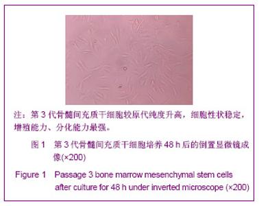
2.1 第3代骨髓间充质干细胞在倒置显微镜下观察 观察可见接种后24 h内,体积较大的单个核细胞贴壁,以后悬浮未贴壁的细胞将不再贴壁。48 h后,已贴壁的细胞开始分裂增殖,细胞呈现出成梭形外观,见图1。72 h后,贴壁细胞体积增大,仍呈梭形,有粗大突起伸出,有些细胞呈三角形或多角形,核居中,有一两个核仁。细胞群聚而形成单独的细胞群。4 d后,细胞铺成单层,可见细胞排列成漩涡状、网状、辐射状。细胞呈梭形、多角形,核圆形或椭圆形。实验所用细胞为第三代骨髓间充质干细胞。"

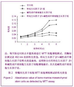
2.2 MTT细胞增殖测试 MTT细胞增殖测试的吸光度值见图2。"
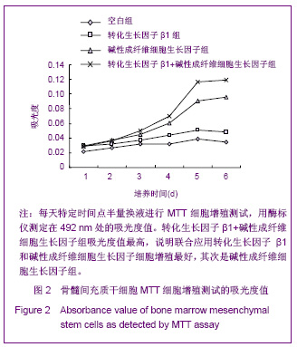
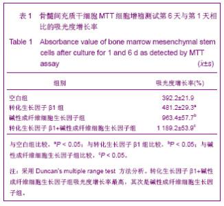
从生长曲线可以看出,一方面,骨髓间充质干细胞生长潜伏期为第1-3天,第3天进入对数增殖期,细胞迅速增殖至第5天开始进入平台期。另一方面,加入生长因子诱导后,出现了不同的增殖情况,转化生长因子β1+碱性成纤维细胞生长因子组细胞到第5天达到生长增殖最高点,并维持在平台期,其次是碱性成纤维细胞生长因子,而转化生长因子β1和空白组细胞增殖能力提高不明显而且提前开始衰减。骨髓间充质干细胞第6天的MTT细胞增殖测试结果见表1。"
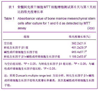

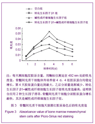
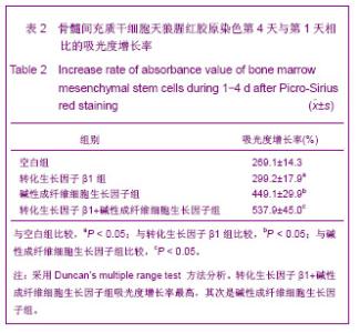
4组中转化生长因子β1+碱性成纤维细胞生长因子组吸光度增长率最高,碱性成纤维细胞生长因子组次之,转化生长因子β1组排第3,空白组最低。转化生长因子β1+碱性成纤维细胞生长因子组和碱性成纤维细胞生长因子组比较、碱性成纤维细胞生长因子组和转化生长因子β1组比较、转化生长因子β1组和空白组比较差异均有显著性意义(P < 0.05)。 2.3 天狼腥红胶原染色结果 天狼腥红胶原染色后的吸光度值见图3。从曲线变化可以看出骨髓间充质干细胞体外培养前4天,4组胶原蛋白均稳定增长,其中转化生长因子β1+碱性成纤维细胞生长因子组增长最快,其次是碱性成纤维细胞生长因子组、转化生长因子β1组、空白组,到第4天胶原蛋白量达到最大,之后分泌量逐渐减少。天狼腥红胶原染色量化峰值第4天结果见表2。 4组中第4天的吸光度值转化生长因子β1+碱性成纤维细胞生长因子组吸光度增长率最高,碱性成纤维细胞生长因子组次之,转化生长因子β1组排第3,空白组最低。转化生长因子β1+碱性成纤维细胞生长因子组和碱性成纤维细胞生长因子组比较、碱性成纤维细胞生长因子组和转化生长因子β1组比较、转化生长因子β1组和空白组比较差异均有显著性意义(P < 0.05)。"
"
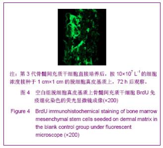
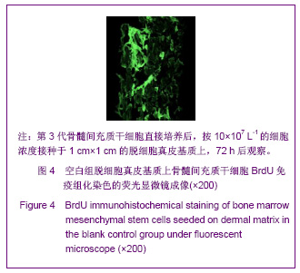
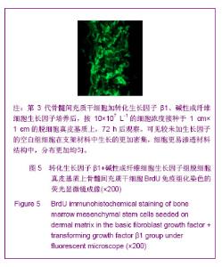
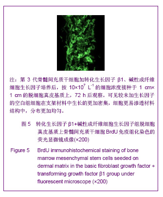
2.4 脱细胞真皮基质上骨髓间充质干细胞细胞BrdU免疫组化染色 脱细胞真皮基质上骨髓间充质干细胞细胞BrdU免疫组化染色后的荧光显微镜成像见图4,5,可以看出碱性成纤维细胞生长因子+转化生长因子β1组较空白组细胞在支架材料中生长的更加密集,细胞更易渗透材料结构中,分布更加均匀。据此可以推断,转化生长因子β1+碱性成纤维细胞生长因子联合使用对骨髓间充质干细胞不仅在细胞增殖上产生明显促进作用,而且在细胞运动爬行排列上起到一定积极作用。"
"
| [1] Nöth U, Rackwitz L, Steinert AF, et al. Cell delivery therapeutics for musculoskeletal regeneration. Adv Drug Deliv Rev. 2010;62(7-8):765-783.[2] Hildebrand KA, Jia F, Woo SL. Response of donor and recipient cells after transplantation of cells to the ligament and tendon. Microsc Res Tech. 2002;58(1):34-38.[3] Watanabe N, Woo SL, Papageorgiou C, et al. Fate of donor bone marrow cells in medial collateral ligament after simulated autologous transplantation. Microsc Res Tech. 2002;58(1):39-44.[4] Liu H, Fan H, Toh SL, et al. A comparison of rabbit mesenchymal stem cells and anterior cruciate ligament fibroblasts responses on combined silk scaffolds. Biomaterials. 2008;29(10):1443-1453.[5] Anitua E, Sánchez M, Orive G, et al. The potential impact of the preparation rich in growth factors (PRGF) in different medical fields. Biomaterials. 2007;28(31):4551-4560.[6] Lee IC, Wang JH, Lee YT, et al. The differentiation of mesenchymal stem cells by mechanical stress or/and co-culture system. Biochem Biophys Res Commun. 2007; 352(1):147-152.[7] Lim JK, Hui J, Li L, et al. Enhancement of tendon graft osteointegration using mesenchymal stem cells in a rabbit model of anterior cruciate ligament reconstruction. Arthroscopy. 2004;20(9):899-910.[8] Ge Z, Goh JC, Lee EH. Selection of cell source for ligament tissue engineering. Cell Transplant. 2005;14(8):573-583.[9] Van Eijk F, Saris DB, Riesle J, et al. Tissue engineering of ligaments: a comparison of bone marrow stromal cells, anterior cruciate ligament, and skin fibroblasts as cell source. Tissue Eng. 2004;10(5-6):893-903.[10] Schmidt CC, Georgescu HI, Kwoh CK, et al. Effect of growth factors on the proliferation of fibroblasts from the medial collateral and anterior cruciate ligaments. J Orthop Res. 1995; 13(2):184-190.[11] Tang Y, Chen HH, Li SM, et al. Zhongguo Xiufu Chongjian Waike Zazhi. 2001;15(3):158-161. 唐毅,陈鸿辉,李斯明等.透明质酸联合碱性成纤维细胞生长因子对韧带细胞增殖的影响[J].中国修复重建外科杂志,2001, 15(3): 158-161.[12] Kim MK, Niyibizi C. Interaction of TGF-beta1 and rhBMP-2 on human bone marrow stromal cells cultured in collagen gel matrix. Yonsei Med J. 2001;42(3):338-344.[13] Wang YT, Zheng QX, Guo XD, et al. Zhonghua Chongjian Zazhi. 2005;22(3);361-363. 王运涛,郑启新,郭晓东,等.外源性转化生长因子-β1对骨髓间充质干细胞TβRⅠ、TβRⅡ表达的影响[J].中华实验外科杂志, 2005,22(3):361-363.[14] Houghton J, Stoicov C, Nomura S, et al. Gastric cancer originating from bone marrow-derived cells. Science. 2004; 306(5701):1568-1571.[15] Burns TC, Ortiz-González XR, Gutiérrez-Pérez M, et al. Thymidine analogs are transferred from prelabeled donor to host cells in the central nervous system after transplantation: a word of caution. Stem Cells. 2006;24(4):1121-1127.[16] Madhavan K,Belchenko D,Motta A,et al. Evaluation of COB-position and crosslinking effects on collagen-based composite constructs. Acta Biomater. 2010.6(4):1413-1422.[17] Sundararaghavan HG, Monteiro GA, Lapin NA, et al. Genipin-induced changes in collagen gels: correlation of mechanical properties to fluorescence. J Biomed Mater Res A. 2008;87(2):308-320.[18] Xiao J, Duan H, Liu Z, et al. Construction of the recellularized corneal stroma using porous acellular corneal scaffold. Biomaterials. 2011;32(29):6962-6971.[19] Akino K, Mineta T, Fukui M, et al. Bone morphogenetic protein-2 regulates proliferation of human mesenchymal stem cells. Wound Repair Regen. 2003;11(5):354-360.[20] Majima T, Marchuk LL, Sciore P, et al. Compressive compared with tensile loading of medial collateral ligament scar in vitro uniquely influences mRNA levels for aggrecan, collagen type II, and collagenase. J Orthop Res. 2000;18(4): 524-531.[21] Tsutsumi S, Shimazu A, Miyazaki K, et al. Retention of multilineage differentiation potential of mesenchymal cells during proliferation in response to FGF. Biochem Biophys Res Commun. 2001;288(2):413-419.[22] Chan BP, Chan KM, Maffulli N, et al. Effect of basic fibroblast growth factor. An in vitro study of tendon healing. Clin Orthop Relat Res. 1997;(342):239-247.[23] Majumdar MK, Banks V, Peluso DP, et al. Isolation, characterization, and chondrogenic potential of human bone marrow-derived multipotential stromal cells. J Cell Physiol. 2000;185(1):98-106.[24] Marui T, Niyibizi C, Georgescu HI, et al. Effect of growth factors on matrix synthesis by ligament fibroblasts. J Orthop Res. 1997;15(1):18-23.[25] Murphy PG, Loitz BJ, Frank CB, et al. Influence of exogenous growth factors on the expression of plasminogen activators by explants of normal and healing rabbit ligaments. Biochem Cell Biol. 1993;71(11-12):522-529.[26] DesRosiers EA, Yahia L, Rivard CH. Proliferative and matrix synthesis response of canine anterior cruciate ligament fibroblasts submitted to combined growth factors. J Orthop Res. 1996;14(2):200-208.[27] Murphy PG, Hart DA. Influence of exogenous growth factors on the expression of plasminogen activators and plasminogen activator inhibitors by cells isolated from normal and healing rabbit ligaments. J Orthop Res. 1994;12(4):564-575.[28] Lange C, Cakiroglu F, Spiess AN, et al. Accelerated and safe expansion of human mesenchymal stromal cells in animal serum-free medium for transplantation and regenerative medicine. J Cell Physiol. 2007;213(1):18-26.[29] Ma XH, Gao CQ, Li BJ, et al. The empirical study on paracrine communication of bone marrow mesenchymal stem cells. Zhonghua Yi Xue Za Zhi. 2010;90(7):496-498.[30] Emin N, Koç A, Durkut S, et al. Engineering of rat articular cartilage on porous sponges: effects of tgf-beta 1 and microgravity bioreactor culture. Artif Cells Blood Substit Immobil Biotechnol. 2008;36(2):123-137.[31] Chua KH, Aminuddin BS, Fuzina NH, et al. Interaction between insulin-like growth factor-1 with other growth factors in serum depleted culture medium for human cartilage engineering. Med J Malaysia. 2004;59 Suppl B:7-8.[32] Srouji S, Blumenfeld I, Rachmiel A, et al. Bone defect repair in rat tibia by TGF-beta1 and IGF-1 released from hydrogel scaffold. Cell Tissue Bank. 2004;5(4):223-230.[33] Kawaguchi H, Nakamura K, Tabata Y, et al. Acceleration of fracture healing in nonhuman primates by fibroblast growth factor-2. J Clin Endocrinol Metab. 2001;86(2):875-880.[34] Richmon JD, Sage AB, Shelton E, et al. Effect of growth factors on cell proliferation, matrix deposition, and morphology of human nasal septal chondrocytes cultured in monolayer. Laryngoscope. 2005;115(9):1553-1560.[35] Luo Q, Song G, Song Y, et al. Indirect co-culture with tenocytes promotes proliferation and mRNA expression of tendon/ligament related genes in rat bone marrow mesenchymal stem cells. Cytotechnology. 2009;61(1-2):1-10. |
| [1] | Pu Rui, Chen Ziyang, Yuan Lingyan. Characteristics and effects of exosomes from different cell sources in cardioprotection [J]. Chinese Journal of Tissue Engineering Research, 2021, 25(在线): 1-. |
| [2] | Wu Xun, Meng Juanhong, Zhang Jianyun, Wang Liang. Concentrated growth factors in the repair of a full-thickness condylar cartilage defect in a rabbit [J]. Chinese Journal of Tissue Engineering Research, 2021, 25(8): 1166-1171. |
| [3] | Zhang Xiumei, Zhai Yunkai, Zhao Jie, Zhao Meng. Research hotspots of organoid models in recent 10 years: a search in domestic and foreign databases [J]. Chinese Journal of Tissue Engineering Research, 2021, 25(8): 1249-1255. |
| [4] | Wang Zhengdong, Huang Na, Chen Jingxian, Zheng Zuobing, Hu Xinyu, Li Mei, Su Xiao, Su Xuesen, Yan Nan. Inhibitory effects of sodium butyrate on microglial activation and expression of inflammatory factors induced by fluorosis [J]. Chinese Journal of Tissue Engineering Research, 2021, 25(7): 1075-1080. |
| [5] | Wang Xianyao, Guan Yalin, Liu Zhongshan. Strategies for improving the therapeutic efficacy of mesenchymal stem cells in the treatment of nonhealing wounds [J]. Chinese Journal of Tissue Engineering Research, 2021, 25(7): 1081-1087. |
| [6] | Liao Chengcheng, An Jiaxing, Tan Zhangxue, Wang Qian, Liu Jianguo. Therapeutic target and application prospects of oral squamous cell carcinoma stem cells [J]. Chinese Journal of Tissue Engineering Research, 2021, 25(7): 1096-1103. |
| [7] | Xie Wenjia, Xia Tianjiao, Zhou Qingyun, Liu Yujia, Gu Xiaoping. Role of microglia-mediated neuronal injury in neurodegenerative diseases [J]. Chinese Journal of Tissue Engineering Research, 2021, 25(7): 1109-1115. |
| [8] | Li Shanshan, Guo Xiaoxiao, You Ran, Yang Xiufen, Zhao Lu, Chen Xi, Wang Yanling. Photoreceptor cell replacement therapy for retinal degeneration diseases [J]. Chinese Journal of Tissue Engineering Research, 2021, 25(7): 1116-1121. |
| [9] | Jiao Hui, Zhang Yining, Song Yuqing, Lin Yu, Wang Xiuli. Advances in research and application of breast cancer organoids [J]. Chinese Journal of Tissue Engineering Research, 2021, 25(7): 1122-1128. |
| [10] | Wang Shiqi, Zhang Jinsheng. Effects of Chinese medicine on proliferation, differentiation and aging of bone marrow mesenchymal stem cells regulating ischemia-hypoxia microenvironment [J]. Chinese Journal of Tissue Engineering Research, 2021, 25(7): 1129-1134. |
| [11] | Zeng Yanhua, Hao Yanlei. In vitro culture and purification of Schwann cells: a systematic review [J]. Chinese Journal of Tissue Engineering Research, 2021, 25(7): 1135-1141. |
| [12] | Kong Desheng, He Jingjing, Feng Baofeng, Guo Ruiyun, Asiamah Ernest Amponsah, Lü Fei, Zhang Shuhan, Zhang Xiaolin, Ma Jun, Cui Huixian. Efficacy of mesenchymal stem cells in the spinal cord injury of large animal models: a meta-analysis [J]. Chinese Journal of Tissue Engineering Research, 2021, 25(7): 1142-1148. |
| [13] | Hou Jingying, Yu Menglei, Guo Tianzhu, Long Huibao, Wu Hao. Hypoxia preconditioning promotes bone marrow mesenchymal stem cells survival and vascularization through the activation of HIF-1α/MALAT1/VEGFA pathway [J]. Chinese Journal of Tissue Engineering Research, 2021, 25(7): 985-990. |
| [14] | Shi Yangyang, Qin Yingfei, Wu Fuling, He Xiao, Zhang Xuejing. Pretreatment of placental mesenchymal stem cells to prevent bronchiolitis in mice [J]. Chinese Journal of Tissue Engineering Research, 2021, 25(7): 991-995. |
| [15] | Liang Xueqi, Guo Lijiao, Chen Hejie, Wu Jie, Sun Yaqi, Xing Zhikun, Zou Hailiang, Chen Xueling, Wu Xiangwei. Alveolar echinococcosis protoscolices inhibits the differentiation of bone marrow mesenchymal stem cells into fibroblasts [J]. Chinese Journal of Tissue Engineering Research, 2021, 25(7): 996-1001. |
| Viewed | ||||||
|
Full text |
|
|||||
|
Abstract |
|
|||||