Chinese Journal of Tissue Engineering Research ›› 2020, Vol. 24 ›› Issue (35): 5600-5606.doi: 10.3969/j.issn.2095-4344.2919
Previous Articles Next Articles
Hoxa9 silencing promotes tibial fracture healing by regulating osteogenic differentiation
Zhang Chao, Li Xingyong, Ma Guifu, Pu Xingyu, Luo Wenyuan
Department of Orthopedics, Gansu Provincial Hospital, Lanzhou 730000, Gansu Province, China
-
Received:2019-11-19Revised:2019-11-22Accepted:2020-02-12Online:2020-12-18Published:2020-10-16 -
Contact:Luo Wenyuan, Chief physician, Department of Orthopedics, Gansu Provincial Hospital, Lanzhou 730000, Gansu Province, China -
About author:Zhang Chao, Master, Attending physician, Department of Orthopedics, Gansu Provincial Hospital, Lanzhou 730000, Gansu Province, China
CLC Number:
Cite this article
Zhang Chao, Li Xingyong, Ma Guifu, Pu Xingyu, Luo Wenyuan. Hoxa9 silencing promotes tibial fracture healing by regulating osteogenic differentiation[J]. Chinese Journal of Tissue Engineering Research, 2020, 24(35): 5600-5606.
share this article
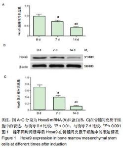
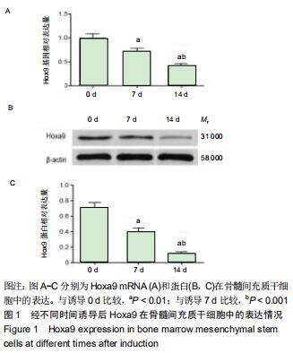
1.5.2 总RNA提取和实时定量PCR(qRT-PCR)检测 按照Trizol试剂盒说明书,提取C3H10T1/2细胞和胫骨中的总RNA,借助Nano核酸分析仪测量提取的RNA的浓度和纯度,将反转录获得的DNA(cDNA)进行qRT-PCR,各组样本设3个复孔。qRT-PCR条件:94 ℃下4 min预变性,94 ℃下30 s变性,60 ℃下30 s退火和延伸,然后进行40个循环。根据相对定量2-??Ct法评价实验中Hoxa9、骨桥蛋白、骨钙素、Runx2及胶原蛋白Ⅰ mRNA的相对表达量。表1列出了实验中所有特异性引物。 "
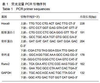
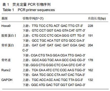
2.1.1 不同诱导时相骨髓间充质干细胞中Hoxa9基因表达情况 实验分别对成骨诱导后0,7及14 d骨髓间充质干细胞中Hoxa9 mRNA及蛋白水平进行检测。结果发现:随着诱导时间的延长Hoxa9 mRNA相对表达在降低,且各个诱导时相的差异有显著性意义(P < 0.01);Western Blot结果显示Hoxa9蛋白相对表达也随着诱导时间的延长呈明显的降低,且各个诱导时相的差异也有显著性意义(P < 0.01),见图1。 "
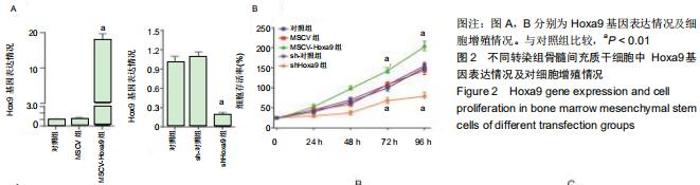
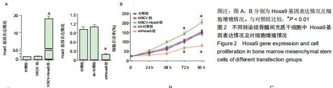
2.1.2 不同转染组骨髓间充质干细胞中Hoxa9 mRNA相对表达及细胞增殖变化 qRT-PCR结果发现:与对照组及MSCV阴性转染组相比较,MSCV-Hoxa9转染组中细胞内Hoxa9 mRNA水平显著上调(P < 0.01);与对照组及sh-对照组相比较,用shHoxa9转染的骨髓间充质干细胞中Hoxa9的表达明显降低(P < 0.01)。CCK8检测结果表明:与对照组相比较,MSCV-Hoxa9组骨髓间充质干细胞的增殖显著增高(P < 0.01);相反,shHoxa9组骨髓间充质干细胞的增殖明显被抑制(P < 0.01),见图2。 "

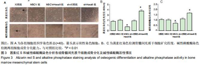
2.1.3 Hoxa9对骨髓间充质干细胞成骨分化的影响 骨髓间充质干细胞分组及处理情况同1.4.1,成骨诱导分化培养基诱导培养14 d,茜素红染色检测骨髓间充质干细胞矿化程度,碱性磷酸酶染色检测两组细胞成骨分化能力。结果发现:MSCV-Hoxa9组骨髓间充质干细胞中钙化结节的数量显著低于对照组及各阴性转染组(P < 0.01);碱性磷酸酶染色染色结果显示MSCV-Hoxa9组成骨分化能力低于对照组及各阴性转染组,碱性磷酸酶活性定量分析结果显示,MSCV-Hoxa9组成骨分化能力明显低于对照组及各阴性转染组(P < 0.01)。而shHoxa9组骨髓间充质干细胞的钙化结节数、成骨分化能力及碱性磷酸酶活性显著高于对照组及各阴性转染组(P < 0.01)。表明Hoxa9对骨髓间充质干细胞的钙化和碱性磷酸酶表达均具有调节作用,见图3。 "

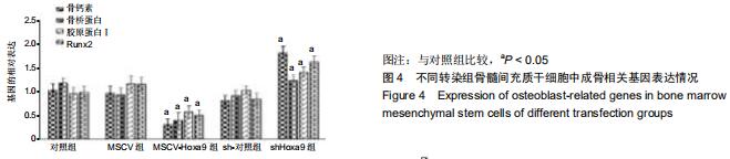
2.1.4 不同转染组骨髓间充质干细胞中成骨分化相关基因及蛋白表达情况 qRT-PCR检测结果显示,与对照组及各阴性转染组相比较,MSCV-Hoxa9组细胞中骨桥蛋白、骨钙素、Runx2和胶原蛋白Ⅰ基因的mRNA相对表达显著下调(P < 0.05);而shHoxa9组骨髓间充质干细胞骨桥蛋白、骨钙素、Runx2和胶原蛋白Ⅰ基因的mRNA相对表达明显高于对照组及各阴性转染组,且差异也具有显著性意义(P < 0.05),见图4。 "
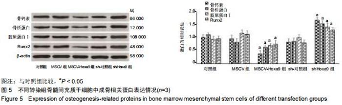
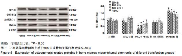
Western blot检测结果显示:在成骨分化过程中,与对照组及各阴性转染细胞相比,shHoxa9转染的骨髓间充质干细胞中成骨相关蛋白骨桥蛋白、骨钙素、Runx2及胶原蛋白Ⅰ的相对表达显著上调(P < 0.05),而MSCV-Hoxa9组细胞中骨桥蛋白、骨钙素、Runx2和胶原蛋白Ⅰ蛋白的相对表达显著下调(P < 0.05),见图5。 "
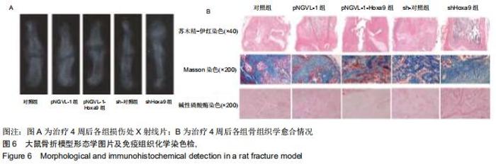
2.2.1 Hoxa9基因对大鼠胫骨骨折愈合的影响 为研究Hoxa9在体内的功效,将大鼠胫骨骨折模型用于实验。X射线片检查显示:对照组大鼠骨折线清晰;与对照组相比,pNGVL-1-Hoxa9组骨折线更明显;shHoxa9组的骨折线在治疗4周后几乎消失,见图6A。 苏木精-伊红染色显示:对照组中纤维性愈合损伤组织和骨性愈合损伤组织增加,而pNGVL-1-Hoxa9组中骨性愈合损伤组织较对照组更加明显,见图6B。治疗4周后,经shHoxa9组大鼠显示骨的大小和数量明显增加,见图6B。用Masson和碱性磷酸酶染色检测组织间胶原纤维和愈合损伤组织内的软骨。如图6B所示,在骨折治疗后28 d,shHoxa9组的胶原纤维面积较大,而软骨面积明显小于对照组,与苏木精-伊红染色结果一致。 "

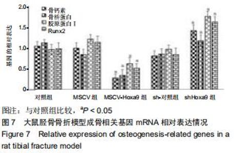
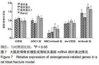
2.2.2 各组体内成骨相关基因表达情况 实验进一步评估了治疗4周后Hoxa9对愈合损伤组织中成骨相关基因mRNA的影响。由愈合损伤组织的RT-PCR结果表明:与对照组及各阴性组相比较,shHoxa9组骨桥蛋白、骨钙素、Runx2和胶原蛋白Ⅰ基因的mRNA相对表达明显上调(P < 0.05);而MSCV-Hoxa9组骨桥蛋白、骨钙素、Runx2和胶原蛋白Ⅰ基因的mRNA相对表达显著下调(P < 0.05),见图7。 "
|
[1] MELING T, HARBOE K, SØREIDE K. Incidence of traumatic long-bone fractures requiring in-hospital management: a prospective age- and gender-specific analysis of 4890 fractures. Injury. 2009;40(11):1212-1219.
[2] DUAN X, AL-QWBANI M, ZENG Y, et al. Intramedullary nailing for tibial shaft fractures in adults. Cochrane Database Syst Rev. 2012;1(1):CD008241.
[3] DAI J, LI L, JIANG C, et al. Bone Morphogenetic Protein for the Healing of Tibial Fracture: A Meta-Analysis of Randomized Controlled Trials. PLoS One. 2015;10(10): e0141670.
[4] 徐磊,夏江.胫骨平台后侧骨折的诊断及手术治疗进展[J].沈阳医学院学报,2016,18(6):494-497.
[5] CARÈ A, FELICETTI F, MECCIA E, et al. HOXB7: a key factor for tumor-associated angiogenic switch.Cancer Res. 2001; 61(17):6532-6539.
[6] SHAH N, SUKUMAR S. The Hox genes and their roles in oncogenesis. Nat Rev Cancer. 2010;10(5):361-371.
[7] ABDEL-FATTAH R, XIAO A, BOMGARDNER D, et al. Differential expression of HOX genes in neoplastic and non-neoplastic human astrocytes. J Pathol. 2006;209(1): 15-24.
[8] GOODMAN FR. Limb malformations and the human HOX genes. Am J Med Genet. 2002;112(3):256-265.
[9] COLLINS C, WANG J, MIAO H, et al. C/EBPα is an essential collaborator in Hoxa9/Meis1-mediated leukemogenesis. Proc Natl Acad Sci U S A. 2014;111(27):9899-9904. [10] POJO M, GONÇALVES CS, XAVIER-MAGALHÃES A, et al. A transcriptomic signature mediated by HOXA9 promotes human glioblastoma initiation, aggressiveness and resistance to temozolomide. Oncotarget. 2015;6(10):7657-7674.
[11] HWANG JA, LEE BB, KIM Y, et al. HOXA9 inhibits migration of lung cancer cells and its hypermethylation is associated with recurrence in non-small cell lung cancer. Mol Carcinog. 2015;54(11):E72-E80.
[12] XU PP, ZHOU D, YAN GJ, et al. Correlation of miR-181a and three HOXA genes as useful biomarkers in acute myeloid leukemia. Int J Lab Hematol. 2019;42(4):16-22.
[13] LIU X, LIU X, LIU W, et al. HOXA9 transcriptionally regulates the EPHB4 receptor to modulate trophoblast migration and invasion. Placenta. 2017;51:38-48.
[14] 马明月.应用胚胎干细胞模型研究环境内分泌干扰物胚胎毒性的展望[J].沈阳医学院学报,2016,18(2):65-67,73.
[15] MOHR S, DOEBELE C, COMOGLIO F, et al. Hoxa9 and Meis1 Cooperatively Induce Addiction to Syk Signaling by Suppressing miR-146a in Acute Myeloid Leukemia. Cancer Cell. 2017;31(4):549-562.
[16] 杨晓红.miR-705对绝经后骨质疏松小鼠的骨髓间充质干细胞分化功能异常的调控作用[D].西安:第四军医大学,2013.
[17] HANDOOL KO, IBRAHIM SM, KAKA U, et al. Optimization of a closed rat tibial fracture model. J Exp Orthop. 2018;5(1):13.
[18] 席梦莹,李汶洋,张琳,等.鸢尾素促进大鼠胫骨骨折愈合的研究[J/OL].重庆医科大学学报,2019:1-7.
[19] 黄弘轩,张姝江,白波.调控骨髓间充质干细胞成软骨分化的研究进展[J].中华关节外科杂志(电子版),2017,11(6):655-660.
[20] 赵成礼.人工关节置换治疗老年股骨转子间骨折的优势与不足[J].中国组织工程研究,2019,23(24):3868-3874.
[21] 陈海涛,安玉光.桡骨骨折模型兔骨折愈合:特异性环氧化酶2抑制剂与非特异性环氧化酶抑制剂的比较[J].中国组织工程研究, 2019,23(15):2385-2390.
[22] 胡博森.矢车菊素-3-O-葡萄糖苷经MAPK信号通路调节成骨细胞增殖和分化的作用研究[D].沈阳:沈阳医学院,2019.
[23] 姜文涛,梅伟,王庆德,等.淫羊藿苷对成骨细胞增殖、分化及矿化的影响[J].中华实验外科杂志,2017,34(3):446-448.
[24] 鲍冲,朱扬,邓洲铭,等.成纤维生长因子受体2对小鼠骨髓间充质干细胞成骨分化能力的影响[J].中华实验外科杂志,2019,36(3): 502-505.
[25] 杨礼庆,董成建,朱姝.成骨分化与修复调控转录因子Runx2在股骨头坏死组织中的表达:非随机平行对照临床观察试验方案[J].中国组织工程研究,2016,20(17):2569-2574.
[26] VIJAY N, BANDANA AV, SU C, et al. Hoxa9 and Hoxa10 induce CML myeloid blast crisis development through activation of Myb expression. Oncotarget. 2017;8(58): 98853-98864.
[27] HUANG Y, SITWALA K, BRONSTEIN J, et al. Identification and characterization of Hoxa9 binding sites in hematopoietic cells. Blood. 2012;119(2):388-398.
[28] KO SY, NAORA H. HOXA9 promotes homotypic and heterotypic cell interactions that facilitate ovarian cancer dissemination via its induction of P-cadherin. Mol Cancer. 2014;13:170.
[29] 李琳.HOXB4、PRDM16及HOXA9基因在急性髓系白血病中的表达及其临床意义[D].青岛:青岛大学,2016.
[30] 何慧慧.斑马鱼造血系统发育过程中Hox基因的表达模式研究[D].上海:上海海洋大学,2012. |
| [1] | Liu Cong, Liu Su. Molecular mechanism of miR-17-5p regulation of hypoxia inducible factor-1α mediated adipocyte differentiation and angiogenesis [J]. Chinese Journal of Tissue Engineering Research, 2021, 25(7): 1069-1074. |
| [2] | Wang Shiqi, Zhang Jinsheng. Effects of Chinese medicine on proliferation, differentiation and aging of bone marrow mesenchymal stem cells regulating ischemia-hypoxia microenvironment [J]. Chinese Journal of Tissue Engineering Research, 2021, 25(7): 1129-1134. |
| [3] | Hou Jingying, Yu Menglei, Guo Tianzhu, Long Huibao, Wu Hao. Hypoxia preconditioning promotes bone marrow mesenchymal stem cells survival and vascularization through the activation of HIF-1α/MALAT1/VEGFA pathway [J]. Chinese Journal of Tissue Engineering Research, 2021, 25(7): 985-990. |
| [4] | Liang Xueqi, Guo Lijiao, Chen Hejie, Wu Jie, Sun Yaqi, Xing Zhikun, Zou Hailiang, Chen Xueling, Wu Xiangwei. Alveolar echinococcosis protoscolices inhibits the differentiation of bone marrow mesenchymal stem cells into fibroblasts [J]. Chinese Journal of Tissue Engineering Research, 2021, 25(7): 996-1001. |
| [5] | Geng Yao, Yin Zhiliang, Li Xingping, Xiao Dongqin, Hou Weiguang. Role of hsa-miRNA-223-3p in regulating osteogenic differentiation of human bone marrow mesenchymal stem cells [J]. Chinese Journal of Tissue Engineering Research, 2021, 25(7): 1008-1013. |
| [6] | Lun Zhigang, Jin Jing, Wang Tianyan, Li Aimin. Effect of peroxiredoxin 6 on proliferation and differentiation of bone marrow mesenchymal stem cells into neural lineage in vitro [J]. Chinese Journal of Tissue Engineering Research, 2021, 25(7): 1014-1018. |
| [7] | Zhu Xuefen, Huang Cheng, Ding Jian, Dai Yongping, Liu Yuanbing, Le Lixiang, Wang Liangliang, Yang Jiandong. Mechanism of bone marrow mesenchymal stem cells differentiation into functional neurons induced by glial cell line derived neurotrophic factor [J]. Chinese Journal of Tissue Engineering Research, 2021, 25(7): 1019-1025. |
| [8] | Pei Lili, Sun Guicai, Wang Di. Salvianolic acid B inhibits oxidative damage of bone marrow mesenchymal stem cells and promotes differentiation into cardiomyocytes [J]. Chinese Journal of Tissue Engineering Research, 2021, 25(7): 1032-1036. |
| [9] | Li Cai, Zhao Ting, Tan Ge, Zheng Yulin, Zhang Ruonan, Wu Yan, Tang Junming. Platelet-derived growth factor-BB promotes proliferation, differentiation and migration of skeletal muscle myoblast [J]. Chinese Journal of Tissue Engineering Research, 2021, 25(7): 1050-1055. |
| [10] | Yang Weiqiang, Ding Tong, Yang Weike, Jiang Zhengang. Combined variable stress plate internal fixation affects changes of bone histiocyte function and bone mineral density at the fractured end of goat femur [J]. Chinese Journal of Tissue Engineering Research, 2021, 25(6): 890-894. |
| [11] | Liu Bo, Chen Xianghe, Yang Kang, Yu Huilin, Lu Pengcheng. Mechanism of DNA methylation in exercise intervention for osteoporosis [J]. Chinese Journal of Tissue Engineering Research, 2021, 25(5): 791-797. |
| [12] | Ma Zetao, Zeng Hui, Wang Deli, Weng Jian, Feng Song. MicroRNA-138-5p regulates chondrocyte proliferation and autophagy [J]. Chinese Journal of Tissue Engineering Research, 2021, 25(5): 674-678. |
| [13] | Wang Yujiao, Liu Dan, Sun Song, Sun Yong. Biphasic calcium phosphate loaded with advanced platelet rich fibrin can promote the activity of rabbit bone marrow mesenchymal stem cells [J]. Chinese Journal of Tissue Engineering Research, 2021, 25(4): 504-509. |
| [14] | Chen Junyi, Wang Ning, Peng Chengfei, Zhu Lunjing, Duan Jiangtao, Wang Ye, Bei Chaoyong. Decalcified bone matrix and lentivirus-mediated silencing of P75 neurotrophin receptor transfected bone marrow mesenchymal stem cells to construct tissue-engineered bone [J]. Chinese Journal of Tissue Engineering Research, 2021, 25(4): 510-515. |
| [15] | Zhou Jihui, Yao Meng, Wang Yansong, Li Xinzhi, Zhou You, Huang Wei, Chen Wenyao. Influence of novel nanoscaffolds on biological behaviors of neural stem cells and the related gene expression [J]. Chinese Journal of Tissue Engineering Research, 2021, 25(4): 532-536. |
| Viewed | ||||||
|
Full text |
|
|||||
|
Abstract |
|
|||||