Chinese Journal of Tissue Engineering Research ›› 2020, Vol. 24 ›› Issue (9): 1464-1470.doi: 10.3969/j.issn.2095-4344.2516
Previous Articles Next Articles
Microscope-assisted versus traditional anterior cervical approach for cervical spondylopathy: a meta-analysis
Luo Haitao, Cheng Zujue, Lü Shigang, Xiao Juexian, He Wei, Huang Kai, Fan Yanghua, Zhu Xingen
- Department of Neurosurgery, Second Affiliated Hospital of Nanchang University, Nanchang 330006, Jiangxi Province, China
-
Received:2019-08-10Revised:2019-08-14Accepted:2019-10-09Online:2020-03-28Published:2020-02-13 -
Contact:Cheng Zujue, Professor, Chief physician, Department of Neurosurgery, Second Affiliated Hospital of Nanchang University, Nanchang 330006, Jiangxi Province, China -
About author:Luo Haitao, Master, Department of Neurosurgery, Second Affiliated Hospital of Nanchang University, Nanchang 330006, Jiangxi Province, China -
Supported by:the Science and Technology Program of Education Department of Jiangxi Province, No. 60159; the Project of Health and Family Planning Commission of Jiangxi Province, No. 20175224; the Project of Development Center for Medical and Health Science and Technology of the Ministry of Health of China, No. W2014ZT268; the Project of Health and Family Planning Commission of Jiangxi Province, No. 20165215
CLC Number:
Cite this article
Luo Haitao, Cheng Zujue, Lü Shigang, Xiao Juexian, He Wei, Huang Kai, Fan Yanghua, Zhu Xingen. Microscope-assisted versus traditional anterior cervical approach for cervical spondylopathy: a meta-analysis[J]. Chinese Journal of Tissue Engineering Research, 2020, 24(9): 1464-1470.
share this article
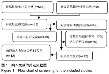
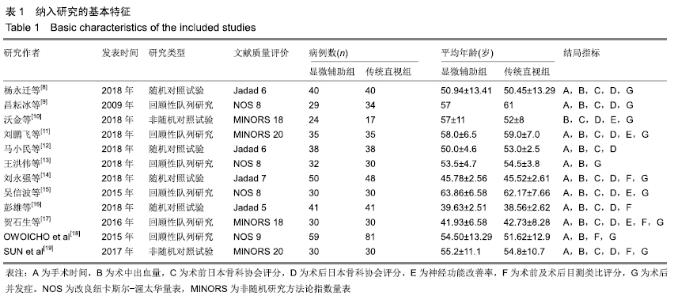
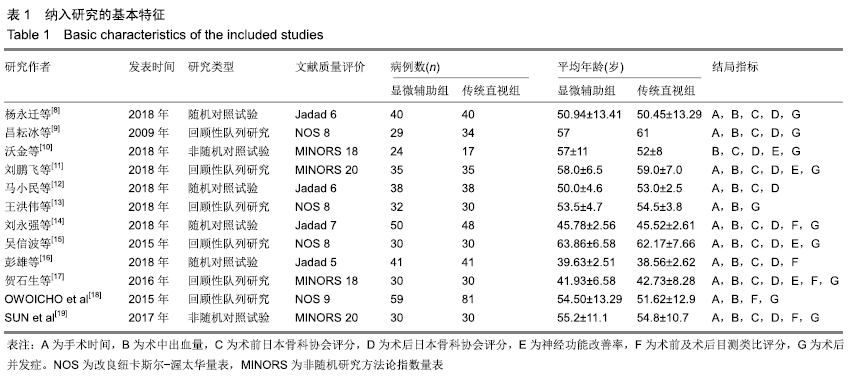
2.1 检索结果 根据上述检索策略检索出相关文献667篇,阅读题目及摘要后筛选出461篇,经2名研究者反复阅读全文后复筛选出42篇,排除综述、个案报告、重复发表的研究、数据不规范的研究,最终纳入文献12篇[8-19],共收集病例892例,其中显微辅助组438例,传统直视组454例,各样本量从17-81例不等,见图1。纳入文献的基本情况详见表1。 "
"
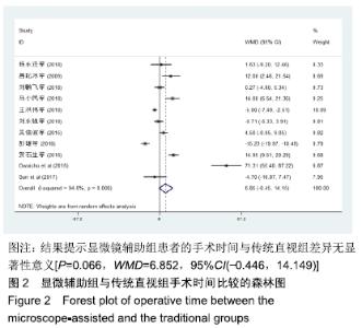
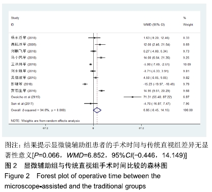
2.2 Meta分析结果 2.2.1 手术时间 共11篇文献报道了手术时间[8-9,11-19],经异质性检验各项研究异质性明显(P < 0.001,I2=94.6%),故采用随机效应模型分析。其中显微镜辅助组414例,传统直视组437例,结果提示显微镜辅助组患者的手术时间与传统直视组差异无显著性意义[P=0.066,WMD=6.852,95%CI(-0.446,14.149)],见图2。 "
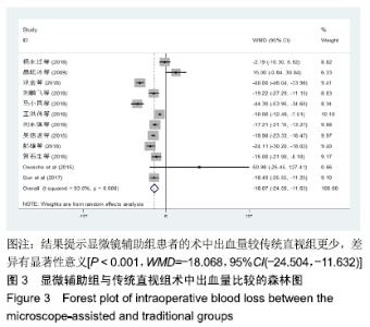
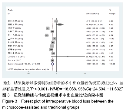
2.2.2 术中出血量 12篇文献均报道术中出血量[8-19],经异质性检验各项研究异质性明显(P < 0.001,I2=93.0%),故采用随机效应模型分析。其中显微镜辅助组438例,传统直视组454例,结果显示与传统直视组相比,显微镜辅助组中患者的术中出血量更少,且差异有显著性意义[P < 0.001,WMD=-18.068,95%CI(-24.504,-11.632)],见图3。 "
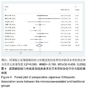
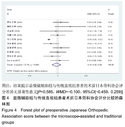
2.2.3 术前日本骨科协会评分 10篇文献报道术前日本骨科协会评分[8-12,14-17,19],经异质性检验各项研究异质性明显(P < 0.001,I2=72.3%),故采用随机效应模型分析。其中显微镜辅助组347例,传统直视组343例,结果显示显微辅助组与传统直视组患者的术前日本骨科协会评分差异无显著性意义,提示2组患者术前日本骨科协会评分同质性好[P=0.585,WMD=-0.100,95%CI(-0.459,0.259)],见图4。 "
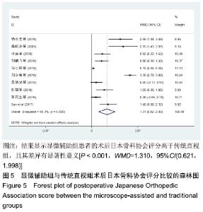
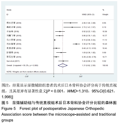
2.2.4 术后日本骨科协会评分 10篇文献报道术后日本骨科协会评分[8-12,14-17,19],经异质性检验各项研究异质性明显(P < 0.001,I2=95.3%),说明各研究间存在统计学异质性,故采用随机效应模型进行分析。其中显微辅助组患者347例,传统直视组343例,结果显示与传统直视组相比,显微辅助组更能提高患者的术后日本骨科协会评分,且差异有显著性意义[P < 0.001,WMD=1.310,95%CI(0.621,1.998)],见图5。 "
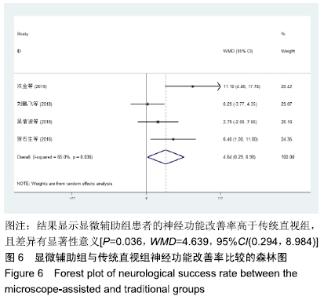
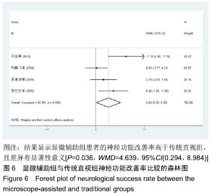
2.2.5 神经功能改善率 4篇文献报道了神经功能改善率的情况[10-11,15,17],经异质性检验各项研究存在明显异质性(P < 0.036,I2=65.0%),故采用随机效应模型分析。其中显微镜辅助组119例,传统直视组112例,结果显示与传统直视组相比,显微辅助组能显著提高患者的神经功能改善率,且差异有显著性意义[P=0.036,WMD=4.639,95%CI(0.294,8.984)],见图6。 "
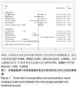
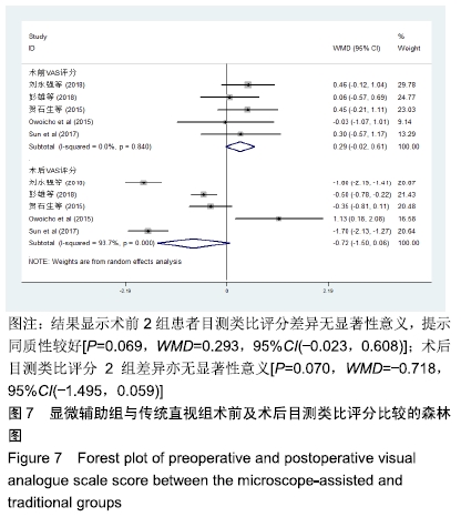
2.2.6 术前及术后目测类比评分 5篇文献报道了术前及术后目测类比评分情况[14,16-19],经异质性检验证明存在明显异质性(P < 0.001,I2=93.7%),故采用随机效应模型分析。其中显微镜辅助组患者210例,传统直视组230例,结果显示术前2组患者目测类比评分差异无显著性意义,提示同质性较好[P=0.069,WMD=0.293,95%CI(-0.023,0.608)];术后目测类比评分2组差异亦无显著性意义[P=0.070,WMD=-0.718,95%CI(-1.495,0.059)],见图7。 "
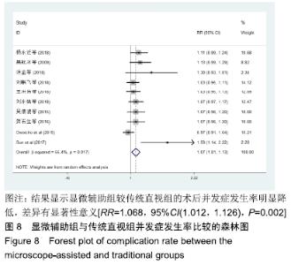
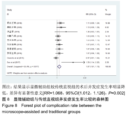
2.2.7 并发症发生率 10篇文献报道了并发症发生情 况[8-11,13-15,17-19],经异质性检验各项研究有明显异质性(P < 0.017,I2=55.4%),故采用随机效应模型分析。其中显微镜辅助组359例,传统直视组375例,结果显示与传统直视组相比,显微辅助组患者的术后并发症发生率明显降低,且差异有显著性意义[RR=1.068,95%CI(1.012,1.126),P=0.002],见图8。 "
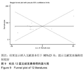
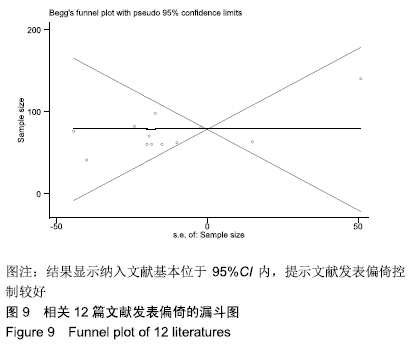
2.3 敏感性分析 敏感性分析时,对单项文献进行逐一排除,对剩余文献进行合并,其结果相对整体合并时未见大的变动,提示排除任一文献对整体结果无明显影响。 2.4 发表偏倚 使用Begg’s检验对所纳入文献术中出血量的发表偏倚做以简要分析,根据漏斗图形状及散点分布情况,显示纳入文献基本位于95%CI内,提示文献发表偏倚控制较好,见图9。 "
| [1] HOFFMAN H, LEE SI, GARST JH, et al. Use of multivariate linear regression and support vector regression to predict functional outcome after surgery for cervical spondylotic myelopathy. J Clin Neurosci. 2015;22 (9): 1444-1449. [2] EDWARDS CC 2ND, RIEW KD, ANDERSON PA, et al. Cervical myelopathy. current diagnostic and treatment strategies. Spine J. 2003; 3(1): 68-81. [3] CLOWARD RB. The anterior approach for removal of ruptured cervical disks. J Neurosurg. 1958;15(6): 602-617. [4] KORINTH MC. Treatment of cervical degenerative disc disease- current status and trends. Zentralbl Neurochir. 2008; 69(3): 113-124. [5] HANKINSON HL, WILSON CB. Use of the operating microscope in anterior cervical discectomy without fusion. J Neurosurg. 1975; 43(4):452-456. [6] JADAD AR, MOORE RA, CARROLL D, et al. Assessing the quality of reports of randomized cIinical trials: is blinding necessary. Control Clin Trials. 1996;17(1):1-12. [7] SLIM K, NINI E, FORESTIER D, et al. Methodological index for non-randomized studies (MINORS): developmen and validation of a new instrument. ANZ J Surg. 2003;73 (9): 712-716. [8] 杨永迁,刘展亮,张惠城.手术显微镜在脊柱外科颈椎前路手术中的应用[J].中国城乡企业卫生,2018,33(1):5-7. [9] 昌耘冰,刘晖,尹庆水,等.显微镜下颈椎前路减压术与常规开放手术的比较研究[J].实用医学杂志,2009,25(15):2487-2489. [10] 沃金,袁峰.显微镜下前路减压置入椎间融合器修复骨性压迫型退行性颈椎病[J].中国组织工程研究,2018,22(23):3621-3627. [11] 刘鹏飞,张文志,贺瑞,等.显微镜辅助下颈前精细化减压治疗脊髓型颈椎病的临床研究[J].中华解剖与临床杂志, 2018,23(3): 203-208. [12] 马小民,刘明,原文琦,等.显微镜与直视下ACDF治疗单节段脊髓型颈椎病的临床效果研究[J].宁夏医学杂志, 2018,40(5): 393-396. [13] 王洪伟,段洪凯,高飞,等.显微镜辅助颈前路减压植骨融合内固定术治疗颈椎病的效果分析[J].中国骨与关节损伤杂志, 2018, 33(7):676-678. [14] 刘永强,吴玉鹏,李晓华,等.显微镜辅助下颈前路椎间盘切除植骨融合术治疗单节段颈椎病临床观察[J].河北医学, 2018,24(8): 1329-1332. [15] 吴信波,范国鑫,虞舜志,等.显微镜辅助下颈前路椎间盘切除植骨融合术治疗多节段脊髓型颈椎病[J].脊柱外科杂志, 2015,13(5): 267-271. [16] 彭雄,陈嘉联.显微镜行颈前路融合手术治疗单节段神经根型颈椎病的近期疗效观察[J].颈腰痛杂志,2018,39(4):455-457. [17] 吴信波,范国鑫,顾昕,等.显微镜辅助下行颈前路椎间盘切除植骨融合术(ACDF)治疗神经根型颈椎病的疗效分析[J].中国矫形外科杂志,2016,24(19):1740-1744. [18] ADOGWA O, ELSAMADICY A, REISER E, et al. Comparison of surgical outcomes after anterior cervical discectomy and fusion: does the intra-operative use of a microscope improve surgical outcomes. J Spine Surg. 2016; 2(1): 25-30. [19] SUN M, KONG L, JIANG Z, et al. Microscope enhanced the efficacy and safety of anterior cervical surgery for managing cervical ossification of the posterior longitudinal ligament. Med Sci Monit Sci. 2017;24(23): 3088-3094. [20] 侯增涛,赵爱琳,郭传友,等.多节段脊髓型颈椎病治疗方式选择与疗效评价[J].中国组织工程研究,2014,18(40):6444-6450. [21] 章波,唐龙,杨波,等.多节段脊髓型颈椎病的手术治疗:三种手术方法的初期临床疗效比较[J].中国矫形外科杂志, 2015,23(1): 5-11. [22] MCAFEE PC, CUNNINGHAM B, DMITRIEV A, et al. Cervical disc replacement-porous coated motion prosthesis: a comparative biomechanical analysis showing the key role of the posterior longitudinal ligament. Spine. 2003; 28(20): 176-185. [23] KIM B, YOON DH, SHIN HC, et al. Surgical outcome and prognostic factors of anterior decompression and fusion for cervical compressive myelopathy due to ossification of the posterior longitudinal ligament. Spine J. 2015;15(5): 875-884. |
| [1] | Chen Junming, Yue Chen, He Peilin, Zhang Juntao, Sun Moyuan, Liu Youwen. Hip arthroplasty versus proximal femoral nail antirotation for intertrochanteric fractures in older adults: a meta-analysis [J]. Chinese Journal of Tissue Engineering Research, 2021, 25(9): 1452-1457. |
| [2] | Chen Jinping, Li Kui, Chen Qian, Guo Haoran, Zhang Yingbo, Wei Peng. Meta-analysis of the efficacy and safety of tranexamic acid in open spinal surgery [J]. Chinese Journal of Tissue Engineering Research, 2021, 25(9): 1458-1464. |
| [3] | Hu Kai, Qiao Xiaohong, Zhang Yonghong, Wang Dong, Qin Sihe. Treatment of displaced intra-articular calcaneal fractures with cannulated screws and plates: a meta-analysis of 15 randomized controlled trials [J]. Chinese Journal of Tissue Engineering Research, 2021, 25(9): 1465-1470. |
| [4] | Huang Dengcheng, Wang Zhike, Cao Xuewei. Comparison of the short-term efficacy of extracorporeal shock wave therapy for middle-aged and elderly knee osteoarthritis: a meta-analysis [J]. Chinese Journal of Tissue Engineering Research, 2021, 25(9): 1471-1476. |
| [5] | Wang Yongsheng, Wu Yang, Li Yanchun. Effect of acute high-intensity exercise on appetite hormones in adults: a meta-analysis [J]. Chinese Journal of Tissue Engineering Research, 2021, 25(8): 1305-1312. |
| [6] | Kong Desheng, He Jingjing, Feng Baofeng, Guo Ruiyun, Asiamah Ernest Amponsah, Lü Fei, Zhang Shuhan, Zhang Xiaolin, Ma Jun, Cui Huixian. Efficacy of mesenchymal stem cells in the spinal cord injury of large animal models: a meta-analysis [J]. Chinese Journal of Tissue Engineering Research, 2021, 25(7): 1142-1148. |
| [7] | Huang Dengcheng, Wang Zhike, Cao Xuewei. Intravenous, topical tranexamic acid alone or their combination in total knee arthroplasty: a meta-analysis of randomized controlled trials [J]. Chinese Journal of Tissue Engineering Research, 2021, 25(6): 948-956. |
| [8] | Li Yan, Wang Pei, Deng Donghuan, Yan Wei, Li Lei, Jiang Hongjiang. Electroacupuncture for pain control after total knee arthroplasty: a meta-analysis [J]. Chinese Journal of Tissue Engineering Research, 2021, 25(6): 957-963. |
| [9] | He Xiangzhong, Chen Haiyun, Liu Jun, Lü Yang, Pan Jianke, Yang Wenbin, He Jingwen, Huang Junhan. Platelet-rich plasma combined with microfracture versus microfracture in the treatment of knee cartilage lesions: a meta-analysis [J]. Chinese Journal of Tissue Engineering Research, 2021, 25(6): 964-969. |
| [10] | Hua Haotian, Zhao Wenyu, Zhang Lei, Bai Wenbo, Wang Xinwei. Meta-analysis of clinical efficacy and safety of antibiotic artificial bone in the treatment of chronic osteomyelitis [J]. Chinese Journal of Tissue Engineering Research, 2021, 25(6): 970-976. |
| [11] | Zhan Fangbiao, Cheng Jun, Zou Xinsen, Long Jie, Xie Lizhong, Deng Qianrong. Intraoperative intravenous application of tranexamic acid reduces perioperative bleeding in multilevel posterior spinal surgery: a meta-analysis [J]. Chinese Journal of Tissue Engineering Research, 2021, 25(6): 977-984. |
| [12] | Zhong Yuanming, Wan Tong, Zhong Xifeng, Wu Zhuotan, He Bingkun, Wu Sixian. Meta-analysis of the efficacy and safety of percutaneous curved vertebroplasty and unilateral pedicle approach percutaneous vertebroplasty in the treatment of osteoporotic vertebral compression fracture [J]. Chinese Journal of Tissue Engineering Research, 2021, 25(3): 456-462. |
| [13] | Li Yang, Zhang Mingyong. Meta-analysis of the effect of double Endobutton and clavicular hook plate on the treatment of acromioclavicular dislocation [J]. Chinese Journal of Tissue Engineering Research, 2021, 25(3): 463-470. |
| [14] | Li Yanle, Yue Xiaohua, Wang Pei, Nie Weizhi, Zhang Junwei, Tan Yonghai, Jiang Hongjiang. Intramedullary nail fixation versus plate fixation in the treatment of displaced midshaft clavicular fractures in adults: a meta-analysis [J]. Chinese Journal of Tissue Engineering Research, 2021, 25(3): 471-476. |
| [15] | Liu Chang, Han Shufeng. Interlocking intramedullary nail for proximal femur versus proximal femoral anti-rotation intramedullary nail or proximal femoral anti-rotation intramedullary nail of Asian for intertrochanteric fractures in older adults: a meta-analysis [J]. Chinese Journal of Tissue Engineering Research, 2021, 25(3): 477-485. |
| Viewed | ||||||
|
Full text |
|
|||||
|
Abstract |
|
|||||