[1] FRANCISRM,ASPRAY TJ,HIDE G,et al.Back pain in osteoporotic vertebral fractures.Osteoporos Int. 2008;19(7): 895-903.
[2] KIM DH, VACCARO AR.Osteoporotic compression fractures of the spine:current options and considerations for treatment. Spine J.2006;6(5):479-487.
[3] EDIDIN AA, ONG KL, LAU E, et al.Morbidity and mortality after vertebral fractures: comparison of vertebral augmentation and nonoperative management in the medicare population.Spine(Phila Pa 1976).2015;40(15):1228-1241.
[4] HA YC, BAEK JH, KO YB, et al. High mortality and poor morbidity after hip fracture in patients with previous vertebral fractures.J Bone Miner Metab.2015;33(5):547-552.
[5] ISMAIL AA, COOPER C, FELSENBERG D, et al.Number and type of vertebral deformities: epidemiological characteristics and relation to back pain and height loss. European Vertebral Osteoporosis Study Group. Osteoporos Int. 1999;9(3): 206-213.
[6] KANCHIKU T, TAGUCHI T, KAWAI S. Magnetic resonance imaging diagnosis and new classification of the osteoporotic vertebral fracture. J Orthop Sci.2003;8(4):463-466.
[7] SUGITA M, WATANABE N, MIKAMI Y, et al.Classification of vertebral compression fractures in the osteoporotic spine.J Spinal Disord Tech.2005;18(4):376-381.
[8] WU CT, LEE SC, LEE ST, et al.Classification of symptomatic osteoporotic compression fractures of the thoracic and lumbar spine.J Clin Neurosci.2006;13(1):31-38.
[9] BARR JD, BARR MS, LEMLEY TJ, et al.Percutaneous vertebroplasty for pain relief and spinal stabilization. Spine (Phila Pa 1976).2000;25(8):923-928.
[10] LIANG D, YE LQ, JIANG XB, et al.Biomechanical effects of cement distribution in the fractured area on osteoporotic vertebral compression fractures: a three-dimensional finite element analysis.J Surg Res.2015;195(1):246-256.
[11] 江晓兵,莫凌,梁德,等.骨水泥在椎体骨折线内弥散情况对椎体成形术治疗效果的影响[J].中国脊柱脊髓杂志,2014,24(2): 144-149.
[12] GILL JB, KUPER M, CHIN PC, et al.Comparing pain reduction following kyphoplasty and vertebroplasty for osteoporotic vertebral compression fractures.Pain Physician. 2007;10(4):583-590.
[13] CHEN B, LI Y, XIE D, et al.Comparison of unipedicular and bipedicular kyphoplasty on the stiffness and biomechanical balance of compression fractured vertebrae.Eur Spine J. 2011;20(8):1272-1280.
[14] CHEN L, YANG H, TANG T.Unilateral versus bilateral balloon kyphoplasty for multilevel osteoporotic vertebral compression fractures: a prospective study.Spine (Phila Pa 1976). 2011; 36(7):534-540.
[15] LIU JT, LIAO WJ, TAN WC, et al.Balloon kyphoplasty versus vertebroplasty for treatment of osteoporotic vertebral compression fracture: a prospective, comparative, and randomized clinical study.Osteoporos Int.2010;21(2):359-364.
[16] ITSHAYEK E, MILLER P, BARZILAY Y, et al.Vertebral augmentation in the treatment of vertebral compression fractures: review and new insights from recent studies.J Clin Neurosci.2012;19(6):786-791.
[17] GARFIN SR, YUAN HA, REILEY MA.New technologies in spine: kyphoplasty and vertebroplasty for the treatment of painful osteoporotic compression fractures.Spine (Phila Pa 1976).2001;26(14):1511-1515.
[18] OGUT T, AYHAN E, KANTARCI F, et al.Medial fracture line significance in calcaneus fracture. J Foot Ankle Surg. 2011; 50(5):517-521.
[19] OZDEN H, DEMIR A, GUVEN G, et al.The relation of sulcus nervi radialis with the fracture line of humerus fracture and radial nerve injury.Surg Radiol Anat.2009;31(4):283-287.
[20] MEMARSADEGHI M, BREITENSEHER MJ, SCHAEFER-PROKOP C, et al. Occult scaphoid fractures: comparison of multidetector CT and MR imaging-initial experience.Radiology.2006;240(1):169-176.
[21] PHAM T, AZULAY-PARRADO J, CHAMPSAUR P, et al. "Occult" osteoporotic vertebral fractures: vertebral body fractures without radiologic collapse.Spine (Phila Pa 1976). 2005;30(21):2430-2435.
[22] PARK SY, LEE SH, SUH SW, et al. Usefulness of MRI in determining the appropriate level of cement augmentation for acute osteoporotic vertebral compression fractures.J Spinal Disord Tech.2013;26(3):E80-85.
[23] LINN J, BIRKENMAIER C, HOFFMANN RT, et al.The intravertebral cleft in acute osteoporotic fractures: fluid in magnetic resonance imaging-vacuum in computed tomography?Spine (Phila Pa 1976).2009;34(2):E88-93.
[24] HUTCHINS TA, WIGGINS RH, STEIN JM, et al.Acute traumatic intraosseous fluid sign predisposes to dynamic fracture mobility.Emerg Radiol.2017;24(2):149-155.
[25] PIAZZOLLA A, SOLARINO G, LAMARTINA C, et al.Vertebral Bone Marrow Edema (VBME) in Conservatively Treated Acute Vertebral Compression Fractures (VCFs): Evolution and Clinical Correlations.Spine (Phila Pa 1976). 2015;40(14): E842-848.
[26] EUSTACE S, KEOGH C, BLAKE M, et al.MR imaging of bone oedema: mechanisms and interpretation.Clin Radiol. 2001; 56(1):4-12.
[27] YU WE, LIANG D, JIANG X, et al.Efficacy and safety of the target puncture technique for treatment of osteoporotic vertebral compression fractures with intravertebral clefts.J Neurointerv Surg.2017;9(11):1113-1117.
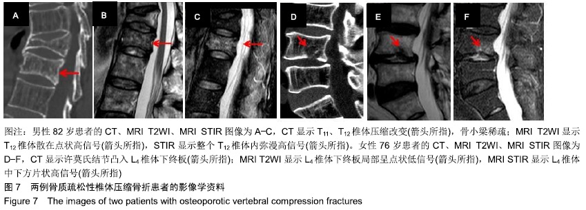
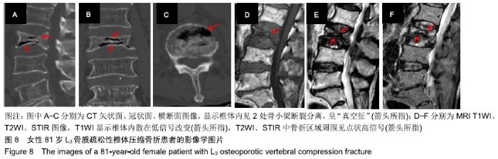
|