Chinese Journal of Tissue Engineering Research ›› 2020, Vol. 24 ›› Issue (6): 821-826.doi: 10.3969/j.issn.2095-4344.2445
Design and clinical application of personalized antibiotic cement spacer of knee joint
Wang Hong1, Wu Quan1, Tang Geng1, Zhang Gong2
- 1School of Mechanical & Electrical Engineering, Guizhou Normal University, Guiyang 550025, Guizhou Province, China; 2Graduate School of Zunyi Medical University, Zunyi 563000, Guizhou Province, China
-
Received:2019-07-09Revised:2019-07-10Accepted:2019-08-20Online:2020-02-28Published:2020-01-16 -
Contact:Wu Quan, PhD, Associate professor, Master’s supervisor, School of Mechanical & Electrical Engineering, Guizhou Normal University, Guiyang 550025, Guizhou Province, China -
About author:Wang Hong, Master candidate, School of Mechanical & Electrical Engineering, Guizhou Normal University, Guiyang 550025, Guizhou Province, China -
Supported by:the Guizhou Normal University Graduate Innovation Foundation, No. YC[2018]027; the Foundation for Excellent Young Talents of Guizhou Province, No. [2015]05; the Science and Technology Program of Guizhou Province, No. [2016]7221; the Doctoral Foundation of Guizhou Normal University in 2014
CLC Number:
Cite this article
Wang Hong, Wu Quan, Tang Geng, Zhang Gong. Design and clinical application of personalized antibiotic cement spacer of knee joint[J]. Chinese Journal of Tissue Engineering Research, 2020, 24(6): 821-826.
share this article
Add to citation manager EndNote|Reference Manager|ProCite|BibTeX|RefWorks
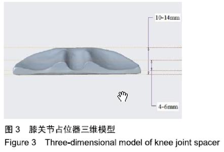
2.1 有限元模型的建立 将设计后的占位器模型与膝关节股骨、胫骨在三维软件中进行装配,将模型按照膝关节屈曲0°,30°,60°,90°时,股骨和胫骨的相对位置进行装配,装配后的膝关节占位器三维模型见图3。以STP格式导入ANSYS 17.0高级有限元分析软件,膝关节和占位器分别定义骨、骨水泥2种材料属性。采用ANSYS软件的自动划分网格功能,将装配体三维模型划分成10 039个单元,18 962个节点。在建立的有限元模型中,软件会自动识别接触区域,生成接触对,发生接触的区域有3部分,包括股骨与股骨侧占位器、胫骨与胫骨侧占位器、上下占位器之间。设置股骨与股骨侧占位器、胫骨与胫骨侧占位器相互接触的面为绑定面,绑定界面默认不会有相对运动,股骨、胫骨侧占位器设定为有限滑动相互作用的面。同时定义接触面为常态行为、无摩擦。在股骨轴向加载垂直向下的力350 N(假设被研究患者的体质量为70 kg),股骨不受约束,胫骨底端设置成固定约束。分析膝关节在运动过程中屈曲(0°-90°)时占位器的受力情况。"

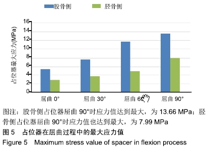
2.2 结果 膝关节在屈曲0°,30°,60°,90°时占位器的受力情况见图4,总体呈缓慢上升趋势,随着屈曲角度增大,占位器配合部分接触面积减少,应力值也随之增加。该过程中股骨侧占位器最大应力从5.42 MPa缓慢增加到 13.66 MPa;胫骨侧占位器最大应力从2.89 MPa缓慢增加到7.99 MPa。胫骨侧占位器应力值小于股骨侧占位器,说明在屈曲过程中应力主要分布在股骨侧占位器。"
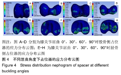

膝关节屈曲过程中占位器在各个角度的应力分布见图5。从云图上可以看出,应力主要分布在占位器相接触的部分,随着角度的变化,接触面积减少,应力也随着角度的变化从占位器的前端移动到占位器的后端,最大应力值出现在接触面的中间部分。股骨侧占位器屈曲90°时应力值达到最大,为13.66 MPa;胫骨侧占位器屈曲90°时应力值也达到最大,为7.99 MPa。骨水泥的抗压强度为96.25 MPa[31-36],行走过程中该结构占位器的应力值远小于骨水泥的最大应力值。应力分布均匀,没有应力集中,患者在屈曲运动时占位器不容易发生破坏,可以满足清除感染期间关节活动的力学要求。"
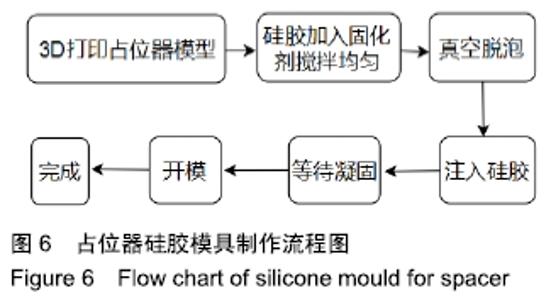
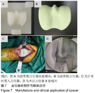
3.1 占位器的制作 采用光固化3D打印成形系统(Object 30,Stratasys,USA)制作占位器实物模型。将设计好的个性化占位器三维模型以stl格式导出,并导入到object studio软件里进行切片、定位等处理,然后打印成形。取出打印后的模型,用高压水枪去除支撑,得到高精度、表面光滑的占位器实体模型。 采用医用硅胶制作占位器模具。首先制备型框,并将占位器实体模型作为母模置于型框内。为保证硅胶模具强度,型框边缘与占位器距离20 mm以上。将适量的医用硅胶加入固化剂搅拌均匀,然后注入型框,并将其置于真空浇注机内进行真空脱泡处理,排除硅胶内的空气,使其表面光滑、组织密实。待硅胶完全固化后,拆除型框,分模并取出占位器母模,得到该占位器硅胶模具,制作流程见图6。 "
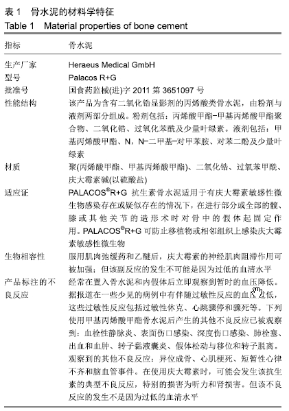
3.2 临床应用 制作占位器之前应对模具进行消毒处理,制作过程中用40 g骨水泥(Heraeus Medical GmbH,批号:国食药监械(进)字2011第3651097号)与4 g万古霉素(礼来苏州制药有限公司,批号:H20080356)搅拌均匀置入硅胶模具中,搅拌均匀有助于抗生素浓度的释放。骨水泥的材料学参数见表1。有关文献表示骨水泥混入的万古霉素小于8 g,对骨水泥的力学强度没有影响[37-41]。将硅胶模具的上下模紧密贴合,待骨水泥凝固后取出用于临床手术,见图7。"
"


此次研究数据来源贵州省人民医院收治的12例人工膝关节置换后感染患者,其中男3例,女9例;年龄43-85岁,平均64岁。 纳入标准:全膝关节置换后感染患者,其诊断标准符合骨肌系统感染协会提出的假体周围感染诊断指南;患者对治疗及试验方案知情同意。 排除标准:未感染的患者,感染但无法接受手术的患者。 临床结果表明,占位器置入后伤口愈合良好,可有效控制膝关节慢性感染,明显缓解关节疼痛,改善关节功能,提高患者生活质量,无并发症发生。术前膝关节活动度平均为20°(0°,80°),术前活动度为20°,伸0°,屈80°,平均屈24°;置入临时占位器后膝关节活动度提升至平均40°(20°,0°),伸20°,屈90°,平均屈55°。术前美国特种外科医院膝关节评分平均为36分(33,42)分,间隔期提升至平均47分(45,53)分。美国特种外科医院评分满分100分,评估项目包括疼痛、功能、活动度、肌力、屈曲畸形及稳定性。同时占位器置入间隔期未发生松动、脱落、破裂、占位器周围发生骨折、伸膝装置破损等情况。二期手术时占位器可顺利取出,且未造成进一步骨缺损。 提示个性化占位器不仅可以提高二次翻修期间关节活动能力,还可以增加患者关节活动时的稳定性,减少患者痛苦,有助于患者术后恢复。采用硅胶模型制作个性化占位器方法简单,关节面匹配度高,手术过程简便,成本小,使用方便。 "

| [1] LIMA AL, OLIVEIRA PR, CARVALHO VC, et al. Periprosthetic joint infections. Interdiscip Perspect Infect Dis. 2013;2013(7):1-7. [2] PARVIZI J, PAWASARAT IM, AZZAM KA, et al. Periprosthetic joint infection the economic impact of methicillin-resistant infections. J Arthroplasty. 2010;25(6):103-107. [3] 王龙超,彭慧明,林进,等.全膝关节置换术后假体周围感染的手术方式及预后[J].中华骨科杂志,2018,38(3): 129-136. [4] WINDISCH C, BRODT S, ROEHNER E, et al. C-reactive protein course during the first 5 days after total knee arthroplasty cannot predict early prosthetic joint infection. Arch Orthop Trauma Surg. 2017;137(8):1115-1119. [5] CHIANG CC, CHIU FY. Cefuroxime-impregnated cement and systemic cefazolin for 1 week in primary total knee arthroplasty: An evaluation of 2700 knees. J Chin Med Assoc. 2012;75(4): 167-170. [6] KOTELNICKI J, MITTS K. Surgical treatments for patients with an infected total knee arthroplasty. JAAPA. 2009;22(11):40, 43-46. [7] TOMS AD. The management of peri-prosthetic infection in total joint arthroplasty. J Bone Joint Surg Br. 2006;88(2):149-155. [8] VIKTOR J, WASSILEW GI, PERKA CF, et al. Cerclages after femoral osteotomy are at risk for bacterial colonization during two-stage septic total hip arthroplasty revision. J Bone Joint Infect. 2018;3(3):138-142. [9] COLLEONI JL, RIBEIRO FN, MOS PAC, et al. Venous thromboembolism prophylaxis after total knee arthroplasty (TKA): aspirin, vs. rivaroxaban. Rev Bras Ortop. 2017;53(1):22-27. [10] 于强,田京.抗生素骨水泥占位器在膝关节假体周围感染二期翻修中的应用[J].中国组织工程研究, 2018,22(31):5048-5055. [11] GEORGE J, MAHMOOD B, SULTAN AA, et al. How Fast Should A Total Knee Arthroplasty Be Performed? An Analysis of 140,199 Surgeries. J Arthroplasty. 2018;33(8):2616-2622. [12] TIBBO ME, CHALMERS BP, BERRY DJ, et al. Primary total knee arthroplasty in patients with neuropathic (charcot) arthropathy: contemporary results. J Arthroplasty. 2018;31(9):2815-2820. [13] 付君,李想,倪明,等. 髋臼加强型抗生素骨水泥占位器在假体周围感染髋臼骨缺损翻修中的应用[J].中华骨科杂志,2018,38(3):143-149. [14] 宗航帆,刘瑞宇,王坤正,等. 未使用占位器的二期翻修治疗髋关节置换术后感染[J].中华关节外科杂志(电子版),2018,12(2):163-167. [15] 佚名. 部分旷置二期翻修治疗全髋关节置换术后假体周围感染的早期疗效[J]. 中华骨科杂志,2019,39(7):422-428. [16] 孙海滨,吕尚军,朱书朝,等.3D打印技术在制作人工髋关节置换术后感染临时间隔器中的临床应用[J]. 中国矫形外科杂志,2016,24(13): 1239-1241. [17] 王卫红,谷振光,杨子峰,等.自制含抗生素骨水泥占位器治疗人工髋关节置换术后感染[J].中国伤残医学,2015,23(13): 42-43. [18] 张浩伟,宋科官.髋关节置换术后假体周围感染机制及治疗[J]. 中国组织工程研究,2015,19(48):7833-7833. [19] 宋兴桂,李昕,宋俊雷,等.保留假体清创术治疗人工关节置换术后假体周围感染的临床研究[J].协和医学杂志,2019,10(4):370-374. [20] WHITTAKER JP, WARREN RE, JONES RS, et al. Is prolonged systemic antibiotic treatment essential in two-stage revision hip replacement for chronic Gram-positive infection? J Bone Joint Surg Br. 2009;91(1):44-51. [21] 胡孔足(译). 抗生素骨水泥髋臼顶可增强人工髋关节感染翻修骨水泥占位器的稳定性[J]. 临床骨科杂志,2016,19(3):354-354. [22] 李军伟,汝强,韩文龙,等. 采用可活动关节间隔垫的二期翻修术治疗人工膝关节术后感染[J].中国实用医刊, 2013, 40(17):16-18. [23] LOVALLO J, HELMING J, JAFARI SM, et al. Intraoperative intra-articular injection of gentamicin: will it decrease the risk of infection in total shoulder arthroplasty. J Shoulder Elbow Surg. 2014;23(9):1272-1276. [24] MCPHERSON EJ, LEWONOWSKI K, DORR LD. Techniques in arthroplasty. Use of an articulated PMMA spacer in the infected total knee arthroplasty. J Arthroplasty. 1995; 10(1):87-89. [25] 张强,周勇刚,陈继营,等. 术中自制临时关节型抗生素骨水泥占位器治疗人工膝关节置换术后感染[J].中国骨伤,2013,26(2):119-123. [26] 李锦华,邵琴,赵宙,等. 抗生素骨水泥占位器在髋关节置换后感染翻修术中的应用[J].临床外科杂志,2013,21(5):383-384. [27] 林志炯,高大伟,董月珍,等. 骨水泥占位器在髋关节感染二期翻修术的临床疗效[J]. 中华关节外科杂志(电子版), 2017,11(4):10-15. [28] 王利,哈巴西•卡肯,殷剑,等.自制活动型抗生素骨水泥间隔器:在全膝关节表面置换后感染翻修中的应用[J].中国组织工程研究,2015, 19(13):2017-2022. [29] GARG P, BANDYOPADHYAY U, MITRA S, et al. Antibiotic- impregnated articulating cement spacer for infected total knee arthroplasty. Indian J Orthop. 2011;45(6):535 -540. [30] 张强,金志刚,周勇刚,等.抗生素骨水泥占位器治疗髋和膝关节置换术后感染的研究进展[J].中华关节外科杂志(电子版), 2008,2(5): 572-577. [31] 张岚峰. 骨水泥型髋关节假体的生物力学特性及其摩擦学研究[D].徐州:中国矿业大学,2017. [32] PAHLEVANZADEH F, BAKHSHESHI-RAD HR, HAMZAH E. In-vitro biocompatibility, bioactivity, and mechanical strength of PMMA-PCL polymer containing fluorapatite and graphene oxide bone cements. J Mech Behav Biomed Mater. 2018;82(6):257-267. [33] ZHANG X, YANG HL, LI S, et al. Natural diatomite particles: Size-, dose-and shape-dependent cytotoxicity and reinforcing effect on injectable bone cement. J Mater Sci Technol. 2018;34(6):1044-1053. [34] BOELCH SP, JORDAN MC, ARNHOLDT J, et al. Antibiotic elution and compressive strength of gentamicin/vancomycin loaded bone cements are considerably influenced by immersion fluid volume. J Mater Sci.2019;30(2):23-29. [35] LIAO J, LI Y, LI H, et al. Preparation, bioactivity and mechanism of nano-hydroxyapatite/sodium alginate/chitosan bone repair material. J Appl Biomater Funct Mater. 2018;16(1):28-35. [36] SOPCAK T, MEDVECKY L, GIRETOVA M, et al. Hydrolysis, setting properties and in vitro characterization of wollastonite/ newberyite bone cement mixtures. J Biomater Appl. 2018;32(7): 871-885. [37] SPRINGER BD, LEE GC, OSMON D, et al. Systemic safety of high-dose antibiotic-loaded cement spacers after resection of an infected total knee arthroplasty. Clin Orthop Relat Res. 2004;427: 47-51. [38] 唐江安,张岩,应辉. 抗生素骨水泥剂量差异对行关节置换术患者康复效果及再发感染风险的影响[J].临床和实验医学杂志,2019,18(2): 70-73. [39] ZHAO QH, ZHU FB, CAI XZ, et al. Effects of low-frequency pulsed wave ultrasound on the shear properties of the interface of vancomycin-loaded acrylic bone cement-stem. Zhonghua Yi Xue Za Zhi. 2017;97(7):545-550. [40] HSU YH, HU CC, HSIEH PH, et al. Vancomycin and ceftazidime in bone cement as a potentially effective treatment for knee periprosthetic joint infection. J Bone Joint Surg. 2017;99(3): 223-231. [41] HAASE K, ROUHI G. A discussion on plating factors that affect stress shielding using finite element analysis. J Biomed Sci. 2010; 5(2):129-141. [42] HA CW. A technique for intraoperative construction of antibiotic spacers. Clin Orthop Related Res. 2006;445(445):204-209. |
| [1] | Wang Jinjun, Deng Zengfa, Liu Kang, He Zhiyong, Yu Xinping, Liang Jianji, Li Chen, Guo Zhouyang. Hemostatic effect and safety of intravenous drip of tranexamic acid combined with topical application of cocktail containing tranexamic acid in total knee arthroplasty [J]. Chinese Journal of Tissue Engineering Research, 2021, 25(9): 1356-1361. |
| [2] | Chen Xinmin, Li Wenbiao, Xiong Kaikai, Xiong Xiaoyan, Zheng Liqin, Li Musheng, Zheng Yongze, Lin Ziling. Type A3.3 femoral intertrochanteric fracture with augmented proximal femoral nail anti-rotation in the elderly: finite element analysis of the optimal amount of bone cement [J]. Chinese Journal of Tissue Engineering Research, 2021, 25(9): 1404-1409. |
| [3] | Wang Debin, Bi Zhenggang. Related problems in anatomy mechanics, injury characteristics, fixed repair and three-dimensional technology application for olecranon fracture-dislocations [J]. Chinese Journal of Tissue Engineering Research, 2021, 25(9): 1446-1451. |
| [4] | Zhang Xiumei, Zhai Yunkai, Zhao Jie, Zhao Meng. Research hotspots of organoid models in recent 10 years: a search in domestic and foreign databases [J]. Chinese Journal of Tissue Engineering Research, 2021, 25(8): 1249-1255. |
| [5] | Yuan Jun, Yang Jiafu. Hemostatic effect of topical tranexamic acid infiltration in cementless total knee arthroplasty [J]. Chinese Journal of Tissue Engineering Research, 2021, 25(6): 873-877. |
| [6] | Song Chengjie, Chang Hengrui, Shi Mingxin, Meng Xianzhong. Research progress in biomechanical stability of lateral lumbar interbody fusion [J]. Chinese Journal of Tissue Engineering Research, 2021, 25(6): 923-928. |
| [7] | Liu Zhao, Xu Xilin, Shen Yiwei, Zhang Xiaofeng, Lü Hang, Zhao Jun, Wang Zhengchun, Liu Xuzhuo, Wang Haitao. Guiding role and prospect of staging and classification combined collapse prediction method for osteonecrosis of femoral head [J]. Chinese Journal of Tissue Engineering Research, 2021, 25(6): 929-934. |
| [8] | Cai Qunbin, Zou Xia, Hu Jiantao, Chen Xinmin, Zheng Liqin, Huang Peizhen, Lin Ziling, Jiang Ziwei. Relationship between tip-apex distance and stability of intertrochanteric femoral fractures with proximal femoral anti-rotation nail: a finite element analysis [J]. Chinese Journal of Tissue Engineering Research, 2021, 25(6): 831-836. |
| [9] | Zhao Zhongyi, Li Yongzhen, Chen Feng, Ji Aiyu. Comparison of total knee arthroplasty and unicompartmental knee arthroplasty in treatment of traumatic osteoarthritis [J]. Chinese Journal of Tissue Engineering Research, 2021, 25(6): 854-859. |
| [10] | Chen Lu, Zhang Jianguang, Deng Changgong, Yan Caiping, Zhang Wei, Zhang Yuan. Finite element analysis of locking screw assisted acetabular cup fixation [J]. Chinese Journal of Tissue Engineering Research, 2021, 25(3): 356-361. |
| [11] | Shu Qihang, Liao Yijia, Xue Jingbo, Yan Yiguo, Wang Cheng. Three-dimensional finite element analysis of a new three-dimensional printed porous fusion cage for cervical vertebra [J]. Chinese Journal of Tissue Engineering Research, 2021, 25(24): 3810-3815. |
| [12] | Zhu Yun, Chen Yu, Qiu Hao, Liu Dun, Jin Guorong, Chen Shimou, Weng Zheng. Finite element analysis for treatment of osteoporotic femoral fracture with far cortical locking screw [J]. Chinese Journal of Tissue Engineering Research, 2021, 25(24): 3832-3837. |
| [13] | Liu Jinyu, Ding Yiwei, Lu Zhengcao, Gao Tianjun, Cui Hongpeng, Li Wen, Du Wei, Ding Yu. Finite element biomechanical study of full endoscopic fenestration decompression for cervical spondylotic myelopathy [J]. Chinese Journal of Tissue Engineering Research, 2021, 25(24): 3850-3854. |
| [14] | Qin Wan’an, Cai Zhouyu, Wei Gejin, Lin Zhoudan. Finite element analysis of absorbable screws and ethibond sutures for the treatment of humerus shaft fractures caused by grenade throwing [J]. Chinese Journal of Tissue Engineering Research, 2021, 25(24): 3855-3859. |
| [15] | Deng Zhibo, Li Zhi, Wu Yahong, Mu Yuan, Mu Yuexi, Yin Liangjun. Local infiltration anesthesia versus femoral nerve block for pain control and safety after total knee arthroplasty: a meta-analysis [J]. Chinese Journal of Tissue Engineering Research, 2021, 25(21): 3401-3408. |
| Viewed | ||||||
|
Full text |
|
|||||
|
Abstract |
|
|||||