Chinese Journal of Tissue Engineering Research ›› 2020, Vol. 24 ›› Issue (28): 4429-4436.doi: 10.3969/j.issn.2095-4344.2315
Different material factors affect the proliferation and osteogenic differentiation of bone marrow mesenchymal stem cells
Wang Yanghao, Wang Weizhou, Duan Hao, Zhong Zongyu, Li Xiaozhuang, Tang Zhihong, He Fei
First Affiliated Hospital of Kunming Medical University, Kunming 650032, Yunnan Province, China
-
Received:2019-12-07Revised:2019-12-10Accepted:2020-02-12Online:2020-10-08Published:2020-08-29 -
Contact:He Fei, Chief physician, First Affiliated Hospital of Kunming Medical University, Kunming 650032, Yunnan Province, China -
About author:Wang Yanghao, Master candidate, First Affiliated Hospital of Kunming Medical University, Kunming 650032, Yunnan Province, China -
Supported by:the National Natural Science Foundation of China, No. 31460244 ; Major Science and Technology Projects of Yunnan Province (Biomedicine), No. 2019ZF002; the Applied Basic Research Project of Yunnan Science and Technology Department Foundation, No. 2015FA002
CLC Number:
Cite this article
Wang Yanghao, Wang Weizhou, Duan Hao, Zhong Zongyu, Li Xiaozhuang, Tang Zhihong, He Fei. Different material factors affect the proliferation and osteogenic differentiation of bone marrow mesenchymal stem cells[J]. Chinese Journal of Tissue Engineering Research, 2020, 24(28): 4429-4436.
share this article
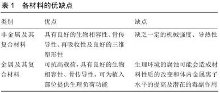
2.1 骨髓间充质干细胞的生物学特性 骨髓间充质干细胞的生物学特性包括以下几点:取材方便,间充质干细胞可以取自于骨髓[1-3];取自于自体,不存在自体配型及免疫排斥问题[1-3];广泛的分化特性,理论上能分化成软骨、脂肪、肌肉、肌腱、神经、肝脏、胰腺、表皮细胞等[2-4];具有免疫调节功能,表面抗原不明显,异体移植排异较轻[1-3];体外培养骨髓间充质干细胞的生长曲线近似“S”形,在前期细胞生长缓慢,前中期细胞进入快速增殖期,中期细胞增长迅速,中后期增长趋势逐渐减慢,后期进入平台期,细胞数基本趋于稳定[8];传代后的细胞具有原代的细胞特性,第一代细胞的生物年龄越小,细胞传代时间越短,经过一定次数的传代之后骨髓间充质干细胞的各项分化能力逐步减弱,其中成骨分化能力第3代最强,从第3-7代内无明显差异[9]。 2.1.1 骨髓间充质干细胞的培养方法 目前,骨髓间充质干细胞的培养方法大致有4种[10-11]:贴壁筛选法、密度梯度离心法、免疫磁珠分选培养法、流式细胞仪分选法,其中贴壁筛选法及密度梯度离心法是目前使用最多的2种培养方式,其过程相对简单、耗时少,两者效果无明显差别[12];而免疫磁珠分选培养法及流式细胞仪分选法因为设备及具体操作相对繁琐则使用相对较少[13];4种培养方法培养出的骨髓间充质干细胞没有明显的差异性[12-13]。 2.1.2 骨髓间充质干细胞的鉴定方法 目前骨髓间充质干细胞的鉴定方法大致有:①通过标准培养方法的骨髓间充质干细胞必须具有对塑料底物黏附特性,该方法在培养及筛选过程中就已体现其作用,是骨髓间充质干细胞培养的基础;②经使用流式细胞仪检测后,骨髓间充质干细胞群体(五阴三阳)阴性表达CD45、CD34、CD9、CD79及人白细胞相关抗原且阳性表达CD3、CD05、CD90[14-15],该方法是骨髓间充质干细胞的直接定性诊断,也是最常用、最直观的一种方法;③骨髓间充质干细胞经体外诱导后必须具有多能分化潜能;该种方法是鉴定骨髓间充质干细胞有无功能的一个重要步骤。 2.2 材料种类 理想的组织工程材料应具有良好的生物相容性、孔隙率、孔径、细胞附着性及足够的机械强度[16];组织工程材料按照材料性质可分为金属及其复合材料类、非金属及其复合材料类两大类,两种类普遍特征见表1。 "

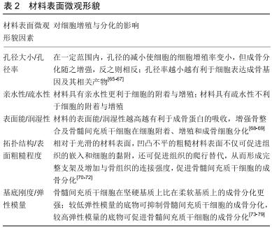
2.2.1 非金属材料 (1)无机材料: 磷酸钙骨水泥:现如今,人工合成的磷酸钙骨水泥由磷酸四钙和无水磷酸二钙按摩尔比1∶3的比例混合成而成;在人体内60%的无机物都含有磷酸钙,因此人工合成的磷酸钙被广泛用于骨材料的研究与临床治疗[17]。XIA等[18]体外实验证明,将离子以液体或粉末的形式结合在一起显著提高了干细胞在磷酸钙骨水泥支架上的性能,掺入物-磷酸钙骨水泥具有良好的生物相容性,且具有更强的碱性磷酸酶活性、骨矿物质合成能力及更好的细胞增殖能力,且液体掺入物优于粉末。 石墨烯:是由紧密堆积的二维蜂窝状晶格的单片碳原子组成。大量研究表明通过产生二维和三维高质量石墨烯衬底可扩展的方法制造的石墨烯,具有体外诱导成骨分化的能力,其可促进成骨基因及其相关表达物质(如:碱性磷酸酶、Runt相关转录因子2、骨钙蛋白等)[19-21],但其中的机制尚未明确。XIE等[22]通过实验分析整合素-FAK机械敏感途径中涉及相关关键蛋白的表达,结果表明石墨烯是通过整合素-FAK轴的调节促进成骨基因的表达。但石墨烯的成骨作用有限,且存在毒性,这一点限制了石墨烯的临床应用[23]。磁性石墨烯氧化物是Fe3O4与氧化石墨烯的新型结合物,具有多种独特的性能,减少了氧化石墨烯原有的细胞毒性作用,但其在骨组织工程中尚未得到研究。HE等[24]发现磁性石墨烯氧化物/成骨诱导培养基0.1 mg/L组矿化程度最高,细胞中β-catenin和Runx 2蛋白水平升高。此外,磁性石墨烯氧化物/成骨诱导培养基对钙结节有明显的加速作用,这些结果证明了磁性石墨烯氧化物促进骨髓间充质干细胞分化的潜在能力。 硅(Si):是人体中重要的微量元素,已有研究在骨骼的活性钙化部位发现硅,预示着硅在骨骼的生长及矿化过程中起着重要作用。首先,硅离子通过Wnt和SHH信号通路显著增强间充质干细胞的矿化、增殖和分化[25];其次,硅离子在促进MG63细胞产生胶原蛋白的同时,可以激活并增强MG63细胞中的ERK途径[26-27]。YANG等[28]体外实验发现二氧化硅纳米颗粒材料对骨髓间充质干细胞无细胞毒性,还能显著增强相关骨蛋白的表达和骨钙结节形成,该结果表明二氧化硅纳米颗粒可以促进间充质干细胞的成骨分化,并且不会影响细胞的增殖。 生物活性玻璃:是一种具有良好骨相容性及一定机械强度的生物材料,受到医学领域广泛的关注。BELLUCCI等[29]制备了一种包含锶和镁的生物活性玻璃颗粒,该颗粒具有较一般生物玻璃较高的结晶温度,更高的生物活性,且在培养环境中可以支持骨髓间充质干细胞的黏附、定植及促进成骨分化。KAZEMI等[30]制备了一种基于锶取代的β-磷酸三钙生物活性玻璃,通过体外实验评估骨髓间充质干细胞在该种材料中所表达出的碱性磷酸酶活性、基质矿化程度、骨性相关基因水平及特异性蛋白水平表达等,发现该材料具有促进成骨潜力。 除上述材料外,其他常见无机材料,如羟基磷灰石、碳纳米管、磷酸三钙等材料都具有各自的优缺点,被广泛用于各种骨组织工程材料研究当中。 (2)有机高分子材料: 聚乳酸:是以乳酸为主要原料聚合得到的聚合物,原料来源充分且可以再生。聚乳酸及其复合材料具有良好的机械性能、物理性能及可生物降解性,因而成为一种非常有研究前景的生物支架材料[31-32];一些研究也指出聚乳酸单体具有较高的脆性及强度,这也限制了其在在临床中的应用性[33-34]。 壳聚糖:是甲壳素经脱乙酰化后得到的多糖产物,同时也是组织工程中的常用支架材料。壳聚糖具有无热源反应、无毒性、低抗原性、生物相容性良好等优点,但其机械性能较差。闫峰等[35]制作出了壳聚糖多孔支架,该支架具有良好的亲水性及细胞吸附性,且骨髓间充质干细胞与该支架融合良好,细胞增殖不会受到抑制。 胶原:是一种天然蛋白质,来源广泛,具有低抗原性及良好的生物相容性、可塑性及降解吸收特性等优点,其缺点是机械性能较差。王莉莉等[36]制备出一种新型具有引导骨组织再生潜能的猪胶原膜,该材料表面疏松多孔,具有良好的亲水性,可明显增加细胞的爬行及迁移能力,促进骨髓间充质干细胞的增殖,以及提高成骨分化相关基因的表达、碱性磷酸酶活性与细胞外基质矿化程度。 水凝胶:是一种来源广泛的高分子网络体系,是具有良好水溶性或亲水性的高分子材料,可通过一定的交联手段获得,其结构与细胞外基质类似,是当前植入材料的研究热门。BI等[37]合成了一种由聚酰胺酰胺树枝状大分子和聚乙二醇原位形成的水凝胶体系,以较高浓度的树枝状聚合物和聚乙二醇形成的水凝胶(硬凝胶)具有相对较短的胶凝时间和较高的强度,同时硬凝胶比较低浓度树枝状聚合物形成的软凝胶显示出更致密和更少的多孔结构。不同硬度的水凝胶都能够促进间充质干细胞细胞在水凝胶基质中的附着、增殖及成骨分化,但硬凝胶的成骨分化作用更为明显。 复合材料:每种材料都有各自的特点,为了达到生物材料最理想化的状态,通过一定的技术手段将几种不同的生物材料进行混合或添加药物、生物活性因子等组合制造成一种新型复合材料,从而更满足于临床治疗中植入材料的要求。 多种材料组合能够让不同材料的优缺点互补,例如由胶原蛋白、羟基磷灰石、地塞米松组合而成的复合材料,可促进骨髓间充质干细胞的增殖能力、碱性磷酸酶活性,并且增强Ⅰ型胶原和骨钙素表达[38]。LIS-BARTOS等[39]将聚乳酸和热塑性聚氨酯溶解在混合的有机溶剂中获得了聚乳酸和热塑性聚氨酯共混物,该材料解决了聚乳酸单体的较高脆性的问题,并具有良好的弹性模量、细胞吸附性、细胞生存及增殖能力。陈娇等[40]运用静电纺丝技术将2种单体即乳酸和乙醇酸随机聚合制作出了聚乳酸-羟基乙酸共聚物支架,该支架具有良好的生物相容性、生物力学、生物降解性且生物降解后无毒副作用,该支架可明显促进骨髓间充质干细胞的黏附增殖、碱性磷酸酶活性、Ⅰ型胶原的合成与分泌及成骨细胞分化。 除各种类型材料互补之外,在材料中加入药物、生物活性因子等也是目前研究的热点方向,能够使得生物材料发挥一些特殊的生物学功能,具有广泛的应用前景。ABAZARIA等[41]通过使用聚乙烯醇、壳聚糖、羟基磷灰石聚合物及富含血小板血浆制备了静电纺丝纳米纤维支架,其具有良好的结构和生物相容性,可提高骨髓间充质干细胞的钙沉积、碱性磷酸酶活性和钙含量,成骨基因表达更为显著,该结果提示该材料可明显促进骨髓间充质干细胞的增殖及成骨分化。硒(Se)具有增强免疫监测、调节细胞氧化应激水平的作用[42-44]。LI等[45]利用纳米技术开发了一种能够缓慢释放有益量硒的多孔Se@SiO2纳米复合材料,该复合材料表现出了体内生物安全性和有效控制Se的低毒性特点[46-47];在体外,多孔Se@SiO2纳米复合材料能促进骨髓间充质干细胞的迁移和成骨分化,保护骨髓间充质干细胞免受H2O2诱导的成骨分化抑制作用;在体内应用多孔Se@SiO2纳米复合材料可促进骨折愈合。 2.2.2 金属材料 金属类及其复合物材料具有抗高载荷、良好的生物相容性、骨传导性,可为植入部位提供生理负荷功能,在临床治疗及相关研究中占有重要地位。 钛合金:目前,钛(Ti)是临床上应用广泛的口腔种植材料,但其机械/拉伸强度不足[48]。众多研究表明,钛锆合金具有比钛更加良好的力学性能和抗拉强度,并且在治疗骨缺损疾病中钛锆合金具有良好的骨整合性及细胞高存活率[49-50]。JIN等[51]发现INcRNA CCL 3-As的过量表达会引起Ⅰ型胶原、骨桥蛋白和Runx2的下调及抑制骨髓间充质干细胞向成骨细胞系的分化,TiO2纳米管通过抑制INcRNA CCL 3-As的过量表达,进一步提高了钛的生物活性,增强骨髓间充质干细胞的活性、增殖能力及成骨分化潜能。 钽金属:具有高孔隙率、低弹性模量、良好的机械强度等特性,有利于黏附细胞的增殖及分化[52-54]。SMITH等[55]发现与同种异体及自体移植使用的临床标准骨替代品相比,将钽小梁金属作为骨科植入物治疗骨缺损病变细的胞增殖速率、成骨基因及相关标志物表达均升高,该结果支持钽小梁金属作为骨科植入物具有更好诱导骨髓间充质干细胞成骨分化的作用。谢辉等[56]以多孔碳化硅为基体材料,采用化学气相沉积技术将钽金属均匀沉积在碳化硅支架材料上形成金属钽涂层,形成新型多孔钽金属支架,将骨髓间充质干细胞与该支架联合培养后发现细胞增殖速率增快,以及大量骨小梁、成骨细胞生成,该结果提示该支架具有促进骨髓间充质干细胞增殖及成骨分化的作用。因此,钽金属及其衍生物是优良的骨生物材料,与其他传统金属材料如钛金属相比是一种更佳的植入材料选择。 纳米级金属材料:HEIDEN等[57]研究发现纳米结构的Fe-Mn和Fe-Mn-Zn金属支架增加了骨髓间充质干细胞的附着力,改善了细胞扩散及多层结构,且该支架相比单纯性Fe-Mn和Fe-Mn-Zn金属支架具有更高的耐腐蚀性及生物相容性。BOSTANCIOGLU等[58]通过将间充质干细胞放置于金属离子(锌、银和铜)掺杂的羟基磷灰石纳米涂层表面,发现含有Zn、Ag和Cu金属离子的羟基磷灰石有助于增强间充质干的成骨分化能力、细胞黏附能力及外源性材料的生物相容性。 除上述材料外,其他的金属材料如氧化铁纳米颗 粒[59]、钴金属、铬金属及其合金等材料[60],也被广泛用于骨组织工程材料研究及临床治疗当中。锶元素目前被用于骨质疏松症治疗,其可增强成骨作用并减少骨吸收,已显示出该元素同时抑制破骨细胞吸收骨并刺激成骨细胞形成骨,与锶元素有机结合的复合材料也是一种潜在的新型骨组织材料[61-62]。 2.3 材料表面微观形貌因素 在细胞增殖分化过程中,即使是同一种材料,但不同孔径率、孔径大小、材料的亲水性、疏水性、表面能/润湿性、表面粗糙程度、基底刚度、拓扑结构、几何形状、弹性模量等都可能会影响骨髓间充质干细胞的增殖及成骨作用;且已有研究提出干细胞可通过一些机械传感器(如细胞骨架、膜通道等)感测机械环境[63-64],提示材料表面微观形貌对于干细胞的增殖及分化具有直接影响作用,具体见表2(均为体外实验)。 2.3.1 孔径率/孔径大小 TEIXEIRA等[65]通过体外实验发现,在不同孔径大小的钛盘中培养细胞,更小孔径的规格会限制细胞的进入,降低细胞增殖率,但其骨标志物基因表达量、成骨细胞表型表达量却提高了。同时还有国内实验发现,孔径率越小,干细胞的成骨分化基因及其产物表达越明显,孔径率越大反而越不利于细胞的成骨分化[66]。COVERDALE等[67]将一定浓度的两性离子磷脂卵磷脂当作表面活性剂掺入静电纺丝聚己内酯支架中,降低了聚己内酯支架的疏水性,减小了支架的孔径,增大了支架的表面积,从而更有利于骨髓间充质干细胞的附着与增殖。 2.3.2 表面能/润湿性 表面能是在恒温、恒压、恒组成情况下可逆地增加物系表面积须对物质所做的非体积功,润湿性是指一种液体在一种固体表面铺展的能力或倾向性。YIN等[68]采用喷砂大颗粒酸蚀或亲水性喷砂大颗粒酸蚀对4级钛和钛锆(13%-17%钛)底物进行改性,得到4种表面微观结构复杂的表面类型,研究发现亲水性喷砂大颗粒酸蚀比喷砂大颗粒酸蚀具有更高的表面能/润湿性,亲水性的钛和钛锆材料具有更合适的理化性质,对骨髓间充质干细胞的生物学反应更具有优越性,说明增加表面能/润湿性可以促进成骨蛋白的吸收,增强骨整合及骨髓间充质干细胞的附着、增殖和成骨细胞分化。潜在机制可能是由于不同的表面能影响黏附蛋白、基质蛋白质沉积,从而影响吸附蛋白质的构象和组成,使细胞表现出不同的生物学行为[69]。 2.3.3 拓扑结构/表面粗糙程度 研究表明基底拓扑结构对于干细胞的增殖和分化过程同样具有调控作用,可能与拓扑结构引起的细胞骨架变化及细胞铺展状态相关[70]。在基底硬度和拓扑结构耦合效应方面,拓扑的特征尺寸及形状对微丝和中间纤维的调控起到了至关重要的作用[71]。在骨植入材料物中相对于光滑表面,通过激光烧结技术造成材料凹凸不平的粗糙表面不仅可促进组织的嵌入和细胞的黏附,而且还可促进组织的爬行替代,从而形成完整支架及增加与骨组织的连接强度[52]。有研究发现,激光烧结钛、喷砂大颗粒酸蚀性钛比单纯钛的表面微观形貌更能促进骨髓间充质干细胞的成骨分化、黏附性能及蛋白质吸附能力,其可能机制涉及了H3K27快速去甲基化及增加成骨相关基因启动子区域H3K4me3的水平[72]。 2.3.4 基底刚度/弹性模量 有研究分析了骨髓间充质干细胞在不同刚度基质上成骨细胞特异性标志物的表达、碱性磷酸酶活性及矿化,发现骨髓间充质干细胞相较于在柔软的基质上,硬的基质更能促进其成骨分化[73]。WITKOWSKA-ZIMNY等[74]研究表明较软凝胶上的间充质干细胞增殖活性低于较硬基质上的增殖活性,较坚硬的底物更有利于成骨分化,并且间充质干细胞所编码α2和β1整合素亚基基因的相对表达更高。这些结果首次证明了基质刚性通过α2整合素介导的机械转导显著影响成骨分化,β1整合素也可一定程度的促进成骨分化。SUN等[75]在62-68 kPa基质刚度条件下证明,该种物理微环境通过整合素α5介导了间充质干细胞的成骨分化,使用阻断整联蛋白α5的抗体将会抑制相关成骨细胞标志物的刚性诱导表达。还有研究表明,黏着斑激酶是整合素介导信号通路中的一个关键信号分子,其转导来自整联蛋白受体的生存信号[76],黏着斑激酶作用过程中还与桩蛋白、β-catenin有直接的相互作用[77-78]。ROWLANDS等[79]证明了在基底刚度范围内同时需要相应的黏附配体才能够有效诱导干细胞的成骨分化。此外,底物的弹性模量越低,越适合用于制备软骨区和软骨的弹性支架,抑制骨髓间充质干细胞的成骨分化,底物的弹性模量越高,越适合用于制作更坚固的支架及成骨细胞培养,促进骨髓间充质干细胞的成骨分化,因此,底物弹性模量在调节细胞的黏附、形态、增殖或分化等方面发挥了重要作用[39]。 "
|
[1] MOHAMMAD MH, AL SHAMMARI AM, AL JUBOORY AA, et al. Characterization of neural stemness status through the neurogenesis process for bone marrow mesenchymal stem cells.Stem Cells Cloning.2016;9:1-15.
[2] 韩振霞,时庆,汪大琨.骨髓源与脐带源间充干细胞的基本生物学特征的比较[J].中国实验血液学杂志,2013,21(5):1248-1255.
[3] ZHAN XS, EL-ASHRAM S, LUO DZ, et al. A Comparative Study of Biological Characteristics and Transcriptome Profiles of Mesenchymal Stem Cells from Different Canine Tissues.Int J Mol Sci.2019;20(6):1485.
[4] MUTLU L, HUFNAGEL D, TAYLOR HS. The endometrium as a source of mesenchymal stem cells for regenerative medicine. Biol Reprod.2015;92(6):138.
[5] IGE O, UMORU L, ARIBO S. Natural products:a minefield of biomaterials.ISRN Mater Sci.2012;2012:1-20.
[6] MORN N, KAPUSETTI G. Piezoelectric material-A promising approachfor bone and cartilage regeneration.Med Hypotheses. 2017;108(10):10-16.
[7] 杨雄峰,姜慧娇,王小义,等.体外负压培养对小鼠骨髓间充质干细胞增殖能力和血管内皮生长因子分泌水平的影响[J].中国组织工程研究,2020,24(1):33-39.
[8] 邹维艳,赵洁,李仪,等.体外培养SD大鼠与BALB/c小鼠骨髓间充质干细胞的生物学特性比较[J].细胞与分子免疫学杂志,2018, 34(12):1099-1104.
[9] 叶含钊,安荣泽,王兆杰.不同代次骨髓间充质干细胞的生物学特性[J].贵州医药,2015,39(1):83-86.
[10] NEUHUBER B, SWANGER SA, HOWARD L, et al. Effects of plating density and culture time on bone marrow stromal cell characteristics.Exp Hematol.2008;36(9):1176-1185.
[11] LI X, ZHANG Y, QI G. Evaluation of isolation methods and culture conditions for rat bone marrow mesenchymal stem cells.Cytotechnology.2013;65(3):323.
[12] 刘贤华,仝青英,白晓东.人骨髓间充质干细胞体外不同分离与培养方法的比较[J].武警医学,2012,23(10):836-839.
[13] MAYLE A, LUO M, JEONG M, et al. Flow cytometry analysis of murine hematopoietic stem cells.ytometry Part A. 2013; 83(1):27-37.
[14] HALL SR, JIANG Y, LEARY E, et al. Identification and isolation of small CD44 negative mesenchymal stem/progenitor cells from human bone marrow using elutriation and polychromatic flow cytometry.Stem Cells Transl Med.2013;2(8):567-578.
[15] SONG K, HUANG M, SHI Q, et al. Cultivation and identification of rat bone marrow derived mesenchymal stem cells. Mole Med Rep.2014;10(2):755.
[16] JANG NK, LEE SE, CHA SR, et al. Osteogenic differentiation of bone marrow stem cell in silk fibroin scaffold.Int J Tissue Reg. 2015;6:56-63.
[17] BARBA A, DIEZ-ESCUDERO A, MAAZOUZ Y, et al. Osteoinduction by foamed and 3D-printed calcium phosphate scaffolds:effect of nanostructure and pore architecture.ACS Appl Mater Interfaces.2017;9(48):41722-41736.
[18] XIA Y, CHEN H, ZHANG F, et al. Gold nanoparticles in injectable calcium phosphate cement enhance osteogenic differentiation of human dental pulp stem cells.Nanomedicine. 2018;14(1):35-45.
[19] XIE H, CHUA M, ISLAM I, et al. CVD-grown monolayer graphene induces osteogenic but not odontoblastic differentiation of dental pulp stem cells. Dent Mater. 2017; 33:e13-e21.
[20] XIE H, CAO T, GOMES JV, et al. Two and three-dimensional graphene substrates to magnify osteogenic differentiation of periodontal ligament stem cells. Carbon.2015;93:266-275.
[21] XIE H, CAO T, RODRÍGUEZ-LOZANO FJ, et al. Graphene for the development of the next-generation of biocomposites for dental and medical applications.Dent Mater.2017;33:765-774.
[22] XIE H, CAO T, FRANCO-OBREGÓN A, et al. Graphene-Induced Osteogenic Differentiation Is Mediated by the Integrin/FAK Axis.Int J Mol Sci.2019;20(3):547.
[23] JIA PP, SUN T, JUNAID M, et al. Pei Nanotoxicity of different sizes of graphene (G) and graphene oxide (GO) in vitro and in vivo.Environ Pollut.2019;247:595-606.
[24] HE Y, LI Y, CHEN G, et al. Concentration-dependent cellular behavior and osteogenic differentiation effect induced in bone marrow mesenchymal stem cells treated with magnetic graphene oxide.J Biomed Mater Res A.2020;108(1):50-60.
[25] HAN P, WU C, XIAO Y. The effect of silicate ions on proliferation, osteogenic differentiation and cell signalling pathways (WNT and SHH) of bone marrow stromal cells. Biomater Sci. 2013;1:379-392.
[26] SHIE MY, DING SJ, CHANG HC. The role of silicon in osteoblast-like cell proliferation and apoptosis.Acta Biomater. 2011;7(6):2604-2614.
[27] LI J, WEI L, SUN J, et al. Effect of ionic products of dicalcium silicate coating on osteoblast differentiation and collagen production via TGF-β1 pathway.J Biomater Appl. 2013;27(5): 595-604.
[28] YANG X, LI Y, LIU X, et al. The stimulatory effect of silica nanoparticles on osteogenic differentiation of human mesenchymal stem cells.Biomed Mater.2016;12(1):015001.
[29] BELLUCCI D, VERONESI E, STRUSI V, et al. Human Mesenchymal Stem Cell Combined with a New Strontium- Enriched Bioactive Glass: An ex-vivo Model for Bone Regeneration.Materials (Basel).2019;12(21):3633.
[30] KAZEMI M, DEHGHAN MM, AZAMI M. Biological evaluation of porous nanocomposite scaffolds based on strontium substituted beta-TCP and bioactive glass: An in vitro and in vivo study.Mater Sci Eng C Mater Biol Appl.2019;105:110071.
[31] 孙朋.聚乳酸基生物医用复合材料制备与性能研究[D].长春:长春工业大学,2019.
[32] 程利,王鑫,赵雄燕.聚乳酸基复合材料的研究进展[J].应用化工, 2019,28(5):1181-1185.
[33] MI HY, SALICK MR, JING X, et al. Characterization of thermoplastic polyurethane/polylactic acid (TPU/PLA) tissue engineering scaffolds fabricated by microcellular injection molding.Mater Sci Eng C Mater Biol Appl.2013;33(8):4767-4776.
[34] MI HY, PALUMBO S, JING X, et al. Thermoplastic polyurethane/ hydroxyapatite electrospun scaffolds for bone tissue engineering: effects of polymer properties and particle size.J Biomed Mater Res B Appl Biomater. 2014;102(7):1434-1444.
[35] 闫峰,张越林,岳伟,等.体外构建纯化骨髓间充质干细胞与壳聚糖生物支架复合体的实验研究[J].中国现代神经疾病杂志,2014, 14(11):1000-1006.
[36] 王莉莉,严佳,李东升,等.两种新型胶原膜引导骨组织再生的体内外性能研究[J].口腔医学,2019,39(6):481-487.
[37] BI X, MATURAVONGSADIT P, TAN Y, et al. Polyamidoamine dendrimer-PEG hydrogel and its mechanical property on differentiation of mesenchymal stem cells.Biomed Mater Eng. 2019;30(1):111-123.
[38] KOOK YJ, LEE DH, SONG JE, et al. Osteogenesis evaluation of duck's feet-derived collagen/hydroxyapatite sponges immersed in dexamethasone.Biomater Res. 2017;21(2): 561-756.
[39] LIS-BARTOS A, SMIESZEK A, FRAŃCZYK K, et al. Fabrication, Characterization, and Cytotoxicity of Thermoplastic Polyurethane/Poly(lactic acid) Material Using Human Adipose Derived Mesenchymal Stromal Stem Cells (hASCs).Polymers (Basel).2018;10(10):1073.
[40] 陈娇,舒莉萍,李轩泽,等.聚乳酸-羟基乙酸共聚物负载人骨髓间充质干细胞构建组织工程骨[J].中国组织工程研究,2019, 23(26):4109-4114.
[41] ABAZARI MF, NEJATI F, NASIRI N, et al. Platelet-rich plasma incorporated electrospun PVA-chitosan-HA nanofibers accelerates osteogenic differentiation and bone reconstruction. Gene.2019;720:144096.
[42] ZENG H, CAO JJ, COMBS GF JR. Selenium in bone health: roles in antioxidant protection and cell proliferation.Nutrients. 2013;5(1):97-110.
[43] KOROWASH SI, BURDZINSKA A, PĘDZISZ P. Selenium- Substituted Hydroxyapatite Nanoparticles and their in Vitro Interaction on Human Bone Marrow- and Umbilical Cord-Derived Mesenchymal Stem Cells. Interceram- International Ceramic Review. 2017;66(6):244-252.
[44] CAO JJ, GREGOIRE BR, ZENG H. Selenium defificiency decreases antioxidative capacity and is detrimental to bone microarchitecture in mice.J Nutr.2012;142(8):1526-1531.
[45] LI C, WANG Q, GU X, et al. Porous Se@SiO2 nanocomposite promotes migration and osteogenic differentiation of rat bone marrow mesenchymal stem cell to accelerate bone fracture healing in a rat model.Int J Nanomedicine.2019;14:3845-3860.
[46] SRIVASTAVA N, MUKHOPADHYAY M. Green synthesis and structural characterization of selenium nanoparticles and assessment of their antimicrobial property.Bioprocess Biosyst Eng. 2015;38(9):1723-1730.
[47] ESTEVEZ H, GARCIA-LIDON JC, LUQUE-GARCIA JL, et al. Effects of chitosan-stabilized selenium nanoparticles on cell proliferation, apoptosis and cell cycle pattern in HepG2 cells: comparison with other selenospecies.Colloids Surf B Biointerfaces. 2014;122:184-193.
[48] AL-NAWAS B, BRÄGGER U, MEIJER HJ, et al. A double-blind randomized controlled trial (RCT) of titanium-13zirconium versus titanium grade IV small-diameter bone level implants in edentulous mandibles—results from a 1-year observation period.Clin Implant Dent Relat Res.2012;14 (6):896-904.
[49] WEN B, ZHU F, LI Z, et al. The osseointegration behavior of titanium-zirconium implants in ovariectomized rabbits.Clin Oral Implants Res.2014;25(7):819-825.
[50] BOLAT G, IZQUIERDO J, SANTANA JJ, et al. Electrochemical characterization of ZrTi alloys for biomedical applications. Electrochim Acta.2013;88(2):447-456.
[51] JIN Z, YAN X, SHEN K, et al. TiO2 nanotubes promote osteogenic differentiation of mesenchymal stem cells via regulation of lncRNA CCL3-AS.Colloids Surf B Biointerfaces. 2019:181:416-425.
[52] JONITZ A, LOCHNER K, LINDNER T, et al. Oxygen consumption, acidification and migration capacity of human primary osteoblasts within a three-dimensional tantalum scaffold.J Mater Sci Mater Med.2011;22(9):2089-2095.
[53] LIU G, WANG J, YANG S, et al. Effect of a porous tantalum rod on early and intermediate stages of necrosis of the femoral head.Biomed Mater.2010;5(6):65-68.
[54] LACHIEWICZ PF, SOILEAU ES. Tantalum components in diffificult acetabular revisions.Clin Orthop Relat Res.2010; 468:454-458.
[55] SMITH JO, SENGERS BG, AARVOLD A, et al. Tantalum trabecular metal - addition of human skeletal cells to enhance bone implant interface strength and clinical application.J Tissue Eng Regen Med.2014;8(4):304-313.
[56] 谢辉,马志杰,王建川,等.新型多孔钽金属支架材料的生物学评价[J].中国组织工程研究,2017,21(18):2839-2845.
[57] HEIDEN M, HUANG S, NAUMAN E, et al. Nanoporous metals for biodegradable implants: Initial bone mesenchymal stem cell adhesion and degradation behavior.J Biomed Mater Res A.2016;104(7):1747-1758.
[58] BOSTANCIOGLU RB, GURBUZ M, AKYUREKLI AG, et al. Adhesion profile and differentiation capacity of human adipose tissue derived mesenchymal stem cells grown on metal ion (Zn, Ag and Cu) doped hydroxyapatite nano-coated surfaces.Colloids Surf B Biointerfaces.2017;155:415-428.
[59] XIA Y, ZHAO Y, ZHANG F, et al. Iron oxide nanoparticles in liquid or powder form enhanced osteogenesis via stem cells on injectable calcium phosphate scaffold.Nanomedicine. 2019;21:102069.
[60] MYLLYMAA S, KAIVOSOJA E, MYLLYMAA K, et al. Adhesion, spreading and osteogenic differentiation of mesenchymal stem cells cultured on micropatterned amorphous diamond, titanium, tantalum and chromium coatings on silicon.J Mater Sci Mater Med.2010;21(1):329-341.
[61] QUERIDO W, ROSSI AL, FARINA M. The effffects of strontium on bone mineral: A review of current knowledge and microanalytical approaches.Micron.2016;80:122-134.
[62] MOGHANIAN A, FIROOZI S, TAHRIRI M. Characterization, in vitro bioactivity and biological studies of sol-gel synthesized SrO substituted 58S bioactive glass.Ceram Int. 2017;43: 4880-4890.
[63] DELAINE-SMITH RM, REILLY GC.The effects of mechanical loading on mesenchymal stem cell differentiation and matrix production.Vitam Horm.2011;87:417-480.
[64] DAVID BG, FUJITA H, YASUDA K, et al. Linking substrate and nucleus via actin cytoskeleton in pluripotency maintenance of mouse embryonic stem cells.Stem Cell Res.2019;41:101614.
[65] TEIXEIRA LN, CRIPPA GE, LEFEBVRE LP, et al. The influence of pore size on osteoblast phenotype expression in cultures grown on porous titanium.Int J Oral Maxillofac Surg. 2012;41(9):1097-1101.
[66] 王敏.人脂肪干细胞在PLGA支架上向肝细胞诱导分化的实验研究[D].北京:中国人民解放军军事医学科学院,2009.
[67] COVERDALE BDM, GOUGH JE, SAMPSON WW, et al. Use of lecithin to control fiber morphology in electrospun poly (ɛ-caprolactone) scaffolds for improved tissue engineering applications. J Biomed Mater Res A.2017;105(10): 2865-2874.
[68] YIN L, CHANG Y, YOU Y, et al. Biological responses of human bone mesenchymal stem cells to Ti and TiZr implant materials.Clin Implant Dent Relat Res.2019;21(4):550-564.
[69] YANG Y, WANG K, GU X, et al. Biophysical Regulation of Cell Behavior-Cross Talk between Substrate Stiffness and Nanotopography.Engineering (Beijing).2017;3(1):36-54
[70] 吕东媛,周吕文,龙勉.干细胞的生物力学研究[J].力学进展, 2017, 47(1):534-585.
[71] LI Z, GONG Y, SUN S, et al. Differential regulation of stiffness, topography, and dimension of substrates in rat mesenchymal stem cells.Biomaterials.2013;34(31):7616-7625.
[72] ZHENG G, GUAN B, HU P, et al. Topographical cues of direct metal laser sintering titanium surfaces facilitate osteogenic differentiation of bone marrow mesenchymal stem cells through epigenetic regulation.Cell Prolif.2018;51(4):e12460.
[73] WITKOWSKA-ZIMNY M, WALENKO K, WROBEL E, et al. Effect of substrate stiffness on the osteogenic differentiation of bone marrow stem cells and bone-derived cells.Cell Biol Int.2013;37:608-616.
[74] WITKOWSKA-ZIMNY M, WROBEL E, MROWKA P. α2β1 integrin-mediated mechanical signals during osteodifferentiation of stem cells from the Wharton's jelly of the umbilical cord. Folia Histochem Cytobiol. 2014;52(4):297-307.
[75] SUN M, CHI G, XU J, et al. Extracellular matrix stiffness controls osteogenic differentiation of mesenchymal stem cells mediated by integrin α5.Stem Cell Res Ther.2018;9(1):52.
[76] HU YL, LU S, SZETO KW, et al. FAK and paxillin dynamics at focal adhesions in the protrusions of migrating cells.Sci Rep. 2014;4:6024.
[77] ZHOU C, ZHANG D, ZOU J, et al. Substrate Compliance Directs the Osteogenic Lineages of Stem Cells from the Human Apical Papilla via the Processes of Mechanosensing and Mechanotransduction.ACS Appl Mater Interfaces.2019; 11(29):26448-26459.
[78] ZHANG D, ZHOU C, WANG Q, et al. Extracellular Matrix Elasticity Regulates Osteocyte Gap Junction Elongation: Involvement of Paxillin in Intracellular Signal Transduction. Cell Physiol Biochem.2018;51(3):1013-1026.
[79] ROWLANDS AS, GEORGE PA, COOPER-WHITE JJ. Directing osteogenic and myogenic differentiation of MSCs: interplay of stiffness and adhesive ligand presentation.Am J Physiol Cell Physiol.2008;295(4):C1037-1044.
[80] TIBBITT MW, ANSETH KS. Dynamic microenvironments: the fourth dimension.Sci Transl Med.2012;4(160):160ps24.
[81] KHETAN S, GUVENDIREN M, LEGANT WR, et al. Degradation-mediated cellular traction directs stem cell fate in covalently crosslinked three-dimensional hydrogels.Nat Mater. 2013;12(5):458-465.
[82] YANG C, TIBBITT MW, BASTA L, et al. Mechanical memory and dosing influence stem cell fate. Nat Mater. 2014;13(6): 645-652.
[83] KLOXIN AM, KASKO AM, SALINAS CN, et al. Photodegradable hydrogels for dynamic tuning of physical and chemical properties.Science.2009;324(5923):59-63.
[84] GU Y, ZHU J, XUE C, et al. Chitosan/silk fibroin-based, Schwann cell-derived extracellular matrix-modified scaffolds for bridging rat sciatic nerve gaps.Biomaterials. 2014;35(7): 2253-2263.
[85] LIU X, ZHANG F, WANG Q, et al. Platelet-inspired multiscaled cytophilic interfaces with high specificity and efficiency toward point-of-care cancer diagnosis.Small.2014;10(22):4677-4683. |
| [1] | Zhang Tongtong, Wang Zhonghua, Wen Jie, Song Yuxin, Liu Lin. Application of three-dimensional printing model in surgical resection and reconstruction of cervical tumor [J]. Chinese Journal of Tissue Engineering Research, 2021, 25(9): 1335-1339. |
| [2] | Wang Zhengdong, Huang Na, Chen Jingxian, Zheng Zuobing, Hu Xinyu, Li Mei, Su Xiao, Su Xuesen, Yan Nan. Inhibitory effects of sodium butyrate on microglial activation and expression of inflammatory factors induced by fluorosis [J]. Chinese Journal of Tissue Engineering Research, 2021, 25(7): 1075-1080. |
| [3] | Wang Shiqi, Zhang Jinsheng. Effects of Chinese medicine on proliferation, differentiation and aging of bone marrow mesenchymal stem cells regulating ischemia-hypoxia microenvironment [J]. Chinese Journal of Tissue Engineering Research, 2021, 25(7): 1129-1134. |
| [4] | Zeng Yanhua, Hao Yanlei. In vitro culture and purification of Schwann cells: a systematic review [J]. Chinese Journal of Tissue Engineering Research, 2021, 25(7): 1135-1141. |
| [5] | Hou Jingying, Yu Menglei, Guo Tianzhu, Long Huibao, Wu Hao. Hypoxia preconditioning promotes bone marrow mesenchymal stem cells survival and vascularization through the activation of HIF-1α/MALAT1/VEGFA pathway [J]. Chinese Journal of Tissue Engineering Research, 2021, 25(7): 985-990. |
| [6] | Liang Xueqi, Guo Lijiao, Chen Hejie, Wu Jie, Sun Yaqi, Xing Zhikun, Zou Hailiang, Chen Xueling, Wu Xiangwei. Alveolar echinococcosis protoscolices inhibits the differentiation of bone marrow mesenchymal stem cells into fibroblasts [J]. Chinese Journal of Tissue Engineering Research, 2021, 25(7): 996-1001. |
| [7] | Geng Yao, Yin Zhiliang, Li Xingping, Xiao Dongqin, Hou Weiguang. Role of hsa-miRNA-223-3p in regulating osteogenic differentiation of human bone marrow mesenchymal stem cells [J]. Chinese Journal of Tissue Engineering Research, 2021, 25(7): 1008-1013. |
| [8] | Lun Zhigang, Jin Jing, Wang Tianyan, Li Aimin. Effect of peroxiredoxin 6 on proliferation and differentiation of bone marrow mesenchymal stem cells into neural lineage in vitro [J]. Chinese Journal of Tissue Engineering Research, 2021, 25(7): 1014-1018. |
| [9] | Zhu Xuefen, Huang Cheng, Ding Jian, Dai Yongping, Liu Yuanbing, Le Lixiang, Wang Liangliang, Yang Jiandong. Mechanism of bone marrow mesenchymal stem cells differentiation into functional neurons induced by glial cell line derived neurotrophic factor [J]. Chinese Journal of Tissue Engineering Research, 2021, 25(7): 1019-1025. |
| [10] | Pei Lili, Sun Guicai, Wang Di. Salvianolic acid B inhibits oxidative damage of bone marrow mesenchymal stem cells and promotes differentiation into cardiomyocytes [J]. Chinese Journal of Tissue Engineering Research, 2021, 25(7): 1032-1036. |
| [11] | Liu Bo, Chen Xianghe, Yang Kang, Yu Huilin, Lu Pengcheng. Mechanism of DNA methylation in exercise intervention for osteoporosis [J]. Chinese Journal of Tissue Engineering Research, 2021, 25(5): 791-797. |
| [12] | Xu Dongzi, Zhang Ting, Ouyang Zhaolian. The global competitive situation of cardiac tissue engineering based on patent analysis [J]. Chinese Journal of Tissue Engineering Research, 2021, 25(5): 807-812. |
| [13] | Chen Junyi, Wang Ning, Peng Chengfei, Zhu Lunjing, Duan Jiangtao, Wang Ye, Bei Chaoyong. Decalcified bone matrix and lentivirus-mediated silencing of P75 neurotrophin receptor transfected bone marrow mesenchymal stem cells to construct tissue-engineered bone [J]. Chinese Journal of Tissue Engineering Research, 2021, 25(4): 510-515. |
| [14] | Wu Zijian, Hu Zhaoduan, Xie Youqiong, Wang Feng, Li Jia, Li Bocun, Cai Guowei, Peng Rui. Three-dimensional printing technology and bone tissue engineering research: literature metrology and visual analysis of research hotspots [J]. Chinese Journal of Tissue Engineering Research, 2021, 25(4): 564-569. |
| [15] | Cheng Jun, Tan Jun, Zhao Yun, Cheng Fangdong, Shi Guojia. Effect of thrombin concentration on the prevention of postoperative cerebrospinal leakage by fibrin glue [J]. Chinese Journal of Tissue Engineering Research, 2021, 25(4): 570-575. |
| Viewed | ||||||
|
Full text |
|
|||||
|
Abstract |
|
|||||