[1] BAROLI B.From Natural Bone Grafts to Tissue Engineering Therapeutics: Brainstorming on Pharmaceutical Formulative Requirements and Challenges. J Pharmaceut Sci.2009;98(4): 1317-1375.
[2] LEE SJ, LEE D, YOON TR, et al.Surface modification of 3D-printed porous scaffolds via mussel-inspired polydopamine and effective immobilization of rhBMP-2 to promote osteogenic differentiation for bone tissue engineering. Acta Biomater.2016;40:182-191.
[3] SARAVANAN S, LEENA RS, SELVAMURUGAN N. Chitosan based biocomposite scaffolds for bone tissue engineering. Int J Biol Macromol. 2016:(16):30115-30119.
[4] KAI D, PRABHAKARAN MP, YU CHAN BQ, et al. Elastic poly(ε-caprolactone)-polydimethylsiloxane copolymer fibers with shape memory effect for bone tissue engineering.Biomed Mater. 2016;11(1):015007.
[5] AMINI AR, LAURENCIN CT, NUKAVARAPU SP.Bone tissue engineering: recent advances and challenges.Crit Rev Biomed Eng. 2012;40(5):363-408.
[6] LIU M, ZENG X, MA C,et al. Injectable hydrogels for cartilage and bone tissue engineering. Bone Res.2017;5:17014.
[7] BAO C, CHONG M S K, QIN L, et al. Effects of tricalcium phosphate in polycaprolactone scaffold for mesenchymal stem cell-based bone tissue engineering. Mater Technol. 2019;(19):1-7.
[8] DASTJERDI MM, FALLAH A, RASHIDI S.An Iterative Method for Estimating Nonlinear Elastic Constants of Tumor in Soft Tissue from Approximate Displacement Measurements.J Healthc Eng. 2019;2019:2374645.
[9] IGNJATOVIC N, TOMIC S, DAKIC M, et al.Synthesis and properties of hydroxyapatite /poly-L-lactide composite biomaterials.Biomaterials.1999;20(9):809-816.
[10] CHEN G,SATO T, USHIDA T, et al.Tissue engineering of cartilage using a hybrid scaffold of synthetic polymer and collagen.Tissue Eng.2004;10(3-4):323-330.
[11] VRBA J, JANCA R, BLAHA M, et al. Modeling of Brain Tissue Heating Caused by Direct Cortical Stimulation for Assessing the Risk of Thermal Damage. IEEE Trans Neural Syst Rehabil Eng. 2019;27(3):440-449.
[12] 苑博,王智巍,唐一钒,等. 3D打印技术构建不同比例聚己内酯-磷酸三钙支架及其体外诱导成骨性能[J].中国组织工程研究,2019, 23(6):7-12.
[13] 柴乐,全仁夫,吕建兰,等.羟基磷灰石复合二氧化锆泡沫陶瓷的理化性能与细胞化性能[J].中国组织工程研究,2019,23(18): 2871-2879.
[14] 陆胜利,祁超.胞外基质材料木糖葡聚糖对HepG2细胞的黏附及相关功能的影响[J].武汉大学学报(医学版),2017,38(4): 525-529.
[15] XU Z, ZHENG YF, ZHAO ZF, et al. The hydrous properties of subcontinental lithospheric mantle: Constraints from water content and hydrogen isotope composition of phenocrysts from Cenozoic continental basalt in North China. Geochimica Et Cosmochimica Acta. 2014;143(2):285-302.
[16] 铁瑛,王冬梅,季彤,等.下颌骨缺损自体骨移植重建的生物力学研究[J].生物医学工程学杂志,2006,23(4):743-747.
[17] YOON CS, JI DS. Effects of In Vitro degradation on the weight loss and tensile properties of PLA/LPCL/HPCL blend fibers. Fibers Polym.2005;6(1):13-18.
[18] GAO J, CHEN S, TANG D, et al. Mechanical Properties and Degradability of Electrospun PCL/PLGA Blended Scaff olds as Vascular Grafts. J Tianjin Univ.2019;25(2):152-160.
[19] MASAKAZU N, TOMOKO T, YOSHIO H,et al.Multi-scale instrumental analyses of plasticized polyhydroxyalkanoates (PHA) blended with polycaprolactone (PCL) and the effects of crosslinkers and graft copolymers. RSC Adv. 2019;9(3):1 551-1561.
[20] THOMAS P, JUNJURI R, JOY NN, et al.Influence of MnCl2 on the properties of an amino acid complex single crystal-l-arginine perchlorate (LAPCl) for optical limiter applications. J Mater Sci.2019;30(9):8407-8421.
[21] CHANG CW, CHIANG CW, JACKSON MB.Fusion pores and their control of neurotransmitter and hormone release.J Gen Physiol.2017;149(3):301-322.
[22] LI G, LU XF, ZHU XL, et al.The defects and microstructure in the fusion zone of multipass laser welded joints with Inconel 52M filler wire for nuclear power plants. Optics Laser Technol. 2017; 94:97-105.
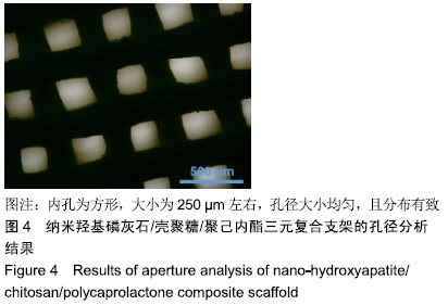
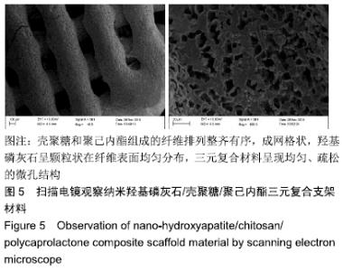
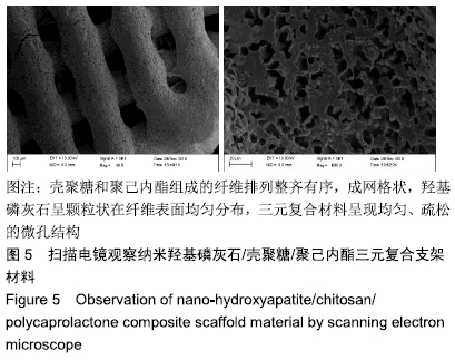
|