Chinese Journal of Tissue Engineering Research ›› 2021, Vol. 25 ›› Issue (3): 438-442.doi: 10.3969/j.issn.2095-4344.2940
Previous Articles Next Articles
Application of 3D printing technology in fracture and deformity of foot and ankle
Huang Youyi1, Yuan Wei2
- 1Medical Department of Nanchang University, Nanchang 330031, Jiangxi Province, China; 2Department of Orthopedics, the First Affiliated Hospital of China Medical University, Shenyang 110001, Liaoning Province, China
-
Received:2020-04-03Revised:2020-04-10Accepted:2020-05-09Online:2021-01-28Published:2020-11-18 -
About author:Huang Youyi, Medical Department of Nanchang University, Nanchang 330031, Jiangxi Province, China -
Supported by:the Liaoning Orthopedic Translational Medicine Research Center and Collaborative Research Network Construction Individual Design and Research & Development Project Based on 3D Printing Technology, No. 2015225014
CLC Number:
Cite this article
Huang Youyi, Yuan Wei. Application of 3D printing technology in fracture and deformity of foot and ankle[J]. Chinese Journal of Tissue Engineering Research, 2021, 25(3): 438-442.
share this article
Add to citation manager EndNote|Reference Manager|ProCite|BibTeX|RefWorks

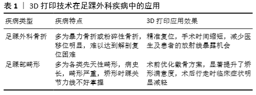
2.1 3D打印技术在足踝外科骨折中的应用 Zhang等[16]回顾了3D打印应用于治疗高能交叉踝关节骨折脱位——“Log-Splitter”损伤中的疗效及预后,发现与常规组相比,3D打印组在手术时间、术中出血量、透视次数、骨折愈合时间、美国足踝外科学会评分功能结果及术后并发症方面,具有更好的安全性和有效性,手术时间短,术中出血量少,所需透视少,功能优良率高。Yao等[17]分析了在应用3D打印技术基础上的跗骨窦入路微创治疗跟骨骨折的疗效,发现在支持钉、跟骨后关节面螺钉和长轴螺钉的定位和放置以及Bohler角、Gissane角和跟骨宽度等参数方面,准确率均有显著提高。一项关于3D打印技术在Pilon骨折中应用的有效性和安全性的Meta分析显示,在手术时间、出血量、术后功能评分、术后目测类比评分、优良率、解剖复位率等方面3D打印辅助手术均明显优于传统手术[18]。穆怀昭等[19]对3D打印应用于正处在发育中的胫骨远端骨折并累及到骺板的青少年进行了评估,得出以下结论:3D打印可制定出个体化的手术治疗方案,具有定位精确、组织损伤小、解剖复位率高、手术时间短、并发症少、射线暴露少、踝关节功能恢复好的优点。Yang 等[20]评价了3D打印治疗三踝骨折的疗效及其在医患沟通中的作用,在手术时间和术中出血量控制、患者的满意度及沟通满意度方面均较对照组显著改善。3D打印可以真实地反映骨折部位的解剖结构,有效地帮助医生规划手术方案,是医患沟通的有效工具。Hamid等[21]使用3D打印的脚手架在严重足踝创伤患者的初步抢救中发挥了重要作用;真实大小的3D打印预成型钢板模型也被Chung等[22]应用于跟骨骨折的微创固定中。 综上所述,踝关节骨折患者接受3D打印技术治疗具有下述显著优势: (1)临床医生根据3D模型个体化标本情况,能够初步避开重要血管、神经,选择手术过程中螺钉置入位置,并且对螺钉的置入长度和方向进行准确观察,最大限度缩短手术过程中测量所需时间,防止反复应用C型臂X射线机观察造成的复杂性,提高了外科医生的信心和把握,操作步骤更加简单且有效,明显减少了患者承受的X射线照射量[15,23]。 (2)传统手术治疗方案在钢板塑形过程中需要经铝板贴合骨面折出预弯雏形,并按照铝板模型进行钢板塑形,这样会明显降低塑形的精确性,延长手术治疗时间,且钢板的置入位置也不稳定。而利用3D打印技术能够早期选取切口、复位方法、内固定方式、内固定材料、钢板放置位置,而后进行术前钢板模拟塑形预弯,使用螺钉长度和置入方向选择提高钢板预弯的精确性,保证钢板置入位置的准确可靠,并提高手术内固定的有效性,明显缩短了手术治疗时间,从而增加手术的成功率[17,24]。 (3)3D打印技术能够对患者的骨折损伤程度、骨折情况和解剖结构进行准确观察,进而直观全面地反映患者的骨折复位前后情况,为患者临床治疗方案的制定提供可靠依据。尤其对于青少年来说,踝关节骨折如出现骺板损伤,若处理不当,术后极易出现肢体发育畸形、关节功能异常及障碍的后遗症[19]。采用3D打印技术,外科医生可以综合分析个体局部的骨折情况, 使用三维物体帮助患者局部骨折骨质恢复正常的生理解剖位置,创造更加微创的手术方式[17,25],可以有效减少术中出血量、术后住院时间及骨折愈合时间,有效减轻术后疼痛程度以及术后的一系列并发症的发生率,获得较好的康复疗效[26-27]。 (4)临床手术均存在一些术后风险,医生在与患者家属沟通时很难做到直观、全面地进行解释。医生利用3D打印模型可以轻松指出患者的问题所在,让患者及其家属对术后风险有一定了解,从而减轻患者的心理压力,提高患者对手术的整体满意度,减少医疗纠纷的发生,提高治疗效率[28-29]。 (5)即使是有着丰富临床经验的外科医师也无法避免失误。而仅仅依靠老一辈的经验方式来培养医生,要达到他们的水平需要非常长的时间,如果有了这种三维数字影像的帮助,培训时间则可以明显缩短[30-32];更重要的是,如果有一个工具能将手术门槛降低,让广大的基层医生也能开展高难度的手术,不需要把患者都集中到大医院来,就可以更好地造福群众,这让人民群众能够广泛地享受到高新科技为个人生命健康带来的福利[33-35]。 2.2 3D打印技术在足踝外科畸形类疾病中的应用 主要回顾了3D打印在高弓足、扁平足、拇外翻患者中的应用。 采用3D打印技术可以对高弓足患者进行个体化术前规划和手术模拟,包括截骨方案设计、内固定材料的选择和预塑形等。有利于手术医生做出最合理、最优化的术前规划,从而设计出更加精准的个体化手术方案。钱辉等[36]对6例高弓足畸形患者采用3D打印技术辅助截骨手术治疗,术后6例患者均足部畸形改善,行走步态稳定,术后目测类比评分、美国足踝外科学会踝-后足及中部足功能评分、Meary角、Hibbs角均优于术前,均未出现骨不愈合等严重并发症。应用 3D打印技术辅助截骨治疗成人高弓足可获得较好的疗效,其在术前规划截骨、内固定材料选择和预塑形、手术模拟操作等方面具有较多优势,可提高术者对手术的熟练度,从而缩短手术时间,提高手术成功率,减少并发症的发生。当然,3D打印技术辅助治疗在高弓足手术中也存在着不足。虽然 3D 打印技术在术前设计截骨方案、模拟骨重建及内固定选择方面有明显优势,但高弓足手术治疗除骨性手术外,还需考虑行跖筋膜松解、肌腱延长或肌腱转位术等软组织手术,不能完全依赖于 3D 打印辅助截骨技术,而忽视肌力平衡在高弓足治疗中的重要作用[37-38]。 高斌等[39]采集了11例复杂扁平足患儿足踝部CT断层扫描数据,并进行数字化三维重建和快速成型,在模型上进行模拟手术,并将其应用于诊治、手术规划和术中参照,平均随访20个月,所有患儿均在3D打印实体模型指导下完成,手术顺利,其中11例患儿术中所见与术前数字化三维重建和3D打印实体模型的畸形结构变化一致。说明数字化重建技术能够准确和直观地反映儿童复杂扁平足畸形的立体形态和解剖结构的空间关系,对临床医生的诊断、手术计划等有较大帮助,并减少手术操作时间,达到更好的手术效果。 张宇航等[40]探讨了3D打印技术制定个体化截骨角在拇外翻截骨矫形术中的临床应用,根据术前设计方案分为计算机截骨组和传统截骨组,结果显示计算机截骨组通过扫描负重状态下的患足获得的立体数据进行建模,更能体现拇外翻病理状态下的立体结构,从图像采集方面减少了相应的误差;2组手术时间、术中失血量、住院时间、术后1周疼痛评分等比较差异无显著性意义。 由此可见,3D打印技术辅助治疗相比于传统手术具有以下优势:见表1。 (1)与传统医学影像学资料(如 X射线片、CT、MRI)相比,3D打印实体模型有助于手术医师在术前更加深入、直观地了解患者病足畸形程度,评估相应的手术风险以及预测术后效果[41-45]。 (2)通过实体打印模型的呈现,术前医患沟通时能使患者及家属更加直观了解手术方案的设定以及存在的手术风险,更好地获得患者及家属的理解,良好的医患沟通能有效地缓解医患矛盾,有利于促进医患关系和谐发展。 (3)有利于制定个体化、精度化的手术方案。3D 打印可以迅速地打印出足部病变部位的实物模型,并可以根据术者需要,在实物模型上进行模拟截骨、模块切除及骨重建等,有利于手术医生做出最合理、最优化的术前规划,从而设计出更加精准的个体化手术方案[46]。 (4) 通过在3D实物模型上进行术前模拟手术操作,可提高术者对手术的熟练度,从而在实际操作中降低手术中出血量,缩短手术时间,减少医护和患者对放射线的暴露时间及剂量。同时术者可以根据手术需求,制作个体化手术器械,提高手术成功率,减少并发症的发生。 (5)3D打印技术除应用于打印实体模型之外,还可以精准制作出各类足部矫形器。相较于传统制作的足部矫形器,3D打印技术可以制作出更加符合人体结构的足部矫形器,对于人体足部矫形,3D打印技术是一个可替代的选择[47-50]。"
| [1] GOOST H, WIMMER MD, BARG A, et al. Fractures of the ankle joint: investigation and treatment options. Dtsch Arztebl Int. 2014;111(21):377-388. [2] EVANSKI PH, WAUGH TR. Management of arthritis of the ankle. An alternative of arthrodesis. Clin Orthop Relat Res. 1977;122: 110-115. [3] DOORNBERG JN, RADEMAKERS MV, VAN DEN BEKEROM MP, et al. Two-dimensional and three-dimensional computed tomography for the classification and characterisation of tibial plateau fractures. Injury. 2011; 42(12): 1416-1425. [4] BLOCH B, SRINIVASAN S, MANGWANI J. Current concepts in the management of ankle osteoarthritis: a systematic review. J Foot Ankle Surg. 2015;54(5):932-939. [5] NWANKWO EC, CHEN F, NETTLES DL, et al. Five-Year Follow-Up of Distal Tibia Bone and Foot and Ankle Trauma Treated with a 3D-Printed Titanium Cage. Case Rep Orthop 2019;2019:7571013. [6] GEORGE E, LIACOURAS P, RYBICKI FJ, et al. Measuring and Establishing the Accuracy and Reproducibility of 3D Printed Medical Models. Radiographics. 2017;37(5):1424-1450. [7] WONG KC, KUMTA SM, GEEL NV, et al. One-step reconstruction with a 3D-printed, biomechanically evaluated custom implant after complex pelvic tumor resection. Comput Aided Surg. 2015;20(1):14-23. [8] ARANDA JL, JIMÉNEZ MF, RODRÍGUEZ M, et al. Tridimensional titanium-printed custom-made prosthesis for sternocostal reconstruction. Eur J Cardiothorac Surg. 2015;48(4):e92-94. [9] WEADOCK WJ, HEISEL CJ, KAHANA A, et al. Use of 3D printed models to create molds for shaping implants for surgical repair of orbital fractures. Acad Radiol. 2020; 27(4):536-542. [10] MORRISON RJ, HOLLISTER SJ, NIEDNER MF, et al. Mitigation of tracheobronchomalacia with 3D-printed personalized medical devices in pediatric patients. Sci Transl Med, 2015;7(285):285ra64. [11] 苏暄, 刘忠军. 3D打印技术带来脊柱外科个体化治疗时代[J]. 中国医药科学, 2015,5(24):1-4. [12] MONTGOMERY SJ, KOONER SS, LUDWIG TE, et al. Impact of 3D printed calcaneal models on fracture understanding and confidence in orthopedic surgery residents. J Surg Educ. 2020;77(2):472-478. [13] LIN KW, HU CJ, YANG WW, et al. Biomechanical Evaluation and Strength Test of 3D-Printed Foot Orthoses. Appl Bionics Biomech. 2019;2019: 4989534. [14] SKELLEY NW, HAGERTY MP, STANNARD JT, et al. Sterility of 3D-printed orthopedic implants using fused deposition modeling. Orthopedics. 2020;43(1): 46-51. [15] MUNOZ-GUIJOSA JM, ZAPATA MARTÍNEZ R, MARTÍNEZ CENDRERO A, et al. Rapid prototyping of personalized articular orthoses by lamination of composite fibers upon 3D-printed molds. Materials. 2020;13(4):939-945. [16] ZHANG YW, XIAO X, XIAO Y, et al. Efficacy and Prognosis of 3D Printing Technology in Treatment of High-Energy Trans-Syndesmotic Ankle Fracture Dislocation - “Log-Splitter” Injury. Med Sci Monit. 2019; 25: 4233-4243. [17] YAO LF, WANG HQ, ZHANG F, et al. Minimally invasive treatment of calcaneal fractures via the sinus tarsi approach based on a 3D printing technique. Math Biosci Eng. 2019; 16(3):1597-1610. [18] BAI J, WANG Y, ZHANG P, et al. Efficacy and safety of 3D print-assisted surgery for the treatment of pilon fractures: a meta-analysis of randomized controlled trials. J Orthop Surg Res. 2018;13(1):283. [19] 穆怀昭, 杨朝昕, 赵佳佳, 等. 常规手术与应用3-D打印技术后手术治疗青少年胫骨远端骺损伤骨折的疗效比较[J]. 海南医学, 2018, 29(22): 3149-3152. [20] YANG L, SHANG XW, FAN JN, et al. Application of 3D printing in the surgical planning of trimalleolar fracture and doctor-patient communication. Biomed Res Int. 2016;2016:2482086. [21] HAMID KS, PAREKH SG, ADAMS SB. Salvage of Severe Foot and Ankle Trauma With a 3D Printed Scaffold. Foot Ankle Int. 2016; 37(4):433-439. [22] CHUNG KJ, HONG DY, KIM YT, et al. Preshaping plates for minimally invasive fixation of calcaneal fractures using a real-size 3D-printed model as a preoperative and intraoperative tool. Foot Ankle Int. 2014; 35(11):1231-1236. [23] DUAN X, HE P, FAN H, et al. Application of 3D-Printed Personalized Guide in Arthroscopic Ankle Arthrodesis. Biomed Res Int. 2018; 2018:3531293. [24] FOO GL, KWEK EBK. Are Three-Dimensional Printed Models Useful for Preoperative Planning of Tibial Plafond Fractures? J Foot Ankle Surg. 2019;58(4):723-729. [25] 房亚峰, 崔国峰, 魏戎. 3D打印辅助跗骨窦微创切口复位内固定治疗跟骨骨折的效果[J].河南医学研究, 2019, 28(17): 3120-3121. [26] 沙鹏. 3D打印个体化模型应用于复杂踝关节骨折治疗中的价值研究[J]. 双足与保健, 2019,28(21): 157-158. [27] 李新春, 康麟, 庞渊. 3D打印技术在Pilon骨折手术治疗中的应用[J]. 新疆医科大学学报, 2015, 38(4):471-473. [28] 胡钧亓, 李耀文, 张叶青, 等. 3D打印技术在临床医学中的应用进展[J]. 山东医药, 2019,59(35):106-109. [29] BIZZOTTO N, TAMI I, SANTUCCI A, et al. 3D Printed replica of articular fractures for surgical planning and patient consent: a two years multi-centric experience. 3D Print Med. 2015;2(1):2. [30] WU AM, WANG K, WANG JS, et al. The addition of 3D printed models to enhance the teaching and learning of bone spatial anatomy and fractures for undergraduate students: a randomized controlled study. Ann Transl Med. 2018; 6(20):403. [31] HASENSTEIN T, PATEL KP, MOORE JL, et al. Printed 3-Dimensional computed tomography scanned ankle fractures as an educational instrument. J Foot Ankle Surg. 2019; 58(6):1081-1084. [32] 李曙明, 李萌, 杨益民, 等. 3D打印及计算机辅助技术在骨科临床教学中的应用探讨——以复杂脊柱畸形治疗为例[J].中国医学教育技术, 2015, 29(2):191-194. [33] MAIER J, WEIHERER M, HUBER M, et al. Optically tracked and 3D printed haptic phantom hand for surgical training system. Quant Imaging Med Surg. 2020;10(2):340-355. [34] ZEMA L, MELOCCHI A, MARONI A, et al. Three-Dimensional Printing of medicinal products and the challenge of personalized therapy. J Pharm Sci. 2017;106(7):1697-1705. [35] PEHDE CE, BENNETT J, LEE PECK B, et al. Development of a 3-D printing laboratory for foot and ankle applications. Clin Podiatr Med Surg. 2020; 37(2):195-213. [36] 钱辉, 刘宗宝, 黄建平, 等. 3D打印辅助截骨治疗成人高弓足6例报告[J]. 广西医学, 2019,41(16):2035-2038. [37] WICART P. Cavus foot, from neonates to adolescents. Orthop Traumatol Surg Res. 2012;98(7):813-828. [38] KARAKIS I, GREGAS M, DARRAS BT, et al. Clinical correlates of Charcot-Marie-Tooth disease in patients with pes cavus deformities. Muscle Nerve. 2013;47(4):488-492. [39] 高斌, 孙巧巧, 杨明, 等. 3D打印技术辅助儿童复杂性扁平足手术临床应用[J]. 中国数字医学, 2015, 10(5):20-22. [40] 张宇航, 毕大卫, 陈亿民, 等. 3D打印技术制定个体化截骨角在拇外翻Chevron截骨矫形术中的应用[J]. 中国骨伤, 2018,31(3):203-207. [41] JOSEPH TN, MROCZEK KJ. Decision making in the treatment of hallux valgus. Bull NYU Hosp Jt Dis. 2007;65(1):19-23. [42] LARHOLT J, KILMARTIN TE. Rotational scarf and akin osteotomy for correction of hallux valgus associated with metatarsus adductus. Foot Ankle Int. 2010;31(3):220-228. [43] FERRER-TORREGROSA J, GARCIA-VICENTE S, FERNÁNDEZ-EHRLING N, et al. Design of a three-dimensional-printed surgical glove for minimal-incision podiatric surgery. J Am Podiatr Med Assoc. 2019; 109(3):207-214. [44] BELVEDERE C, SIEGLER S, FORTUNATO A, et al. New comprehensive procedure for custom-made total ankle replacements: Medical imaging, joint modeling, prosthesis design, and 3D printing. J Orthop Res. 2019; 37(3):760-768. [45] HIRAO M, IKEMOTO S, TSUBOI H, et al. Computer assisted planning and custom-made surgical guide for malunited pronation deformity after first metatarsophalangeal joint arthrodesis in rheumatoid arthritis: a case report. Comput Aided Surg. 2014; 19:13-19. [46] ZHANG YH, BI DW, CHEN YM, et al. Application of three-dimensional printing technology to design individual angle section on Chevron of hallux valgus osteotomy. Zhongguo gu shang. 2018;31(3):203-207. [47] MO S, LEUNG SHS, CHAN ZYS, et al. The biomechanical difference between running with traditional and 3D printed orthoses. J Sports Sci. 2019; 37(19):2191-2197. [48] XU R, WANG Z, REN Z, et al. Comparative Study of the Effects of Customized 3D printed insole and Prefabricated Insole on Plantar Pressure and Comfort in Patients with Symptomatic Flatfoot. Med Sci Monit. 2019;25:3510-3519. [49] WOJCIECHOWSKI E, CHANG AY, BALASSONE D, et al. Feasibility of designing, manufacturing and delivering 3D printed ankle-foot orthoses: a systematic review. J Foot Ankle Res. 2019; 12:11. [50] HOLTKAMP FC, WOUTERS EJM, VAN HOOF J, et al. Use of and satisfaction with ankle foot orthoses. Clin Res Foot Ankle. 2015; 3:1-8. [51] 周济. 智能制造——“中国制造2025”的主攻方向[J]. 中国机械工程, 2015, 26(17):2273-2284. [52] SUN YC, WAN Y, NAM R, et al. 4D-printed hybrids with localized shape memory behaviour: Implementation in a functionally graded structure. Sci Rep. 2019; 9(1):18754. [53] ZHANG Q, KUANG X, WENG S, et al. Rapid volatilization induced mechanically robust shape-morphing structures toward 4D printing. ACS Appl Mater Interfaces. 2020;12(15):17979-17987 [54] TAYLOR DL, IN HET PANHUIS M. Self-Healing Hydrogels. Adv Mater. 2016; 28(41):9060-9093. [55] SHIE MY, SHEN YF, ASTUTI SD, et al. Review of Polymeric Materials in 4D Printing Biomedical Applications. Polymers (Basel). 2019;11(11). pii: E1864. |
| [1] | Pu Rui, Chen Ziyang, Yuan Lingyan. Characteristics and effects of exosomes from different cell sources in cardioprotection [J]. Chinese Journal of Tissue Engineering Research, 2021, 25(在线): 1-. |
| [2] | Xu Feng, Kang Hui, Wei Tanjun, Xi Jintao. Biomechanical analysis of different fixation methods of pedicle screws for thoracolumbar fracture [J]. Chinese Journal of Tissue Engineering Research, 2021, 25(9): 1313-1317. |
| [3] | Jiang Yong, Luo Yi, Ding Yongli, Zhou Yong, Min Li, Tang Fan, Zhang Wenli, Duan Hong, Tu Chongqi. Von Mises stress on the influence of pelvic stability by precise sacral resection and clinical validation [J]. Chinese Journal of Tissue Engineering Research, 2021, 25(9): 1318-1323. |
| [4] | Lu Dezhi, Mei Zhao, Li Xianglei, Wang Caiping, Sun Xin, Wang Xiaowen, Wang Jinwu. Digital design and effect evaluation of three-dimensional printing scoliosis orthosis [J]. Chinese Journal of Tissue Engineering Research, 2021, 25(9): 1329-1334. |
| [5] | Zhang Tongtong, Wang Zhonghua, Wen Jie, Song Yuxin, Liu Lin. Application of three-dimensional printing model in surgical resection and reconstruction of cervical tumor [J]. Chinese Journal of Tissue Engineering Research, 2021, 25(9): 1335-1339. |
| [6] | Zhang Yu, Tian Shaoqi, Zeng Guobo, Hu Chuan. Risk factors for myocardial infarction following primary total joint arthroplasty [J]. Chinese Journal of Tissue Engineering Research, 2021, 25(9): 1340-1345. |
| [7] | Wei Wei, Li Jian, Huang Linhai, Lan Mindong, Lu Xianwei, Huang Shaodong. Factors affecting fall fear in the first movement of elderly patients after total knee or hip arthroplasty [J]. Chinese Journal of Tissue Engineering Research, 2021, 25(9): 1351-1355. |
| [8] | Wang Jinjun, Deng Zengfa, Liu Kang, He Zhiyong, Yu Xinping, Liang Jianji, Li Chen, Guo Zhouyang. Hemostatic effect and safety of intravenous drip of tranexamic acid combined with topical application of cocktail containing tranexamic acid in total knee arthroplasty [J]. Chinese Journal of Tissue Engineering Research, 2021, 25(9): 1356-1361. |
| [9] | Xiao Guoqing, Liu Xuanze, Yan Yuhao, Zhong Xihong. Influencing factors of knee flexion limitation after total knee arthroplasty with posterior stabilized prostheses [J]. Chinese Journal of Tissue Engineering Research, 2021, 25(9): 1362-1367. |
| [10] | Huang Zexiao, Yang Mei, Lin Shiwei, He Heyu. Correlation between the level of serum n-3 polyunsaturated fatty acids and quadriceps weakness in the early stage after total knee arthroplasty [J]. Chinese Journal of Tissue Engineering Research, 2021, 25(9): 1375-1380. |
| [11] | Zhang Chong, Liu Zhiang, Yao Shuaihui, Gao Junsheng, Jiang Yan, Zhang Lu. Safety and effectiveness of topical application of tranexamic acid to reduce drainage of elderly femoral neck fractures after total hip arthroplasty [J]. Chinese Journal of Tissue Engineering Research, 2021, 25(9): 1381-1386. |
| [12] | Wang Haiying, Lü Bing, Li Hui, Wang Shunyi. Posterior lumbar interbody fusion for degenerative lumbar spondylolisthesis: prediction of functional prognosis of patients based on spinopelvic parameters [J]. Chinese Journal of Tissue Engineering Research, 2021, 25(9): 1393-1397. |
| [13] | Lü Zhen, Bai Jinzhu. A prospective study on the application of staged lumbar motion chain rehabilitation based on McKenzie’s technique after lumbar percutaneous transforaminal endoscopic discectomy [J]. Chinese Journal of Tissue Engineering Research, 2021, 25(9): 1398-1403. |
| [14] | Chen Xinmin, Li Wenbiao, Xiong Kaikai, Xiong Xiaoyan, Zheng Liqin, Li Musheng, Zheng Yongze, Lin Ziling. Type A3.3 femoral intertrochanteric fracture with augmented proximal femoral nail anti-rotation in the elderly: finite element analysis of the optimal amount of bone cement [J]. Chinese Journal of Tissue Engineering Research, 2021, 25(9): 1404-1409. |
| [15] | Du Xiupeng, Yang Zhaohui. Effect of degree of initial deformity of impacted femoral neck fractures under 65 years of age on femoral neck shortening [J]. Chinese Journal of Tissue Engineering Research, 2021, 25(9): 1410-1416. |
| Viewed | ||||||
|
Full text |
|
|||||
|
Abstract |
|
|||||