[1] SUN HB. Mechanical loading, cartilage degradation, and arthritis. Ann N Y Acad Sci. 2010;1211:37-50.
[2] LIU X, SUN JQ, HEGGENESS MH, et al. Direct quantification of the rupture force of single hyaluronan/hyaluronan binding protein bonds. FEBS Lett. 2004;563(1-3):23-27.
[3] JI WC, ZHANG XW, QIU YS. Selected suitable seed cell, scaffold and growth factor could maximize the repair effect using tissue engineering method in spinal cord injury. World J Exp Med. 2016;6(3):58-62.
[4] LI X, YANG J, LIU D, et al. Knee loading inhibits osteoclast lineage in a mouse model of osteoarthritis. Sci Rep. 2016;6: 24668.
[5] HOENIG E, WINKLER T, MIELKE G, et al. High amplitude direct compressive strain enhances mechanical properties of scaffold-free tissue-engineered cartilage. Tissue Eng Part A. 2011;17(9-10):1401-1411.
[6] HUANG AH, FARRELL MJ, KIM M, et al. Long-term dynamic loading improves the mechanical properties of chondrogenic mesenchymal stem cell-laden hydrogel. Eur Cell Mater. 2010; 19:72-85.
[7] BONAVENTURE J, KADHOM N, COHEN-SOLAL L, et al. Reexpression of cartilage-specific genes by dedifferentiated human articular chondrocytes cultured in alginate beads. Exp Cell Res. 1994;212(1):97-104.
[8] LONG F, ORNITZ DM. Development of the endochondral skeleton. Cold Spring Harb Perspect Biol. 2013;5(1):a008334.
[9] SHEN G, DARENDELILER MA. The adaptive remodeling of condylar cartilage---a transition from chondrogenesis to osteogenesis. J Dent Res. 2005;84(8):691-699.
[10] NIMS RJ, CIGAN AD, DURNEY KM, et al. Constrained Cage Culture Improves Engineered Cartilage Functional Properties by Enhancing Collagen Network Stability. Tissue Eng Part A. 2017;23(15-16):847-858.
[11] KARAMESINIS K, SPYROPOULOU A, DALAGIORGOU G, et al. Continuous hydrostatic pressure induces differentiation phenomena in chondrocytes mediated by changes in polycystins, SOX9, and RUNX2. J Orofac Orthop. 2017;78(1): 21-31.
[12] HALBWIRTH F, NICULESCU-MORZSA E, ZWICKL H, et al. Mechanostimulation changes the catabolic phenotype of human dedifferentiated osteoarthritic chondrocytes. Knee Surg Sports Traumatol Arthrosc. 2015;23(1):104-111.
[13] YOU B, LI Q, DONG H, et al. Bilayered HA/CS/PEGDA hydrogel with good biocompatibility and self-healing property for potential application in osteochondral defectrepair. J Mater Sci Technol. 2018;34(6):1016-1025.
[14] 程潭,张柳,张英泽,等.大鼠膝关节不稳定骨性关节炎软骨与软骨下骨组织微观结构的变化[J].中国矫形外科杂志, 2012,20(3): 267-270.
[15] CARVER SE, HEATH CA. Semi-continuous perfusion system for delivering intermittent physiological pressure to regenerating cartilage. Tissue Eng. 1999;5(1):1-11.
[16] ASZODI A, HUNZIKER EB, BRAKEBUSCH C, et al. Beta1 integrins regulate chondrocyte rotation, G1 progression, and cytokinesis. Genes Dev. 2003;17(19):2465-2479.
[17] MAWATARI T, NAKAMICHI I, SUENAGA E, et al. Effects of heme oxygenase-1 on bacterial antigen-induced articular chondrocyte catabolism in vitro. J Orthop Res. 2013;31(12): 1943-1949.
[18] NAZEMPOUR A, QUISENBERRY CR, VAN WIE BJ, et al. Nanomechanics of Engineered Articular Cartilage: Synergistic Influences of Transforming Growth Factor-β3 and Oscillating Pressure. J Nanosci Nanotechnol. 2016;16(3):3136-3145.
[19] MALAVIYA P, NEREM RM. Fluid-induced shear stress stimulates chondrocyte proliferation partially mediated via TGF-beta1. Tissue Eng. 2002;8(4):581-590.
[20] DISCHER DE, MOONEY DJ, Zandstra PW. Growth factors, matrices, and forces combine and control stem cells. Science. 2009;324(5935):1673-1677.
[21] TIAN J, ZHANG FJ, LEI GH. Role of integrins and their ligands in osteoarthritic cartilage. Rheumatol Int. 2015;35(5): 787-798.
[22] ARGATOV I, MISHURIS G. Elliptical contact of thin biphasic cartilage layers: exact solution for monotonic loading. J Biomech. 2011;44(4):759-761.
[23] ZHOU Z, YU H, WANG Y, et al. ZNF606 interacts with Sox9 to regulate chondrocyte differentiation. Biochem Biophys Res Commun. 2016;479(4):920-926.
[24] TAO K, REY-RICO A, FRISCH J, et al. Effects of combined rAAV-mediated TGF-β and sox9 gene transfer and overexpression on the metabolic and chondrogenic activities in human bone marrow aspirates. J Exp Orthop. 2017;4(1):4.
[25] HARRISON A, LIN S, POUNDER N, et al. Mode & mechanism of low intensity pulsed ultrasound (LIPUS) in fracture repair. Ultrasonics. 2016;70:45-52.
[26] SIEBERS MC, TER BRUGGE PJ, WALBOOMERS XF, et al. Integrins as linker proteins between osteoblasts and bone replacing materials. A critical review. Biomaterials. 2005;26(2): 137-146.
[27] FRANCESCHI RT, XIAO G. Regulation of the osteoblast- specific transcription factor, Runx2: responsiveness to multiple signal transduction pathways. J Cell Biochem. 2003; 88(3):446-454.
[28] LOESER RF. Integrins and chondrocyte-matrix interactions in articular cartilage. Matrix Biol. 2014;39:11-16.
[29] TAKAHASHI I, MASUDA T, KOHSAKA K, et al. Molecular mechanisms of mechanical stress response during chondrogenesis. J Biomech Sci Eng. 2009;4(3):307-317.
[30] MOUSSA M, LAJEUNESSE D, HILAL G, et al. Platelet rich plasma (PRP) induces chondroprotection via increasing autophagy, anti-inflammatory markers, and decreasing apoptosis in human osteoarthritic cartilage. Exp Cell Res. 2017;352(1):146-156.
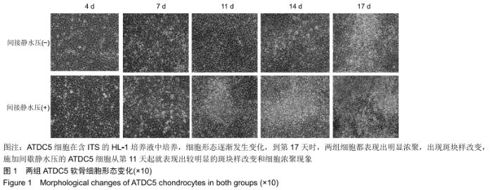
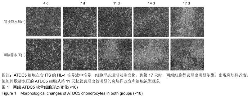
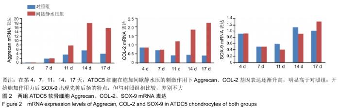
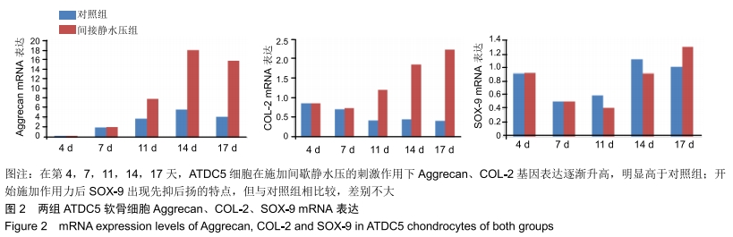
|