Chinese Journal of Tissue Engineering Research ›› 2020, Vol. 24 ›› Issue (7): 1076-1082.doi: 10.3969/j.issn.2095-4344.2019
Previous Articles Next Articles
Electrical stimulation combined with neurotrophin 3 promotes proliferation and differentiation of endogenous neural stem cells after spinal cord injury in rats
Zhang Peigen1, Heng Xiaolai1, Xie Di1, Wang Jin1, Ma Jinglin2, Kang Xuewen2
- 1Sichuan Mianyang 404 Hospital/the Second Affiliated Hospital of North Sichuan Medical College, Mianyang 621000, Sichuan Province, China; 2Key Laboratory of Orthopedics of Gansu Province (Lanzhou University), Lanzhou 730030, Gansu Province, China
-
Received:2019-06-14Revised:2019-06-18Accepted:2019-07-20Online:2020-03-08Published:2020-01-19 -
Contact:Kang Xuewen, Chief physician, Professor, Master’s supervisor, Key Laboratory of Orthopedics of Gansu Province (Lanzhou University), Lanzhou 730030, Gansu Province, China -
About author:Zhang Peigen, Master, Associate chief physician, Sichuan Mianyang 404 Hospital/the Second Affiliated Hospital of North Sichuan Medical College, Mianyang 621000, Sichuan Province, China -
Supported by:the Science and Technology Foundation for the Youth of Gansu Province, No. 145RJYD300
CLC Number:
Cite this article
Zhang Peigen, Heng Xiaolai, Xie Di, Wang Jin, Ma Jinglin, Kang Xuewen. Electrical stimulation combined with neurotrophin 3 promotes proliferation and differentiation of endogenous neural stem cells after spinal cord injury in rats[J]. Chinese Journal of Tissue Engineering Research, 2020, 24(7): 1076-1082.
share this article
2.1 实验动物数量分析 实验中动物无死亡脱落,96只SD大鼠均进入结果分析。 2.2 各组大鼠后肢运动能力 与假手术组相比,脊髓损伤组大鼠的BBB评分明显降低(P < 0.01)。与脊髓损伤组相比,电刺激组、电刺激+NT-3组大鼠后肢功能开始逐渐恢复,电刺激组BBB评分在造模后14,21,28 d高于脊髓损伤组(P < 0.05),电刺激+NT-3组在造模后7,14,21,28 d高于脊髓损伤组(P < 0.05)。电刺激+NT-3组造模后7,14,21,28 d的BBB评分明显高于电刺激组(P < 0.05)。具体BBB评分见表1。 "
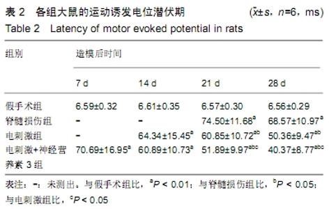
2.3 各组大鼠运动诱发电位潜伏期 脊髓损伤组大鼠7,14 d及电刺激组大鼠7 d时双后肢运动诱发电位潜伏期均未测出;电刺激组14,21,28 d时运动诱发电位潜伏期较假手术组明显延长(P < 0.05),21,28 d时运动诱发电位潜伏期较模型组缩短(P < 0.05);电刺激+NT-3组7,14,21,28 d时运动诱发电位潜伏期较假手术组明显延长(P < 0.05),21,28 d时运动诱发电位潜伏期较模型组缩短(P < 0.05);电刺激+NT-3组21,28 d时运动诱发电位潜伏期较电刺激组缩短(P < 0.05),见表2。 "
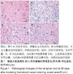
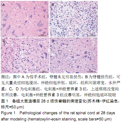
2.4 各组大鼠损伤脊髓的病理变化 假手术组大鼠脊髓未见明显损伤,细胞形态正常、排列整齐,无肿胀和出血;脊髓损伤组可见大量炎症细胞浸润,神经细胞肿胀、破坏,组织间隙增宽,水肿严重;电刺激组、电刺激+NT-3组大鼠上述病理改变均有所改善,电刺激+NT-3组改善明显,神经细胞破坏较轻,见图1。 "
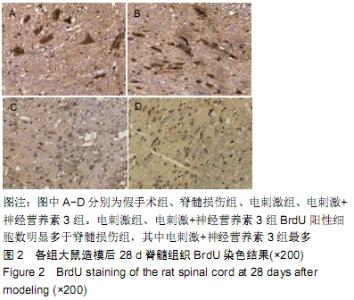
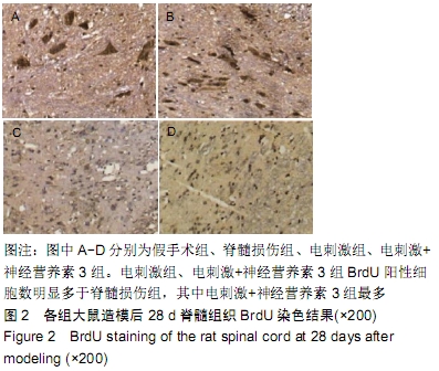
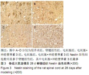
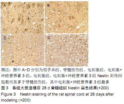
2.5 各组大鼠内源性神经干细胞的增殖情况 假手术组脊髓组织中BrdU阳性细胞和Nestin阳性细胞少见;而脊髓损伤后,脊髓组织中BrdU和Nestin阳性细胞数稍有增多;电刺激组、电刺激+NT-3组BrdU和Nestin阳性细胞数明显多于脊髓损伤组,电刺激+NT-3组最多,见图2,3。"
"
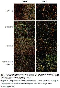
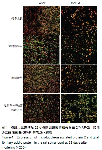
2.6 各组大鼠内源性神经干细胞的分化情况 与假手术组比较,脊髓损伤组微管相关蛋白2的表达增加,电刺激组、电刺激+NT-3组微管相关蛋白2的表达进一步增加,电刺激+ NT-3组最多;与假手术组比较,脊髓损伤组胶质纤维酸性蛋白的表达增加,电刺激组、电刺激+NT-3组胶质纤维酸性蛋白的表达较脊髓损伤组降低,电刺激+NT-3组最少,见图4。"
| [1] WITIW CD, FEHLINGS MG. Acute Spinal Cord Injury. J Spinal Disord Tech. 2015;28(6):202-210. [2] 王涛丽,顾兵,李华南,等.急性脊髓损伤后的炎症反应及其抗炎治疗[J].中国药理学通报,2015,31(4):452-457. [3] LIANG J, DENG G, HUANG H. The activation of BDNF reduced inflammation in a spinal cord injury model by TrkB/p38 MAPK signaling. Exp Ther Med. 2019;17(3): 1688-1696. [4] 王一吉,周红俊,李建军,等.脊髓损伤神经学分类国际标准检查表最新修订及解读[J].中国康复理论与实践,2015,21(8): 879-882. [5] JIN MC, MEDRESS ZA, AZAD TD, et al. Stem cell therapies for acute spinal cord injury in humans: a review. Neurosurg Focus. 2019;46(3):E10. [6] MOHAMADI Y, NOORI MOGHAHI SMH, MOUSAVI M, et al. Intrathecal transplantation of Wharton's jelly mesenchymal stem cells suppresses the NLRP1 inflammasome in the rat model of spinal cord injury. J Chem Neuroanat. 2019;97:1-8. [7] ZHAO Y, ZUO Y, JIANG J, et al. Neural stem cell transplantation combined with erythropoietin for the treatment of spinal cord injury in rats.Exp Ther Med. 2016;12(4): 2688-2694. [8] CHENG Z, ZHU W, CAO K, et al. Anti-Inflammatory Mechanism of Neural Stem Cell Transplantation in Spinal Cord Injury. Int J Mol Sci. 2016;17(9): E1380. [9] RUZICKA J, MACHOVA-URDZIKOVA L, GILLICK J, et al. A Comparative Study of Three Different Types of Stem Cells for Treatment of Rat Spinal Cord Injury. Cell Transplant. 2017; 26(4):585-603. [10] STENUDD M, SABELSTRÖM H, FRISÉN J. Role of endogenous neural stem cells in spinal cord injury and repair. JAMA Neurol. 2015;72(2):235-237. [11] KABATAS S, TENG YD. Potential roles of the neural stem cell in the restoration of the injured spinal cord: review of the literature. Turk Neurosurg. 2010;20(2):103-110. [12] SCHNELL L, SCHNEIDER R, KOLBECK R, et al. Neurotrophin-3 enhances sprouting of corticospinal tract during development and after adult spinal cord lesion. Nature. 1994;367(6459):170-173. [13] 姚立炜,冯世庆,孔晓红,等.大鼠脊髓损伤后脊髓组织中基质细胞衍生因子1α的时空分布[J].中国脊柱脊髓杂志,2013,23(9): 842-848. [14] 王维,马雪,赵晓晨,等.功能电刺激对脊髓损伤大鼠运动功能恢复和神经再生的影响[J].中华神经医学杂志,2014,13(1):17-21. [15] LAKSHMAN N, XU W, MORSHEAD CM. A Neurosphere Assay to Evaluate Endogenous Neural Stem Cell Activation in a Mouse Model of Minimal Spinal Cord Injury. J Vis Exp. 2018; (139). doi: 10.3791/57727. [16] YE J, QIN Y, TANG Y, et al. Methylprednisolone inhibits the proliferation of endogenous neural stem cells in nonhuman primates with spinal cord injury. J Neurosurg Spine. 2018; 29(2):199-207. [17] Ferrucci M, Ryskalin L, Busceti CL. Are there endogenous stem cells in the spinal cord?Arch Ital Biol. 2017;155(4): 118-130. [18] SANDQUIST EJ, SAKAGUCHI DS.Adult neural stem cell plasticity.Neural Regen Res. 2019;14(2):256-257. [19] ZHANG H, FANG X, HUANG D, et al. Erythropoietin signaling increases neurogenesis and oligodendrogenesis of endogenous neural stem cells following spinal cord injury both in vivo and in vitro.Mol Med Rep. 2018;17(1):264-272. [20] WANG H, MEI X, CAO Y, et al. HMGB1/Advanced Glycation End Products (RAGE) does not aggravate inflammation but promote endogenous neural stem cells differentiation in spinal cord injury. Sci Rep. 2017;7(1):10332. [21] AVERBUCH-HELLER L, PRUGININ M, KAHANE N, et al. Neurotrophin 3 stimulates the differentiation of motoneurons from avian neural tube progenitor cells. Proc Natl Acad Sci U S A. 1994;91(8):3247-3251. [22] RAMU J, BOCKHORST KH, GRILL RJ, et al. Cortical reorganization in NT3-treated experimental spinal cord injury: Functional magnetic resonance imaging. Exp Neurol. 2007; 204(1):58-65. [23] HANNA-MITCHELL AT, O'LEARY D, MOBARAK MS, et al. The impact of neurotrophin-3 on the dorsal root transitional zone following injury. Spinal Cord. 2008;46(12):804-810. [24] LI Z, WU H. Effects of human urine-derived stem cells combined with chondroitinase ABC on the expressions of nerve growth factor and brain-derived neurotrophic factor in the spinal cord injury. Zhongguo Xiu Fu Chong Jian Wai Ke Za Zhi. 2017;31(11):1377-1383. [25] HASNAN N, EKTAS N, TANHOFFER AI,et al. Exercise responses during functional electrical stimulation cycling in individuals with spinal cord injury .Med Sci Sports Exerc. 2013;45(6):1131-1138. [26] CAPPELLA P, GASPARRI F, PULICI M, et al. Cell Proliferation Method: Click Chemistry Based on BrdU Coupling for Multiplex Antibody Staining. Curr Protoc Cytom. 2015;72:7.34.1-17. [27] CAPPELLA P, GASPARRI F, PULICI M, et al. A novel method based on click chemistry, which overcomes limitations of cell cycle analysis by classical determination of BrdU incorporation, allowing multiplex antibody staining. Cytometry A. 2008;73(7):626-636. [28] BERNAL A, ARRANZ L. Nestin-expressing progenitor cells: function, identity and therapeutic implications. Cell Mol Life Sci. 2018;75(12):2177-2195. [29] LINDSAY SL, BARNETT SC. Are nestin-positive mesenchymal stromal cells a better source of cells for CNS repair? Neurochem Int. 2017;106:101-107. [30] MOTHE AJ, TATOR CH. Proliferation, migration, and differentiation of endogenous ependymal region stem/progenitor cells following minimal spinal cord injury in the adult rat. Neuroscience. 2005;131(1):177-187. [31] ZHOU L, BAUMGARTNER BJ, HILL-FELBERG SJ, et al. Neurotrophin-3 expressed in situ induces axonal plasticity in the adult injured spinal cord. J Neurosci. 2003;23(4): 1424-1431. [32] CHEN Q, ZHOU L, SHINE HD. Expression of neurotrophin-3 promotes axonal plasticity in the acute but not chronic injured spinal cord. J Neurotrauma. 2006; 23(8):1254-1260. [33] JOHNSON GV, JOPE RS. The role of microtubule-associated protein 2 (MAP-2) in neuronal growth, plasticity, and degeneration. J Neurosci Res. 1992;33(4):505-512. [34] SÁNCHEZ C, DÍAZ-NIDO J, AVILA J. Phosphorylation of microtubule-associated protein 2 (MAP2) and its relevance for the regulation of the neuronal cytoskeleton function. Prog Neurobiol. 2000;61(2):133-168. [35] BRENNER M, KISSEBERTH WC, SU Y, Besnard F, et al. GFAP promoter directs astrocyte-specific expression in transgenic mice.J Neurosci. 1994;14(3 Pt 1):1030-1037. [36] SALOUCI M, ANTOINE N, SHIKH AL SOOK MK, et al. Developmental profiles of GFAP-positive astrocytes in sheep cerebellum. Vet Res Commun. 2014;38(4):279-285. [37] DE MENEZES MF, NICOLA F, VITAL DA SILVA IR, et al.Glial fibrillary acidic protein levels are associated with global histone H4 acetylation after spinal cord injury in rats.Neural Regen Res. 2018;13(11):1945-1952. [38] BENESOVA J, HOCK M, BUTENKO O, et al. Quantification of astrocyte volume changes during ischemia in situ reveals two populations of astrocytes in the cortex of GFAP/EGFP mice. J Neurosci Res. 2009;87(1):96-111. |
| [1] | Min Youjiang, Yao Haihua, Sun Jie, Zhou Xuan, Yu Hang, Sun Qianpu, Hong Ensi. Effect of “three-tong acupuncture” on brain function of patients with spinal cord injury based on magnetic resonance technology [J]. Chinese Journal of Tissue Engineering Research, 2021, 25(在线): 1-8. |
| [2] | Jiang Hongying, Zhu Liang, Yu Xi, Huang Jing, Xiang Xiaona, Lan Zhengyan, He Hongchen. Effect of platelet-rich plasma on pressure ulcers after spinal cord injury [J]. Chinese Journal of Tissue Engineering Research, 2021, 25(8): 1149-1153. |
| [3] | Wan Ran, Shi Xu, Liu Jingsong, Wang Yansong. Research progress in the treatment of spinal cord injury with mesenchymal stem cell secretome [J]. Chinese Journal of Tissue Engineering Research, 2021, 25(7): 1088-1095. |
| [4] | Kong Desheng, He Jingjing, Feng Baofeng, Guo Ruiyun, Asiamah Ernest Amponsah, Lü Fei, Zhang Shuhan, Zhang Xiaolin, Ma Jun, Cui Huixian. Efficacy of mesenchymal stem cells in the spinal cord injury of large animal models: a meta-analysis [J]. Chinese Journal of Tissue Engineering Research, 2021, 25(7): 1142-1148. |
| [5] | Li Cai, Zhao Ting, Tan Ge, Zheng Yulin, Zhang Ruonan, Wu Yan, Tang Junming. Platelet-derived growth factor-BB promotes proliferation, differentiation and migration of skeletal muscle myoblast [J]. Chinese Journal of Tissue Engineering Research, 2021, 25(7): 1050-1055. |
| [6] | Ma Binxiang, He Wanqing, Zhou Guangchao, Guan Yonglin. Triptolide improves motor dysfunction in rats following spinal cord injury [J]. Chinese Journal of Tissue Engineering Research, 2021, 25(5): 701-706. |
| [7] | Sun Jianwei, Yang Xinming, Zhang Ying. Effect of montelukast combined with bone marrow mesenchymal stem cell transplantation on spinal cord injury in rat models [J]. Chinese Journal of Tissue Engineering Research, 2021, 25(25): 3962-3969. |
| [8] | Zhou Wu, Wang Binping, Wang Yawen, Cheng Yanan, Huang Xieshan. Transforming growth factor beta combined with bone morphogenetic protein-2 induces the proliferation and differentiation of mouse MC3T3-E1 cells [J]. Chinese Journal of Tissue Engineering Research, 2021, 25(23): 3630-3635. |
| [9] | Yan Peng, Ma Yufei, Cui Jingfu, Hao Shaofei, Liu Jinhui, Guan Chunlei, Wang Xiaoran, Yang Xiaoyu. Mechanism of anodic block electrical stimulation of sacral nerve root to reconstruct bladder function [J]. Chinese Journal of Tissue Engineering Research, 2021, 25(23): 3684-3689. |
| [10] | Xu Weilong, Zuo Yuan, Xin Daqi, He Chenyang, Zhao Peng, Shi Ming, Zhou Boyuan, Liu Yating, Zhao Yan. Selection of modeling methods for acute compressive spinal cord injury: a network Meta-analysis [J]. Chinese Journal of Tissue Engineering Research, 2021, 25(23): 3767-3772. |
| [11] | Tian Ting, Li Xiaoguang. Problems and challenges in regeneration and repair of spinal cord injury [J]. Chinese Journal of Tissue Engineering Research, 2021, 25(19): 3039-3048. |
| [12] | Ren Bingkai, Zheng Yibin, Huang Leiwen, Wu Fanhui, Yang Dong . Value of respiratory tract management and fiberoptic bronchoscopy in traumatic cervical spinal cord injury [J]. Chinese Journal of Tissue Engineering Research, 2021, 25(18): 2902-2907. |
| [13] | Chen Xingying, Hao Fei, Gao Yudan, Zhao Wen, Duan Hongmei, Yang Chaoyang, Li Xiaoguang. Rehabilitation training combined with neurotrophin 3-chitosan scaffolds enhanced skeletal muscle morphology and functional recovery in rats with spinal cord injury [J]. Chinese Journal of Tissue Engineering Research, 2021, 25(16): 2514-2520. |
| [14] | Wu Cunshu, Zhan Xiaoxuan, Zhao Siyi, Huang Fan, Zhang Yue, Qiu Mingwang, Xia Jingxian, Lu Xiaobo. Gait training for spinal cord injury based on radar plotting: an overview of systematic reviews [J]. Chinese Journal of Tissue Engineering Research, 2021, 25(14): 2287-2296. |
| [15] | Qian Nannan, Zhang Qian, Yang Rui, Ao Jun, Zhang Tao. Mesenchymal stem cells in the treatment of spinal cord injury: cell therapy and combination of new drugs and biomaterials [J]. Chinese Journal of Tissue Engineering Research, 2021, 25(13): 2114-2120. |
| Viewed | ||||||
|
Full text |
|
|||||
|
Abstract |
|
|||||