[1] Kretzer RM. A clinical perspective and definition of spinal cord injury. Spine. 2016;41:S27-S27.
[2] Sekhon LH, Fehlings MG. Epidemiology, demographics, and pathophysiology of acute spinal cord injury. Spine. 2001;26(24S):S2-S12.
[3] 陈星月,陈栋,陈春慧,等.中国创伤性脊髓损伤流行病学和疾病经济负担的系统评价[J].中国循证医学杂志,2018,18(2):143-150.
[4] THURET S, MOON LD, GAGE FH. Therapeutic interventions after spinal cord injury. Nat Rev Neurosci. 2006;7(8):628.
[5] SCHWAB ME. Repairing the injured spinal cord. Science. 2002;295(5557):1029-1031.
[6] LIU D, CHEN J, JIANG T, et al. Biodegradable spheres protect traumatically injured spinal cord by alleviating the glutamate-induced excitotoxicity. Adv Mater 2018;30(14):1706032.
[7] CHERIYAN T, RYAN DJ, WEINREB JH, et al. Errico Spinal cord injury models: a review. Spinal Cord. 2014;52(8):588.
[8] ZHANG N, FANG M, CHEN H, et al. Evaluation of spinal cord injury animal models. Neural Regen Res. 2014;9(22):2008-2012.
[9] Joshi M, Fehlings MG. Development and characterization of a novel, graded model of clip compressive spinal cord injury in the mouse: Part 2. Quantitative neuroanatomical assessment and analysis of the relationships between axonal tracts, residual tissue, and locomotor recovery. J Neurotraum. 2002;19(2):191-203.
[10] Marques SA, Garcez VF, Del Bel EA, et al. A simple, inexpensive and easily reproducible model of spinal cord injury in mice: morphological and functional assessment. J Neurosci Meth. 2009;177(1):183-193.
[11] Kontogeorgakos VA, Voulgaris S, Korompilias AV, et al. The efficacy of erythropoietin on acute spinal cord injury. An experimental study on a rat model. Arch. Orthop Traum Surg. 2009;129(2):189-194.
[12] LV H, YANG J, LIAN Z, et al. NG2 expression in rats with acute T10 spinal cord injury. Neural Regen Res. 2012;7(5):359-362.
[13] 陈匡阳,马彬,王亚楠,等.SYRCLE动物实验偏倚风险评估工具简介[J].中国循证医学杂志,2014,14(10):1281-1285.
[14] 闫加鹏.LY294002对脊髓损伤后胶质瘢痕形成的影响[D].福州:福建医科大学,2012.
[15] 谭亮.SD大鼠脊髓损伤后早期注射BMSCs对小胶质细胞影响的实验研究[D].福州:福建医科大学,2011.
[16] 康健.三种不同途径移植BMSCs对脊髓损伤后脊髓及小胶质细胞功能影响的研究[D].福州:福建医科大学,2012.
[17] 葛杜鹃.截瘫三联针对SD大鼠脊髓损伤神经功能保护及行为学的影响[D].成都:成都中医药大学,2013.
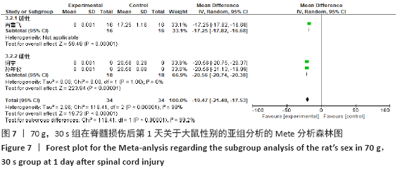
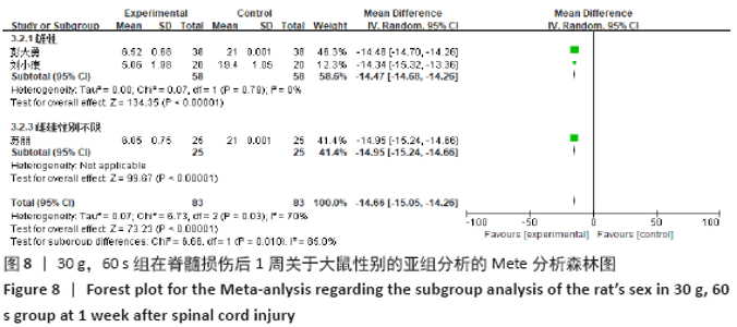
[18] 彭大勇.急性脊髓损伤的生物信息学分析及相关基因HIF-1α的表达分析[D].济南:山东大学,2015.
[19] 刘小康,徐建广,连小峰,等.大鼠钳夹式急性脊髓损伤模型的制备与评价[J].中国矫形外科杂志,2012,20(14):1318-1322.
[20] 苏朋.大鼠脊髓损伤后自噬的变化及高压氧对其影响的实验研究[D].苏州:苏州大学, 2013.
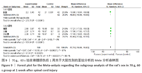
[21] 耿鑫.电针调控Notch信号通路修复大鼠脊髓损伤的实验研究[D].昆明:昆明医科大学, 2016.
[22] 谢勇.电针对大鼠脊髓损伤后Notch1蛋白表达的影响的实验研究[D].昆明:昆明医科大学,2014.
[23] 刘禹.电针对大鼠脊髓损伤后BMP-2/4/7的调控的实验研究[D].昆明:昆明医科大学,2014.
[24] 李经辉,黄辉,吴海鹰,等.钳夹型急性大鼠脊髓损伤模型的建立与评价[J].昆明理工大学学报(自然科学版),2012,37(6):67-71, 75.
[25] 何宇,张安仁,孙年怡,等.隔日限食疗法对脊髓损伤大鼠的保护作用及其机制[J].中国康复理论与实践,2018,24(2):153-159.
[26] 孙年怡,熊兴娟,何宇,等.隔日限食疗法可促进钳夹型脊髓损伤模型大鼠运动功能的恢复[J].中国组织工程研究,2018,22(4):564-569.
[27] 肖雪飞. 天麻素抑制大鼠脊髓损伤后胶质瘢痕形成及其促进神经功能恢复的研究[D]. 昆明:昆明医科大学,2017.
[28] MCDONALD JW, SADOWSKY C. Spinal-cord injury. Lancet. 2002;359(9304):417-425.
[29] DUMONT RJ, OKONKWO DO, VERMA S, et al. Acute spinal cord injury, part I: pathophysiologic mechanisms. Clin Neuropharmacol. 2001;24(5): 254-264.
[30] Datto JP, Bastidas JC, Miller NL, et al. Female rats demonstrate improved locomotor recovery and greater preservation of white and gray matter after traumatic spinal cord injury compared to males. J Neurotraum. 2015; 32(15):1146-1157.
[31] 闫慧博,鲁凯伍,金大地,等.性别对大鼠脊髓全横断模型影响的探讨[J].实验室研究与探索,2007,26(3):19-22.
[32] BASSO DM, BEATTIE MS, BRESNAHAN JC. A sensitive and reliable locomotor rating scale for open field testing in rats. J Neurotraum. 1995;12(1):1-21.
[33] POON PC, GUPTA D, SHOICHET MS, et al. Clip compression model is useful for thoracic spinal cord injuries: histologic and functional correlates. Spine. 2007;32(25):2853-2859.
[34] RIVLIN AS, TATOR CH. Effect of duration of acute spinal cord compression in a new acute cord injury model in the rat. Surg Neurol. 1978; 10(1):38-43.
[35] GAUDET AD, FONKEN LK, AYALA MT, et al. Spinal cord injury in rats disrupts the circadian system. eNeuro. 2018. doi: 10.1523/ENEURO.0328-18.2018.
|