Chinese Journal of Tissue Engineering Research ›› 2020, Vol. 24 ›› Issue (7): 985-990.doi: 10.3969/j.issn.2095-4344.1864
Neuronal differentiation of rat bone marrow mesenchymal stem cells via lentivirus-mediated bone morphogenetic protein 7 transfection
Zhang Wen, Lei Kun, Gao Lei, Li Kuanxin
- Department of Joint and Spine Surgery, the Second Affiliated Hospital of the Medical College of Shihezi University (Xinjiang Production and Construction Corps Hospital), Urumqi 830000, Xinjiang Uygur Autonomous Region, China
-
Received:2019-04-27Revised:2019-05-16Accepted:2019-06-22Online:2020-03-08Published:2020-01-19 -
Contact:Li Kuanxin, MD, Chief physician, Department of Joint and Spine Surgery, the Second Affiliated Hospital of the Medical College of Shihezi University (Xinjiang Production and Construction Corps Hospital), Urumqi 830000, Xinjiang Uygur Autonomous Region, China -
About author:Zhang Wen, Master candidate, Department of Joint and Spine Surgery, the Second Affiliated Hospital of the Medical College of Shihezi University (Xinjiang Production and Construction Corps Hospital), Urumqi 830000, Xinjiang Uygur Autonomous Region, China -
Supported by:the National Natural Science Foundation of China, No. 81560216
CLC Number:
Cite this article
Zhang Wen, Lei Kun, Gao Lei, Li Kuanxin. Neuronal differentiation of rat bone marrow mesenchymal stem cells via lentivirus-mediated bone morphogenetic protein 7 transfection[J]. Chinese Journal of Tissue Engineering Research, 2020, 24(7): 985-990.
share this article
Add to citation manager EndNote|Reference Manager|ProCite|BibTeX|RefWorks
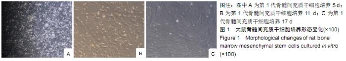
2.1 骨髓间充质干细胞培养形态变化 第1代骨髓间充质干细胞培养11 d,细胞主要表现为长纺锤体;培养17 d,细胞主要表现为梭形、多角形,聚集生长呈漩涡状,同向排列,细胞排列紧密,逐渐融合成片,见图1。"
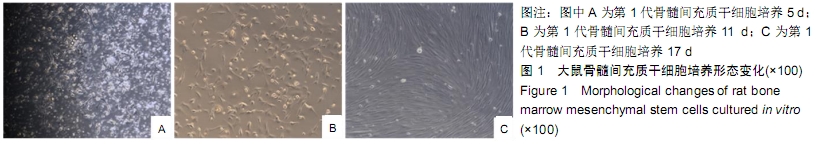
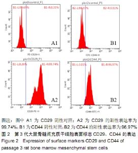
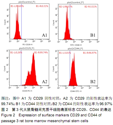
2.2 骨髓间充质干细胞流式细胞仪检测结果 第3代骨髓间充质干细胞高表达CD29、CD44,阳性率分别为99.74%和96.97%,见图2。"
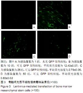
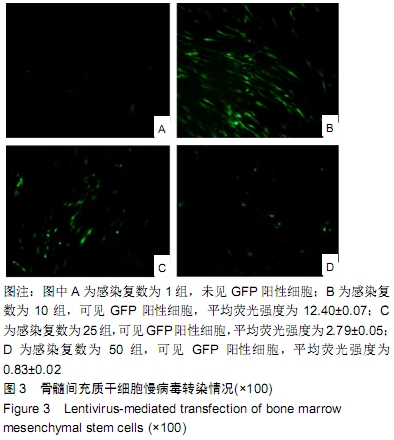
2.3 慢病毒转染情况 LV-GFP转染后3 d,感染复数为1组未见GFP阳性细胞,感染复数为10,25,50组可见GFP阳性细胞,平均荧光强度分别为12.40±0.07,2.79±0.05,0.83±0.02,其中感染复数为10组平均荧光强度高于其余组(P < 0.05),提示感染复数为10时感染效果最好,为最适感染复数;采用感染复数为10进行后续慢病毒转染实验,见图3。"
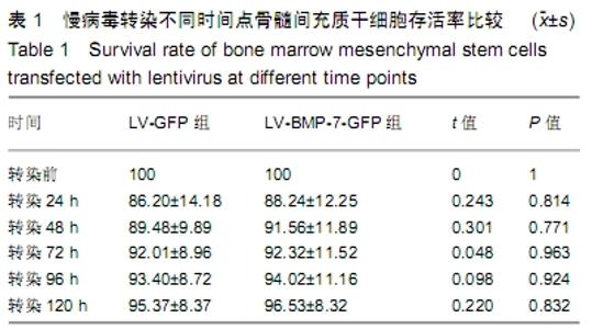
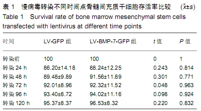
2.4 不同时间点骨髓间充质干细胞存活率 慢病毒转染后,LV-GFP组和LV-BMP-7-GFP组骨髓间充质干细胞存活率与转染前相比,差异无显著性意义(P > 0.05),表明慢病毒对骨髓间充质干细胞的增殖无明显影响,见表1。"
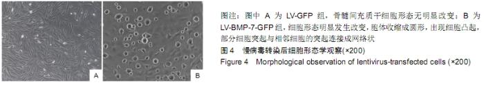
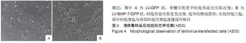
2.5 转染后细胞形态学变化 慢病毒转染骨髓间充质干细胞72 h后观察细胞形态,结果显示:LV-GFP组细胞形态学无明显改变,见图4A;LV-BMP-7-GFP组细胞形态学发生明显变化,胞体收缩成圆形,出现细胞突起,见图4B,呈典型的神经元样细胞。"
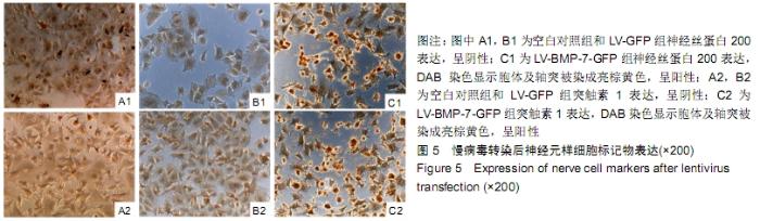
2.6 神经元样细胞标记物神经丝蛋白200、突触素1表达情况 空白对照组、LV-GFP组神经丝蛋白200、突触素1表达呈阴性;LV-BMP-7-GFP组可见神经丝蛋白200、突触素1表达呈阳性,胞体和轴突被染成亮棕黄色,见图5。 "

| [1] IDE C, NAKANO N, KANEKIYO K. Cell transplantation for the treatment of spinal cord injury - bone marrow stromal cells and choroid plexus epithelial cells. Neural Regen Res. 2016; 11(9):1385-1388. [2] XU J, LU H, MIAO ZN, et al. Immunoregulatory effect of neuronal-like cells in inducting differentiation of bone marrow mesenchymal stem cells. Eur Rev Med Pharmacol Sci. 2016; 20(24):5041-5048. [3] 荣为为,韩明子,金世柱,等.骨髓间充质干细胞在组织工程研究中的应用新进展[J].现代生物医学进展, 2017,17(5):982-984. [4] 冯烨军,陈明.骨髓间充质干细胞在神经修复中的研究进展[J].神经损伤与功能重建, 2016,11(5):426-428. [5] LIU F, PLACZEK M. Axon guidance effects of classical morphogens Shh and BMP7 in the hypothalamo-pituitary system. Neurosci Lett. 2014;562:108-113. [6] 李晓峰,赵劲民,苏伟,等.大鼠骨髓间充质干细胞的培养与鉴定[J].中国组织工程研究, 2011,15(10):1721-1725. [7] SANJURJO-RODRÍGUEZ C, MARTÍNEZ-SÁNCHEZ AH, HERMIDA-GÓMEZ T, et al. Differentiation of human mesenchymal stromal cells cultured on collagen sponges for cartilage repair. Histol Histopathol. 2016;31(11):1221-1239. [8] MEAD B, LOGAN A, BERRY M, et al. Concise Review: Dental Pulp Stem Cells: A Novel Cell Therapy for Retinal and Central Nervous System Repair. Stem Cells. 2017;35(1):61-67. [9] AQUINO-MARTÍNEZ R, ANGELO AP, PUJOL FV. Calcium-containing scaffolds induce bone regeneration by regulating mesenchymal stem cell differentiation and migration. Stem Cell Res Ther. 2017;8(1):265. [10] SOLEIMANNEJAD M, EBRAHIMI-BAROUGH S, SOLEIMANI M, et al. Fibrin gel as a scaffold for photoreceptor cells differentiation from conjunctiva mesenchymal stem cells in retina tissue engineering. Artif Cells Nanomed Biotechnol. 2018;46(4): 805-814. [11] WATANABE S, UCHIDA K, NAKAJIMA H, et al. Early transplantation of mesenchymal stem cells after spinal cord injury relieves pain hypersensitivity through suppression of pain-related signaling cascades and reduced inflammatory cell recruitment. Stem Cells. 2015;33(6):1902-1914. [12] OKUDA A, HORII-HAYASHI N, SASAGAWA T, et al. Bone marrow stromal cell sheets may promote axonal regeneration and functional recovery with suppression of glial scar formation after spinal cord transection injury in rats. J Neurosurg Spine. 2017;26(3):388-395. [13] RUZICKA J, MACHOVA-URDZIKOVA L, GILLICK J, et al. A Comparative Study of Three Different Types of Stem Cells for Treatment of Rat Spinal Cord Injury. Cell Transplant. 2017; 26(4): 585-603. [14] SINGH M, KAKKAR A, SHARMA R, et al. Synergistic Effect of BDNF and FGF2 in Efficient Generation of Functional Dopaminergic Neurons from human Mesenchymal Stem Cells. Sci Rep. 2017;7(1):10378. [15] 卞合涛,郝楼,黄卫. NT-3基因修饰骨髓间充质干细胞移植治疗脑梗死及其机制研究[J].中华神经医学杂志, 2017,16(9): 906-910. [16] 康湘萍,陈超,梁超,等.大鼠骨髓间充质干细胞培养、鉴定及神经样细胞分化[J].中国老年学杂志, 2018, 38(1):10-14. [17] HÖGEL F, HOFFMANN S, HUNGERER S, et al. Bone healing of critical size defects of the rat femur after the application of bone marrow aspirate and two different rh-BMP7 concentrations. Eur J Trauma Emerg Surg. 2015;41(5): 557-563. [18] COLE AE, MURRAY SS, XIAO J. Bone Morphogenetic Protein 4 Signalling in Neural Stem and Progenitor Cells during Development and after Injury. Stem Cells Int. 2016; 2016: 9260592. [19] PICKUP MW, OWENS P, MOSES HL. TGF-β, Bone Morphogenetic Protein, and Activin Signaling and the Tumor Microenvironment. Cold Spring Harb Perspect Biol. 2017;9(5):a022285. [20] LEUNG VYL, ZHOU L, TAM WK, et al. Bone morphogenetic protein-2 and -7 mediate the anabolic function of nucleus pulposus cells with discrete mechanisms. Connect Tissue Res. 2017;58(6):573-585. [21] DETTMAN RW, BIRCH D, FERNANDO A, et al. Targeted Knockdown of Bone Morphogenetic Protein Signaling within Neural Progenitors Protects the Brain and Improves Motor Function following Postnatal Hypoxia-Ischemia. Dev Neurosci. 2018;40(1):23-38. [22] YANG L, XU J, WANG Y, et al. Progress in the study on bone morphogenetic protein 2/4 in central nervous system. Zhong Nan Da Xue Xue Bao Yi Xue Ban. 2018;43(2):222-228. [23] COURTER LA, SHAFFO FC, GHOGHA A, et al. BMP7-induced dendritic growth in sympathetic neurons requires p75(NTR) signaling. Dev Neurobiol. 2016;76(9):1003-1013. [24] CHANDRASEKARAN V, LEA C, SOSA JC, et al. Reactive oxygen species are involved in BMP-induced dendritic growth in cultured rat sympathetic neurons. Mol Cell Neurosci. 2015; 67:116-125. [25] GUAN J, LI H, LV T, et al. Bone morphogenetic protein-7 (BMP-7) mediates ischemic preconditioning-induced ischemic tolerance via attenuating apoptosis in rat brain. Biochem Biophys Res Commun. 2013;441(3):560-566. [26] OKUDA A, HORII-HAYASHI N, SASAGAWA T, et al. Bone marrow stromal cell sheets may promote axonal regeneration and functional recovery with suppression of glial scar formation after spinal cord transection injury in rats. J Neurosurg Spine. 2017;26(3):388-395. [27] DONG Y, YANG L, YANG L, et al. Transplantation of neurotrophin-3-transfected bone marrow mesenchymal stem cells for the repair of spinal cord injury.Neural Regen Res. 2014;9(16): 1520-1524. [28] GRANSEE HM, ZHAN WZ, SIECK GC, et al. Localized delivery of brain-derived neurotrophic factor-expressing mesenchymal stem cells enhances functional recovery following cervical spinal cord injury. J Neurotrauma. 2015; 32(3):185-193. [29] MANTHOU M, ABDULLA DS, PAVLOV SP, et al. Whole body vibration (WBV) following spinal cord injury (SCI) in rats: Timing of intervention. Restor Neurol Neurosci. 2017;35(2): 185-216. [30] YIN YM, LU Y, ZHANG LX, et al. Bone marrow stromal cells transplantation combined with ultrashortwave therapy promotes functional recovery on spinal cord injury in rats. Synapse. 2015;69(3): 139-147. |
| [1] | Hou Jingying, Yu Menglei, Guo Tianzhu, Long Huibao, Wu Hao. Hypoxia preconditioning promotes bone marrow mesenchymal stem cells survival and vascularization through the activation of HIF-1α/MALAT1/VEGFA pathway [J]. Chinese Journal of Tissue Engineering Research, 2021, 25(7): 985-990. |
| [2] | Liang Xueqi, Guo Lijiao, Chen Hejie, Wu Jie, Sun Yaqi, Xing Zhikun, Zou Hailiang, Chen Xueling, Wu Xiangwei. Alveolar echinococcosis protoscolices inhibits the differentiation of bone marrow mesenchymal stem cells into fibroblasts [J]. Chinese Journal of Tissue Engineering Research, 2021, 25(7): 996-1001. |
| [3] | Geng Yao, Yin Zhiliang, Li Xingping, Xiao Dongqin, Hou Weiguang. Role of hsa-miRNA-223-3p in regulating osteogenic differentiation of human bone marrow mesenchymal stem cells [J]. Chinese Journal of Tissue Engineering Research, 2021, 25(7): 1008-1013. |
| [4] | Lun Zhigang, Jin Jing, Wang Tianyan, Li Aimin. Effect of peroxiredoxin 6 on proliferation and differentiation of bone marrow mesenchymal stem cells into neural lineage in vitro [J]. Chinese Journal of Tissue Engineering Research, 2021, 25(7): 1014-1018. |
| [5] | Zhu Xuefen, Huang Cheng, Ding Jian, Dai Yongping, Liu Yuanbing, Le Lixiang, Wang Liangliang, Yang Jiandong. Mechanism of bone marrow mesenchymal stem cells differentiation into functional neurons induced by glial cell line derived neurotrophic factor [J]. Chinese Journal of Tissue Engineering Research, 2021, 25(7): 1019-1025. |
| [6] | Pei Lili, Sun Guicai, Wang Di. Salvianolic acid B inhibits oxidative damage of bone marrow mesenchymal stem cells and promotes differentiation into cardiomyocytes [J]. Chinese Journal of Tissue Engineering Research, 2021, 25(7): 1032-1036. |
| [7] | Li Cai, Zhao Ting, Tan Ge, Zheng Yulin, Zhang Ruonan, Wu Yan, Tang Junming. Platelet-derived growth factor-BB promotes proliferation, differentiation and migration of skeletal muscle myoblast [J]. Chinese Journal of Tissue Engineering Research, 2021, 25(7): 1050-1055. |
| [8] | Wang Shiqi, Zhang Jinsheng. Effects of Chinese medicine on proliferation, differentiation and aging of bone marrow mesenchymal stem cells regulating ischemia-hypoxia microenvironment [J]. Chinese Journal of Tissue Engineering Research, 2021, 25(7): 1129-1134. |
| [9] | Chen Junyi, Wang Ning, Peng Chengfei, Zhu Lunjing, Duan Jiangtao, Wang Ye, Bei Chaoyong. Decalcified bone matrix and lentivirus-mediated silencing of P75 neurotrophin receptor transfected bone marrow mesenchymal stem cells to construct tissue-engineered bone [J]. Chinese Journal of Tissue Engineering Research, 2021, 25(4): 510-515. |
| [10] | Sun Jianwei, Yang Xinming, Zhang Ying. Effect of montelukast combined with bone marrow mesenchymal stem cell transplantation on spinal cord injury in rat models [J]. Chinese Journal of Tissue Engineering Research, 2021, 25(25): 3962-3969. |
| [11] | Zhang Lishu, Liu Anqi, He Xiaoning, Jin Yan, Li Bei, Jin Fang. Alpl gene affects the therapeutic effect of bone marrow mesenchymal stem cells on ulcerative colitis [J]. Chinese Journal of Tissue Engineering Research, 2021, 25(25): 3970-3975. |
| [12] | Hao Xiaona, Zhang Yingjie, Li Yuyun, Xu Tao. Bone marrow mesenchymal stem cells overexpressing prolyl oligopeptidase on the repair of liver fibrosis in rat models [J]. Chinese Journal of Tissue Engineering Research, 2021, 25(25): 3988-3993. |
| [13] | Jiang Tao, Ma Lei, Li Zhiqiang, Shou Xi, Duan Mingjun, Wu Shuo, Ma Chuang, Wei Qin. Platelet-derived growth factor BB induces bone marrow mesenchymal stem cells to differentiate into vascular endothelial cells [J]. Chinese Journal of Tissue Engineering Research, 2021, 25(25): 3937-3942. |
| [14] | Zhou Wu, Wang Binping, Wang Yawen, Cheng Yanan, Huang Xieshan. Transforming growth factor beta combined with bone morphogenetic protein-2 induces the proliferation and differentiation of mouse MC3T3-E1 cells [J]. Chinese Journal of Tissue Engineering Research, 2021, 25(23): 3630-3635. |
| [15] | Mo Jianling, He Shaoru, Feng Bowen, Jian Minqiao, Zhang Xiaohui, Liu Caisheng, Liang Yijing, Liu Yumei, Chen Liang, Zhou Haiyu, Liu Yanhui. Forming prevascularized cell sheets and the expression of angiogenesis-related factors [J]. Chinese Journal of Tissue Engineering Research, 2021, 25(22): 3479-3486. |
| Viewed | ||||||
|
Full text |
|
|||||
|
Abstract |
|
|||||