Chinese Journal of Tissue Engineering Research ›› 2017, Vol. 21 ›› Issue (34): 5481-5486.doi: 10.3969/j.issn.2095-4344.2017.34.012
Previous Articles Next Articles
Experimental research on bletilla carrying exogenous basic fibroblast growth factor that promotes wound healing
- 1Department of Medical Cosmetology, 2Department of Oncology, 3Office of Medical Affairs, 4Room for Dressing, Hebei General Hospital, Shijiazhuang 050051, Hebei Province, China
-
Received:2017-10-13Online:2017-12-08Published:2018-01-04 -
About author:Wang Xiao, Master, Associate chief physician, Department of Medical Cosmetology, Hebei General Hospital, Shijiazhuang 050051, Hebei Province, China
CLC Number:
Cite this article
Wang Xiao, Zhao Jing, Du Meng-ying, Sun Hai-juan, Gao Xiang-wei, Wu Ran.
share this article
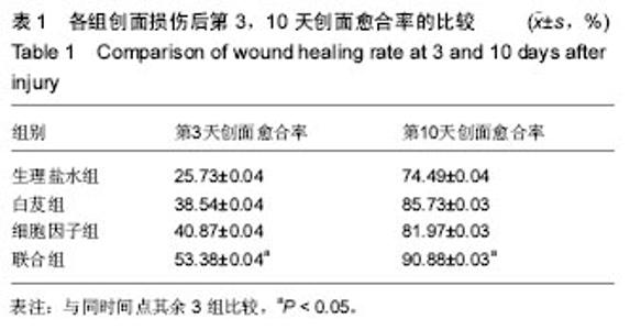
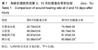
2.1 实验动物数量分析 纳入的40只新西兰大白兔均进入结果分析,无死亡和感染,无脱失。 2.2 各组创面愈合时间 生理盐水组、白芨组、细胞因子组和联合组的创面愈合时间分别为(17.50±0.59),(15.80±0.89),(15.60±0.56),(12.80±0.33) d。联合组创面愈合的时间比生理盐水组平均提前了4.5 d(P=0.038 4),比白芨组平均提前了3.0 d(P=0.046 7),比细胞因子组平均提前了2.8 d(P=0.025 0)。 2.3 各组创面愈合率 从表1可见,创面损伤后第3天,联合组伤口愈合率最高,生理盐水组最低,白芨组和细胞因子组相近,联合组创面愈合率高于细胞因子组(P=0.043 6)、白芨组(P=0.035 6)和生理盐水组(P=0.039 4);创面损伤后第10天,联合组创面愈合率最高,生理盐水组最低,白芨组和细胞因子组相近,联合组创面愈合率高于细胞因子组(P=0.039 2)、白芨组(P=0.037 2)和生理盐水组(P=0.043 5)。"

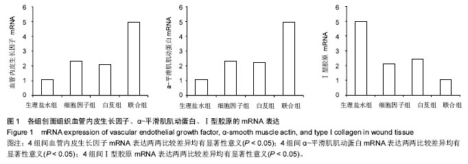
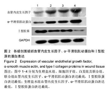
2.4 各组血管内皮生长因子、α-平滑肌肌动蛋白、Ⅰ型胶原的mRNA及蛋白表达水平 血管内皮生长因子、α-平滑肌肌动蛋白、Ⅰ型胶原的荧光定量PCR检测与Western blot结果基本一致。生理盐水组、细胞因子组、白芨组、联合组的血管内皮生长因子mRNA表达量(图1A)分别为:1.050±0.063、2.292±0.092、2.080±0.065、4.950±0.037,组间两两比较差异均有显著性意义(P < 0.05)。生理盐水组、细胞因子组、白芨组、联合组的α-平滑肌肌动蛋白mRNA表达量(图1B)分别为:1.046±0.075、2.347±0.049、2.200±0.081、4.956±0.075,组间两两比较差异均有显著性意义(P < 0.05)。生理盐水组、细胞因子组、白芨组、联合组的Ⅰ型胶原mRNA表达量(图1C)分别为:4.966± 0.045、2.125±0.079、2.444±0.038、1.035±0.064,组间两两比较差异均有显著性意义(P < 0.05)。各组血管内皮生长因子、α-平滑肌肌动蛋白、Ⅰ型胶原蛋白表达情况见图2,表达趋势与荧光定量PCR检测结果一致。"
"

| [1]Werner S,Grose R.Regulation of wound healing by growth factors and cytokines.Physiol Rev.2003;83(3):835-870.[2]程飚,刘宏伟,陈葵,等.碱性成纤维细胞生长因子对深Ⅱ°烫伤大鼠创面再上皮化过程中表皮干细胞的烫伤大鼠创面再上皮化过程中表皮干细胞的生物学作用[J].中华实验外科杂志,2007,24(8):1008-1011.[3]Brown AC,Adams D,de Caestecker M,et al.FGF/EGF signaling regulates the renewal of early nephron progenitors during embryonic development.Development. 2011;138(23):5099-5112.[4]Mie M,Sasaki S,Kobatake E.Construction of a bFGF-tethered multi-functional extracellular matrix protein through coiled-coil structures for neurite outgrowth induction.Biomed Mater.2014;9(1):015004.[5]Zhou W,Zhao M,Zhao Y,et al.A fibrin gel loaded with chitosan nanoparticles for local delivery of bFGF:preparation and in vitro release studies.J Mater Sci Mater Med.2011;22(5):1221-1230.[6]Bennett NT,Schultz GS.Growth factor and wound healing:PartⅡ.Role in normal and chronic wound healing.Am J Surg.1993;166(1):74-81.[7]Thalmann-Goetsch A,Engelmann K,Bednarz J.Comparative study on the effects of different growth factor on migration of bovine corneal endothelial cells during wound healing.Acta Ophthalmol Scand.1997; 75(5):490-495.[8]Eming SA,Medalie DA,Tompkins RG,et al.Genetically modified human keratinocytes overexpressing PDGF-A enhance the performance of a composite skin graft.Huu Gene Ther.1998;9(4):529-539.[9]常菲,吴杭庆,张毅,等.基因枪转人碱性成纤维细胞生长因子基因促进深Ⅱ度烧伤创面的愈合[J].中国组织工程研究与临床康复,2009,13(24):4611-4615.[10]曹永清,何春梅,陆金根,等.温脾健胃方对大鼠慢性创面愈合的影响[J].中西医结合学报,2005,3(3):220-224.[11]Lau TW,Lam FF,Lau KM,et al.Pharmacologicalinvestigationon the wound healingeffects of Radix Rehmanniae in an animal model of diabetic foot ulcer.J Ethnopharmacol.2009;123(1):155-162. [12]杨素清,张瀚月.白芨在常见皮肤疾病外治法中的应用[J].中国临床研究, 2014,6(10):33-35.[13]邱红梅,张颖,周岐新,等.白芨多糖对小鼠免疫功能的调节作用[J].中国生物制品学杂志,2011,24(6):676-678.[14]董莉,董永喜,刘星星,等.白芨多糖对大鼠血小板聚集、疑血功能及TXB2、6-keto-PGF1α表达的影响[J].贵阳医学院学报,2014,39(4):459-462.[15]任华忠,何毓敏,杨丽.白及化学成分及其药理活性研究进展[J].亚太传统医药,2009,5(2):134-140.[16]彭锐,李胜利,邹阳,等.明胶白芨胶/纳米血竭多孔材料对大鼠创面组织血管内皮细胞生长因子表达的影响[J].中国中医骨伤科杂志, 2007,15(6): 15-16.[17]王红英.白及甘露聚糖抗胃溃疡及抗炎、镇痛作用的实验研究[J].浙江中医药大学学报,2009,33(1):119-121.[18]悦随士,田河林,李丽鸣,等.白芨甘露聚糖胶浆治疗上消化道出血的临床观察[J].中国现代医学杂志, 2007,17(19):2375-2377.[19]马培耕,廖建中,庄雪芬.白芨胶载高分子神经生长因子治疗深度压疮的研究[J].中国现代医生,2014,52(14):50-52.[20]吕经纬.VEGF、bFGF浓度梯度对猪血管内皮细胞、成纤维细胞增殖移行的影响[D].南京医科大学鼓楼临床医学院:烧伤整形外科,2015.[21]宋瑞,卞徽宁,赖文,等.碱性成纤维细胞生长因子对增生性瘢痕与正常皮肤成纤维细胞胶原、纤维连接蛋白表达的影响[J].中国组织工程研究与临床康复,2010,14(41):7784-7789.[22]Jia MM,Li YS,Pei LJ,et al. Effect of San-huang-sheng-fu oil on wounds of full-thickness scald in rabbits.Zhonghua Shao Shang Za Zhi.2013; 29(1):50-54.[23]侯量,吴旭,王武军. 蜕皮甾酮对伤口促愈作用的初步研究[J].南方医科大学学报,2007,27(3):312-314.[24]王志刚,窦科峰,李海民,等.致康胶囊促进大鼠伤口愈合及与转化生长因子-β表达的关系[J].中华实验外科杂志, 2004,21(10):1197-1198.[25]Yu N,Zhai X,Xin C.An experimental study of rabbits' wound repair by amniotic carrier complex membrane containing bFGF and vitamin C and loaded with BMSCs.Zhongguo Xiu Fu Chong Jian Wai Ke Za Zhi. 2008;22(12):1495-1500.[26]Hemmati AA,Aghel N,Rashidi I,et al.Topical grape (Vitis vinifera) seed extract promotes repair of full thickness wound in rabbit.Int Wound J.2011;8(5):514-520.[27]林子洪,许环亲,胡婧晔,等.复方中药促进家兔皮肤创伤愈合的实验研究[J].实用医学杂志,2007,23(14):2129-2131.[28]付小兵,孙同柱,盛志勇.几种用于创伤修复研究的动物模型[J].中华实验外科杂志,1999,16(5):479-480.[29]宁浩勇,孟宇宏,王大鹏,等.重组人碱性成纤维细胞生长因子(Bfgf)促进海水浸泡兔软组织创伤愈合的实验研究[J].中国医药导刊, 2010,12(6): 1000-1001. [30]邱彦,鲁毅,段靖,等.血余炭纳米纤维膜促进家兔创面愈合的实验研究[J].药学实践杂志,2013,31(6):438-441,458.[31]Zimny S,Schatz H,Pfohl U.The effects of applied felted foam on wound healing and healing times in the therapy of neuropathic diabetic foot ulcers.Diabet Med. 2003;20(8):622-625.[32]Jessup RL.What is the best method for assessing the rate of wound healing? A comparison of 3 mathematical formulas.Adv Skin Wound Care.2006;19(3):138-147.[33]吴婷,陈金梅,李莹.实时荧光定量PCR技术研究进展及应用[J].大家健康,2014,8(4):12-13.[34]杨丕波.bFGF和EGF在大鼠皮肤及移植口腔粘膜创伤修复中的表达[D].华中科技大学:口腔临床医学,2013.[35]Xiao YF,Liu SX,Wu DD,et al.Inhibitory effect of arsenic trioxide on angiogenesis and expression of vascular endothelial growth factor in gastric cancer.World Journal of Gastroenterol.2006;12(36):5780-5786.[36]付小兵.现代创伤修复学[M].北京:人民军医出版社,1999.[37]陆树良.烧双创面愈合和新技术[M].北京:人民军医出版社,2003.[38]Spvingor I,Ninehoff P,Aacil YB.MP-2 and bFGF in an irradiated bone mode.Craniomaxillofac Surg.2008;4:210-217.[39]Chua KB,Aminuddin BS,Fuzina NH,et al.Basic fibroblast growth factor with human serum supplementation:enhancement of human chondrocyte proliferation and promotion of cattilage regeneration. Singapore Med J.2007;4:324-325.[40]程飚,刘宏伟,付小兵,等.碱性成纤维细胞生长因子对深Ⅱ度烫伤大鼠创面再上皮化过程中表皮干细胞的生物学作用[J].中华实验外科杂志, 2007, 24(8):1008-1011.[41]Pacicca DM,Patel N,Lee C,et al.Expression of angiogenic factors during distraction osteogenesis.Bone.2003;33:889-898.[42]Robinson CJ.Growth factors:threapeutic advance-s in wound healing.Ann Med.1993;25(6):535-538.[43]Claffey KP,Robins-on GS.Regulation of VEGF/VPF expression in tumor cells,consequences for tumor growth and metastasis.Cancer Metastasis Rev.1996;15(2):165-176.[44]杨登齐,李秀丽,孙进华,等.aFGF与bFGF治疗Ⅲ度烧伤的多中心随机Ⅳ期双盲临床阳性药物对照试验[J].中外医学研究, 2014,12(29):1-2.[45]Galeano M,Deodato B,Altavilla D,el at.Effect of recombinant adeno-associated virus vector-mediated vascular endothelial growth factor gene transfer on wound healing after burn injury.Crit Care Med.2003;31(4):1017-1025.[46]Deodhar AK,Rana RE,Surgical physiology of wound healing:a review.J Postgrad Med.1997;43:52-56.[47]Moulin V,Auger FA,Garrel D,et al.Role of wound healing myofibroblasts on re-epithelialization of human skin.Burns.2000;26(1):3-12.[48]Darby I,Skalli O,Gabbiani G..Alpha-smooth miscle actin is transiently expressed by myofibroblasts during experimental wound healing.Lab Invest.1990;63(1):21-29.[49]Betz P,Nerlich A,Wilske J,et al.Time-dependent appearance of myofibroblasts in granulation tissue of human skin wounds.Int J Legal Med.1992;105:99-103.[50]Badid C,Mounier N,Costa AM,et al.Role of myofibroblasts during normal tissue repair and excessive scaring:interest of their assessment in nephropathies.Histol Histopathol.2000;15:269-280.[51]Messadi DV,Le A,berg S,et al.Expression of apoptosis-associated genes by human dermal scar fibroblasts.Wound Repair Regen. 1999;7(6):511-517.[52]漆雪梅,刘柳.肌成纤维细胞与创伤的关系[J].中华医学美容杂志, 2001,7(2):109-111.[53]Funato N,Moriyama K,Shimokawa H,et al.Basic fibroblast growth factor induces apoptosis in myofibroblastic cells isolated from rat palatal mucosa.Biochem Biophys Res Commun.1997;240:21-26.[54]程飚,付小兵,盛志勇,等.碱性成纤维细胞生长因子对肌成纤维细胞作用及其对创面愈合的影响[J].中华医学杂志, 2002,82(17):1187-1191.[55]林丽,林邵强,王晓玉,等.bFGF对人脐带间充质干细胞增殖及胶原产生的影响[J].中国病理生理杂志,2013,29(3):509-514.[56]何威,刘荣卿,钟白玉.瘢痕疙瘩成纤维细胞胶原合成的实验研究[J].中国皮肤科杂志,1999,32(6):373-375. |
| [1] | Yao Xiaoling, Peng Jiancheng, Xu Yuerong, Yang Zhidong, Zhang Shuncong. Variable-angle zero-notch anterior interbody fusion system in the treatment of cervical spondylotic myelopathy: 30-month follow-up [J]. Chinese Journal of Tissue Engineering Research, 2022, 26(9): 1377-1382. |
| [2] | Zhang Jinglin, Leng Min, Zhu Boheng, Wang Hong. Mechanism and application of stem cell-derived exosomes in promoting diabetic wound healing [J]. Chinese Journal of Tissue Engineering Research, 2022, 26(7): 1113-1118. |
| [3] | An Weizheng, He Xiao, Ren Shuai, Liu Jianyu. Potential of muscle-derived stem cells in peripheral nerve regeneration [J]. Chinese Journal of Tissue Engineering Research, 2022, 26(7): 1130-1136. |
| [4] | Li Weiming, Xu Qingwen, Li Yijun, Sun Yanbo, Cui Jin, Xu Pengyuan . Deep seawater promotes wound healing in diabetic mice by activating PI3K/Akt pathway [J]. Chinese Journal of Tissue Engineering Research, 2022, 26(5): 724-729. |
| [5] | He Yunying, Li Lingjie, Zhang Shuqi, Li Yuzhou, Yang Sheng, Ji Ping. Method of constructing cell spheroids based on agarose and polyacrylic molds [J]. Chinese Journal of Tissue Engineering Research, 2022, 26(4): 553-559. |
| [6] | He Guanyu, Xu Baoshan, Du Lilong, Zhang Tongxing, Huo Zhenxin, Shen Li. Biomimetic orientated microchannel annulus fibrosus scaffold constructed by silk fibroin [J]. Chinese Journal of Tissue Engineering Research, 2022, 26(4): 560-566. |
| [7] | Chen Xiaoxu, Luo Yaxin, Bi Haoran, Yang Kun. Preparation and application of acellular scaffold in tissue engineering and regenerative medicine [J]. Chinese Journal of Tissue Engineering Research, 2022, 26(4): 591-596. |
| [8] | Kang Kunlong, Wang Xintao. Research hotspot of biological scaffold materials promoting osteogenic differentiation of bone marrow mesenchymal stem cells [J]. Chinese Journal of Tissue Engineering Research, 2022, 26(4): 597-603. |
| [9] | Shen Jiahua, Fu Yong. Application of graphene-based nanomaterials in stem cells [J]. Chinese Journal of Tissue Engineering Research, 2022, 26(4): 604-609. |
| [10] | Zhang Tong, Cai Jinchi, Yuan Zhifa, Zhao Haiyan, Han Xingwen, Wang Wenji. Hyaluronic acid-based composite hydrogel in cartilage injury caused by osteoarthritis: application and mechanism [J]. Chinese Journal of Tissue Engineering Research, 2022, 26(4): 617-625. |
| [11] | Li Hui, Chen Lianglong. Application and characteristics of bone graft materials in the treatment of spinal tuberculosis [J]. Chinese Journal of Tissue Engineering Research, 2022, 26(4): 626-630. |
| [12] | Gao Cangjian, Yang Zhen, Liu Shuyun, Li Hao, Fu Liwei, Zhao Tianyuan, Chen Wei, Liao Zhiyao, Li Pinxue, Sui Xiang, Guo Quanyi. Electrospinning for rotator cuff repair [J]. Chinese Journal of Tissue Engineering Research, 2022, 26(4): 637-642. |
| [13] | Guan Jian, Jia Yanfei, Zhang Baoxin , Zhao Guozhong. Application of 4D bioprinting in tissue engineering [J]. Chinese Journal of Tissue Engineering Research, 2022, 26(3): 446-455. |
| [14] | Liu Jiali, Suo Hairui, Yang Han, Wang Ling, Xu Mingen. Influence of lay-down angles on mechanical properties of three-dimensional printed polycaprolactone scaffolds [J]. Chinese Journal of Tissue Engineering Research, 2022, 10(16): 2612-2617. |
| [15] | Huang Bo, Chen Mingxue, Peng Liqing, Luo Xujiang, Li Huo, Wang Hao, Tian Qinyu, Lu Xiaobo, Liu Shuyun, Guo Quanyi . Fabrication and biocompatibility of injectable gelatin-methacryloyl/cartilage-derived matrix particles composite hydrogel scaffold [J]. Chinese Journal of Tissue Engineering Research, 2022, 10(16): 2600-2606. |
| Viewed | ||||||
|
Full text |
|
|||||
|
Abstract |
|
|||||