Chinese Journal of Tissue Engineering Research ›› 2017, Vol. 21 ›› Issue (34): 5487-8492.doi: 10.3969/j.issn.2095-4344.2017.34.013
Previous Articles Next Articles
Preparation of porcine small intestinal submucosa sponge and observation of cell adhesion
- 1Center for Medical Education Research, 2Department of Human Anatomy, Shenyang Medical College, Shenyang 110034, Liaoning Province, China; 3Shenyang Medical College, Shenyang 110034, Liaoning Province, China
-
Received:2017-10-30Online:2017-12-08Published:2018-01-04 -
About author:Sun Hui-zhe, Master, Associate professor, Center for Medical Education Research, Shenyang Medical College, Shenyang 110034, Liaoning Province, China -
Supported by:the Science Research General Project of Liaoning Provincial Department of Education in 2014, No. L2014416
CLC Number:
Cite this article
Sun Hui-zhe, Tian Wei, Zeng Liang, Qiu Jin-yun, Zhang Qian.
share this article
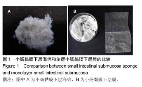
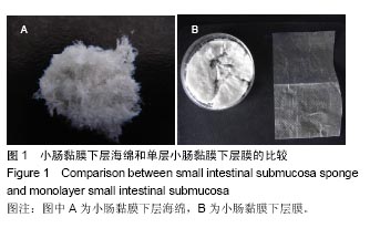
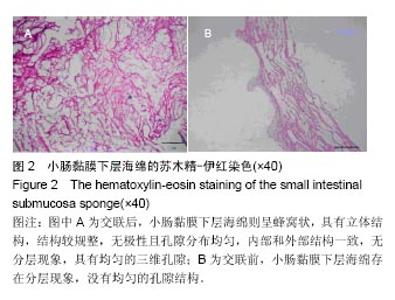
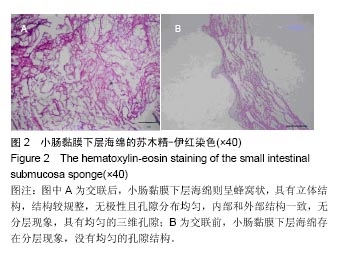
2.1 小肠黏膜下层海绵的苏木精-伊红染色观察结果 小肠黏膜下层海绵与小肠黏膜下层膜的大体结构见图1。苏木精-伊红染色显示,交联前,小肠黏膜下层海绵存在分层现象,没有均匀的孔隙结构;交联后,小肠黏膜下层海绵则呈蜂窝状,具有立体结构,结构较规整,无极性且孔隙分布均匀,内部和外部结构一致,无分层现象,具有均匀的三维孔隙(与小肠黏膜下层膜对照),并未发现蓝染的核物质和细胞碎片,不同质量浓度的小肠黏膜下层海绵结构无差别,见图2。"
"
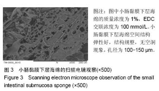
2.2 小肠黏膜下层海绵的扫描电镜观察结果 随着小肠黏膜下层海绵质量浓度的增高,其孔径密度与大小出现了变化,当其质量浓度在1%时,在100 mmol/L EDC作用下的空间结构弹性好,结构规整,无空洞现象,孔径为100-150 µm;当其质量浓度2%时,孔径大小为150- 200 µm,此后随其质量浓度的增高,孔径值逐渐下降,并且结构出现松散现象,见图3。随着EDC浓度的增加,海绵内部逐渐从松散状态转变为互相交织的现象,形成大量较均匀的三维孔隙,尤其以100 mmol/L显著,此后随着EDC浓度升高,在孔隙交界处出现了空洞。"


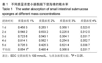
2.3 小肠黏膜下层海绵吸水率的测定结果 质量浓度1%小肠黏膜下层海绵(100 mmol/L EDC交联)的吸水率最高可达到80%,见表1,这样的变化可能与小肠黏膜下层海绵交联后的亲水能力增加,即羧基增加有关,并且多孔样结构的增加导致吸水能力的变化。 从平均值看,质量浓度1%小肠黏膜下层海绵的吸水能力比2%高出0.35倍,质量浓度3%与4%小肠黏膜下层海绵的吸水能力相差不明显,说明质量浓度1%小肠黏膜下层海绵的结构更利于水分的流动与变化,结果可得出如下结论:小肠黏膜下层海绵的浓度大小决定着吸水性,在一定质量浓度下,孔径大小和吸水力呈正比,但是当质量浓度到了4%时吸水力已没有了明显改变。"
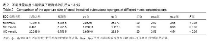
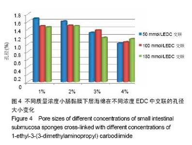
2.4 小肠黏膜下层海绵孔径大小的测量结果 冷冻干燥法制出的小肠黏膜下层海绵是多孔连通性分布的,质量浓度1%组支架的孔径为100-150 μm,质量浓度2%组的孔径为150-200 μm,其他两质量浓度组的孔径逐渐下降,此外,以质量浓度1%组支架各层之间的连接多数是较细的纤维般结构,导致整个支架内部结构较粗糙,利于细胞黏附(表2)。实验观察结果显示,在100 mmol/L EDC交联下1%质量分数的小肠黏膜下层海绵效果最好。 在50 mmol/L EDC交联下,质量浓度1%-2%组小肠黏膜下层海绵的孔径减少较小,2%-3%组孔径大小减小增大,3%-4%组孔径依旧减小,但无2%-3%组变化大。在100 mmol/L EDC交联下,质量浓度1%-2%组小肠黏膜下层海绵的孔径几乎无明显差异,2%-4%组孔径缩小趋势变大。在150 mmol/L EDC交联下,质量浓度1%-2%组肠黏膜下层海绵的孔径有细微增大,2%-3%组孔径明显缩小,3%-4%组孔径缩小程度减小(图4)。"

"
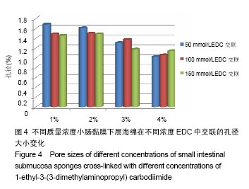
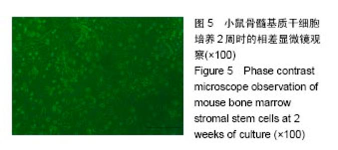
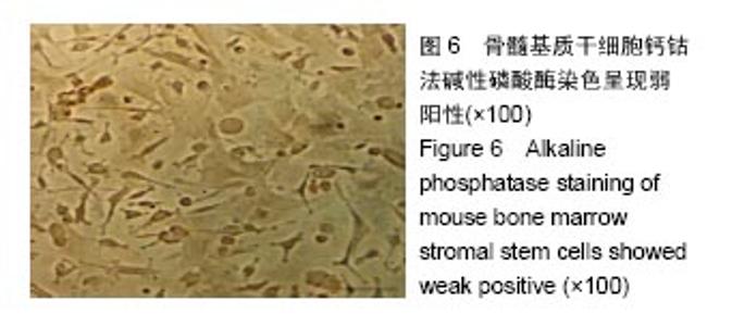
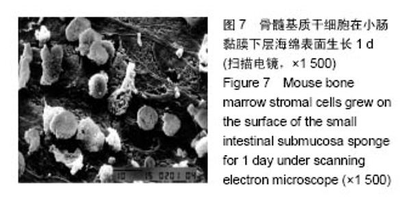
随着EDC交联浓度的升高,质量浓度1%组小肠黏膜下层海绵的孔径逐渐减少,2%组的孔径逐渐减少;3%组的孔径在交联浓度50-100 mmol/L时增大,在交联浓度100-150 mmol/L时明显缩小;4%组的孔径逐渐增大。 2.5 骨髓间充质干细胞培养结果 刚刚接种时,骨髓细胞悬液中的细胞大小不一,呈圆形,无法辨认其细胞核,2 d之后细胞开始贴壁,以多角形为主,还有梭形与三角形,悬浮血细胞在换液后逐渐被清除,纯化过程在2次换液后基本完成。细胞集落约在7 d后形成,没有接触抑制,大多数层次不清,形状不规则,2周后细胞则长满全层,见图5。 细胞鉴定:将1周的细胞传代,培在养瓶中放入多聚赖氨酸包被的载玻片,铺满后,将其取出,钙钴法碱性磷酸酶染色呈弱阳性,见图6。扫描电镜观察,小肠黏膜下层海绵上的骨髓基质干细胞多为星形和多角形,与光镜观察结果一致,见图7。"
"

"

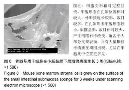
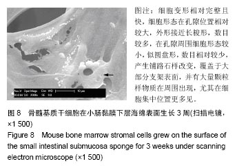
2.6 骨髓基质干细胞与小肠黏膜下层海绵的联合培养结果 培养1周时,细胞在海绵中都表现为圆形,无明显变形和贴服,于支架内分布均匀且大小一致,局部出现细胞集中现象和重叠分布,细胞生成许多凸起及接触;培养2周时,细胞形态由圆形变成长梭形和星形,重叠现象在孔隙附近更为多见,产生铺路石样改变,覆盖于小部分支架表面,并有颗粒样物质在周围出现,尤其在细胞集中位置;培养3周时,细胞变形相对完整且快,细胞形态在孔隙位置相对较大,外形接近长梭形,数目较多,在孔隙周围细胞形态较小,似圆盘形,数目相对较少,产生铺路石样改变,覆盖于大部分支架表面,并有大量颗粒样物质在周围出现,尤其在细胞集中位置更多见(图8)。"
| [1]Yang K,Zhang Y,Zhang N,et al.Recent progress of small intestinal submucosa in application research of tissue repair and reconstruction.Zhongguo Xiu Fu Chong Jian Wai Ke Za Zhi.2013;27(9):1138-1143.[2]Lu HK,Ren AJ,Sun XL,et al.Compound graft of porcine small intestinal submucosa with Schwann cells to reconstruct injured cavernous nerves and restore erectile function. Zhonghua Nan Ke Xue.2010;16(9):834-839.[3]Shi L,Ronfard V.Biochemical and biomechanical characterization of porcine small intestinal submucosa (SIS): a mini review.Int J Burns Trauma.2013;3(4):173-179.[4]Horiguchi A.Editorial comment to outcome of small intestinal submucosa graft for repair of anterior urethral strictures.Int J Urol.2013;20(6):629-630.[5]Villoldo GM,Loresi M,Giudice C,et al.Histologic changes after urethroplasty using small intestinal submucosa unseeded with cells in rabbits with injured urethra.Urology.2013; 81(6): 1380-1381.[6]孙慧哲,田伟,曾亮,等.参考猪小肠黏膜下基质海绵的制备[J].中国组织工程研究,2016,20(21):3110-3116.[7]董鸿鸣,柏树令.小鼠骨髓基质干细胞的培养及常见的组化染色方法[J].中国医科大学学报,2004,33(4):294-298.[8]卢洪凯,任安吉,孙晓璐,等.复合雪旺细胞的猪小肠粘膜下层对大鼠海绵体神经损伤勃起功能恢复的实验研究[J].中华男科学杂志,2010(9):834-839.[9]张开刚,曾炳芳,张长青.小肠粘膜下层的制备及细胞相容性的实验研究[J].中华创伤骨科杂志,2005,7(4):344-348.[10]Ding JX,Zhang XY,Chen LM,et al.Vaginoplasty using acellular porcine small intestinal submucosa graft in two patients with Meyer-von-Rokitansky-Kuster-Hauser syndrome: a prospective new technique for vaginal reconstruction.Gynecol Obstet Invest.2013;75(2):93-96.[11]苏琰,张长青,张开刚,等.小肠粘膜下层与雪旺细胞生物相容性的研究[J].中华创伤骨科杂志,2007,9(2):153-156.[12]Lin HK,Godiwalla SY,Palmer B,et al.Understanding roles of porcine small intestinal submucosa in urinary bladder regeneration: identification of variable regenerative characteristics of small intestinal submucosa.Tissue Eng Part B Rev.2014;20(1):73-83. [13]Roos S,Wyder M,Candi A,et al.Binding studies on isolated porcine small intestinal mucosa and in vitro toxicity studies reveal lack of effect of C.perfringens beta-toxin on the porcine intestinal epithelium.Toxins(Basel).2015;7(4):1235-1252.[14]Ramos CM,Francisco JC,Olandoski M,et al.Myocardial regeneration after implantation of porcine small intestinal submucosa in the left ventricle.Rev Bras Cir Cardiovasc.2014; 29(2):202-213. [15]Winskel H,Ratitamkul T,Charoensit A.The role of tone and segmental information in visual-word recognition in Thai.Q J Exp Psychol(Hove).2017;70(7):1282-1291. [16]Yi JS,Lee HJ,Lee HJ,et al.Rat peripheral nerve regeneration using nerve guidance channel by porcine small intestinal submucosa.J Korean Neurosurg Soc.2013;53(2):65-71.[17]Mondalek FG,Lawrence BJ,Kropp BP,et al.Author manuscript; available in PMC 2010 September 30. Published in final edited form as: Biomaterials.Biomaterials. 2008;29(9): 1159-1166.[18]Iwasaki J,Hata T,Uemoto S,et al.Portocaval shunt for hepatocyte package: challenging application of small intestinal graft in animal models.Organogenesis.2013;9(4):273-279.[19]Liu Z,Feng X,Wang H,et al.Carbon nanotubes as VEGF carriers to improve the early vascularization of porcine small intestinal submucosa in abdominal wall defect repair.Int J Nanomedicine.2014;9:1275-1286. [20]Hoeppner J,Marjanovic G,Helwig P,et al.Extracellular matrices for gastrointestinal surgery: ex vivo testing and current applications.World J Gastroenterol.2010;16(32):4031-4038.[21]Song Z,Peng Z,Liu Z,et al.Reconstruction of abdominal wall musculofascial defects with small intestinal submucosa scaffolds seeded with tenocytes in rats.Tissue Eng Part A. 2013;19(13-14):1543-1553. [22]Lin HK,Godiwalla SY,Palmer B,et al.Understanding roles of porcine small intestinal submucosa in urinary bladder regeneration: identification of variable regenerative characteristics of small intestinal submucosa.Tissue Eng Part B Rev.2014;20(1):73-83. [23]Mondalek FG,Lawrence BJ,Kropp BP,et al.Author manuscript; available in PMC 2010 September 30.Published in final edited form as: Biomaterials.Biomaterials.2008;29(9):1159-1166. [24]Nakatsu H,Ueno T,Oga A,et al.Influence of mesenchymal stem cells on stomach tissue engineering using small intestinal submucosa.Tissue Eng Regen Med. 2015;9(3): 296-304. [25]Orlando G,Wood KJ,De Coppi P,et al.Author manuscript; available in PMC 2013 May 1. Published in final edited form as: Ann Surg.Ann Surg.2012;255(5):867-880. [26]Lam MT,Wu JC.Author manuscript;available in PMC 2013 September 1. Published in final edited form as: Expert Rev Cardiovasc Ther.Expert Rev Cardiovasc Ther. 2012;10(8): 1039-1049. [27]Patil PB,Chougule B, Kumar VK,et al.Recellularization of acellular human small intestine using bone marrow stem cells.Stem Cells Transl Med.2013;2(4):307-315. [28]Shimazu T,Villena J,Tohno M,et al.Immunobiotic Lactobacillus jensenii elicit anti-inflammatory activity in porcine intestinal epithelial cells by modulating negative regulators of the toll-like receptor signaling pathway.Infect Immun.2012;80:276-288. [29]Villena J,Suzuki R,Fujie H,et al.Immunobiotic Lactobacillus jensenii modulates toll-like receptor 4-induced inflammatory response via negative regulation in porcine antigen presenting cells.Clin Vaccine Immunol.2012;19:1038-1053.[30]Villena J,Kitazawa H.Role of Toll-like Receptors in the Modulation of Intestinal Inflammation by Immunobiotics.Probiotics: Immunobiotics and Immunogenics,2013:89-127.[31]Villena J,Aso H,Alvarez S,et al.Porcine Toll-Like Receptors and Their Crosstalk with Immunobiotics: Impact in the Regulation of Gut Inflammatory Immunity.Probiotics: Sources, Types and Health Benefits,NOVA Science Publishers,Inc., New York,2012:53-84.[32]Chiba E,Tomosada Y,Vizoso-Pinto MG,et al.Immunobiotic Lactobacillus rhamnosus improves resistance of infant mice against respiratory syncytial virus infection.Int Immunopharmacol. 2013;17:373-382. |
| [1] | Yao Xiaoling, Peng Jiancheng, Xu Yuerong, Yang Zhidong, Zhang Shuncong. Variable-angle zero-notch anterior interbody fusion system in the treatment of cervical spondylotic myelopathy: 30-month follow-up [J]. Chinese Journal of Tissue Engineering Research, 2022, 26(9): 1377-1382. |
| [2] | An Weizheng, He Xiao, Ren Shuai, Liu Jianyu. Potential of muscle-derived stem cells in peripheral nerve regeneration [J]. Chinese Journal of Tissue Engineering Research, 2022, 26(7): 1130-1136. |
| [3] | Zhou Ying, Zhang Huan, Liao Song, Hu Fanqi, Yi Jing, Liu Yubin, Jin Jide. Immunomodulatory effects of deferoxamine and interferon gamma on human dental pulp stem cells [J]. Chinese Journal of Tissue Engineering Research, 2022, 26(7): 1012-1019. |
| [4] | Zhang Jinglin, Leng Min, Zhu Boheng, Wang Hong. Mechanism and application of stem cell-derived exosomes in promoting diabetic wound healing [J]. Chinese Journal of Tissue Engineering Research, 2022, 26(7): 1113-1118. |
| [5] | Chen Shuo, Xiao Dongqin, Li Xingping, Ran Bin, Shi Feng, Zhang Chengdong, Deng Li, Huang Nanxiang, Liu Kang, Feng Gang, Duan Ke. Preparation and characterization of tantalum functional coating on titanium implant [J]. Chinese Journal of Tissue Engineering Research, 2022, 26(4): 546-552. |
| [6] | He Yunying, Li Lingjie, Zhang Shuqi, Li Yuzhou, Yang Sheng, Ji Ping. Method of constructing cell spheroids based on agarose and polyacrylic molds [J]. Chinese Journal of Tissue Engineering Research, 2022, 26(4): 553-559. |
| [7] | He Guanyu, Xu Baoshan, Du Lilong, Zhang Tongxing, Huo Zhenxin, Shen Li. Biomimetic orientated microchannel annulus fibrosus scaffold constructed by silk fibroin [J]. Chinese Journal of Tissue Engineering Research, 2022, 26(4): 560-566. |
| [8] | Chen Xiaoxu, Luo Yaxin, Bi Haoran, Yang Kun. Preparation and application of acellular scaffold in tissue engineering and regenerative medicine [J]. Chinese Journal of Tissue Engineering Research, 2022, 26(4): 591-596. |
| [9] | Kang Kunlong, Wang Xintao. Research hotspot of biological scaffold materials promoting osteogenic differentiation of bone marrow mesenchymal stem cells [J]. Chinese Journal of Tissue Engineering Research, 2022, 26(4): 597-603. |
| [10] | Shen Jiahua, Fu Yong. Application of graphene-based nanomaterials in stem cells [J]. Chinese Journal of Tissue Engineering Research, 2022, 26(4): 604-609. |
| [11] | Zhang Tong, Cai Jinchi, Yuan Zhifa, Zhao Haiyan, Han Xingwen, Wang Wenji. Hyaluronic acid-based composite hydrogel in cartilage injury caused by osteoarthritis: application and mechanism [J]. Chinese Journal of Tissue Engineering Research, 2022, 26(4): 617-625. |
| [12] | Li Hui, Chen Lianglong. Application and characteristics of bone graft materials in the treatment of spinal tuberculosis [J]. Chinese Journal of Tissue Engineering Research, 2022, 26(4): 626-630. |
| [13] | Gao Cangjian, Yang Zhen, Liu Shuyun, Li Hao, Fu Liwei, Zhao Tianyuan, Chen Wei, Liao Zhiyao, Li Pinxue, Sui Xiang, Guo Quanyi. Electrospinning for rotator cuff repair [J]. Chinese Journal of Tissue Engineering Research, 2022, 26(4): 637-642. |
| [14] | Guan Jian, Jia Yanfei, Zhang Baoxin , Zhao Guozhong. Application of 4D bioprinting in tissue engineering [J]. Chinese Journal of Tissue Engineering Research, 2022, 26(3): 446-455. |
| [15] | Huang Bo, Chen Mingxue, Peng Liqing, Luo Xujiang, Li Huo, Wang Hao, Tian Qinyu, Lu Xiaobo, Liu Shuyun, Guo Quanyi . Fabrication and biocompatibility of injectable gelatin-methacryloyl/cartilage-derived matrix particles composite hydrogel scaffold [J]. Chinese Journal of Tissue Engineering Research, 2022, 10(16): 2600-2606. |
| Viewed | ||||||
|
Full text |
|
|||||
|
Abstract |
|
|||||