Chinese Journal of Tissue Engineering Research ›› 2017, Vol. 21 ›› Issue (28): 4454-4461.doi: 10.3969/j.issn.2095-4344.2017.28.005
Previous Articles Next Articles
Influence of staring at mobile phone on static balance, plantar pressure gait characteristics and lower limb joints in young men
Jin Zong-xue1, He Hui1, Ruan Bin2, Guo Hui3, Xiong Kai-yu1
- 1Teaching Experiment Center, 2Department of Rehabilitation, Beijing Sport University, Beijing 100084, China; 3Sports Department, Shenyang University of Technology, Shenyang 110870, Liaoning Province, China
-
Revised:2017-04-30Online:2017-10-08Published:2017-11-10 -
Contact:Xiong Kai-yu, M.D., Professor, Doctoral supervisor, Teaching Experiment Center, Beijing Sport University, Beijing 100084, China -
About author:Jin Zong-xue, Studying for doctorate, Teaching Experiment Center, Beijing Sport University, Beijing 100084, China -
Supported by:the Fundamental Research Funds for the Central Universities, No. 2016BS011; the Project of the National Experimental Teaching Demonstration Center; the General Research Project of Department of Education of Liaoning Province, No. WGD2016014
CLC Number:
Cite this article
Jin Zong-xue, He Hui, Ruan Bin, Guo Hui, Xiong Kai-yu. Influence of staring at mobile phone on static balance, plantar pressure gait characteristics and lower limb joints in young men[J]. Chinese Journal of Tissue Engineering Research, 2017, 21(28): 4454-4461.
share this article
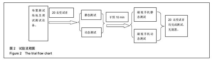
2.1 参与者数量分析 按意向性处理,纳入20名男性青年受试者均按照试验要求完成静立和自然行走状态下凝视手机的足底测试试验,无脱落,试验流程见图2。"

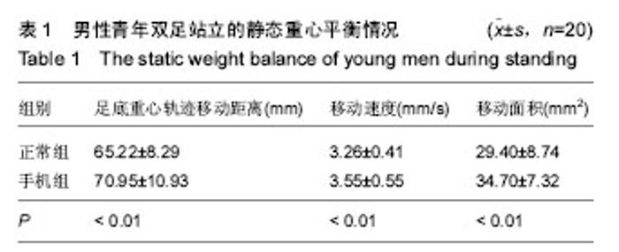
2.2 男性青年双足站立静态平衡及各区域参数情况 2.2.1 男性青年双足站立静态平衡情况 足底重心轨迹是作用于支撑面上全部力量的分布中心,相对于支撑面的控制反映了人体的稳定性,也是评定人体平衡功能的有效的临床应用方法[11]。应用测力平台监测静态平衡能力主要应用的相关参数如轨迹前后摆动范围、轨迹左右摆动范围、轨迹曲线的面积、摆动曲线的总长度以及扩散速度等可从不同角度上反映平衡功能[12]。 由表1可知,男性青年凝视于手机站立20 s整个身体的重心轨迹移动距离、速度和移动面积非常明显高于自然站立时的参数,因此可知,男性青年凝视于手机站立20 s的平衡能力要明显差于自然站立的平衡能力。"


2.2.2 男性青年双足站立各区域参数情况 足底各区域的接触面积、压力峰值以及压强均是反映足底压力分布的重要指标,其大小对踝关节及膝关节甚至髋关节的损伤具有重要的影响[13]。人体站立或行走过程中,足底压力区域大小的分布比例主要是由下肢关节过度旋转的影响的[14]。 由表2可知,男性青年在站立20 s过程中,凝视于手机的左侧足底的T1、M1、M2和M3的区域接触面积明显大于正常站立时,LH的接触面积明显小于正常站立时;凝视于手机右侧足底的M1、M2的区域面积明显大于正常站立时,其他足底压力区域差异无显著性意义。 由表3可知,男性青年在站立20 s过程中,凝视于手机的左右两侧足底的T1、M1、M2和M3的区域压力峰值明显大于正常站立时,其他足底压力区域差异无显著性意义。 由表4可知,男性青年再站立20 s过程中,凝视于手机的两侧足底各区域的压强峰值与正常站立的值差异无显著性意义。 由表5可知,男性青年在站立20 s过程中,凝视于手机的左右两侧足底的T1、M1、M2和M3的区域平均压强明显大于正常站立时,其他足底压力区域差异无显著性意义。"
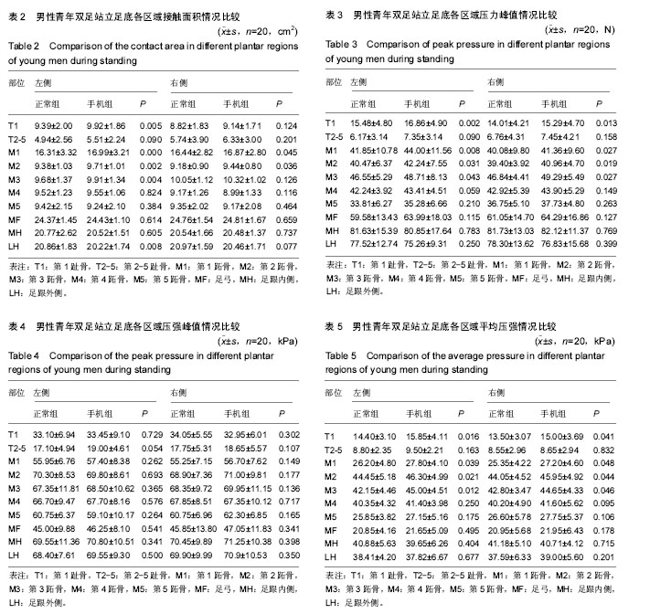
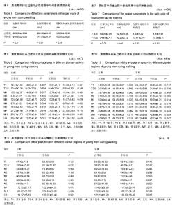
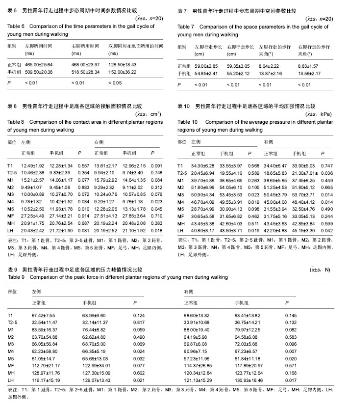
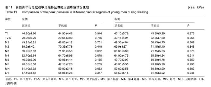
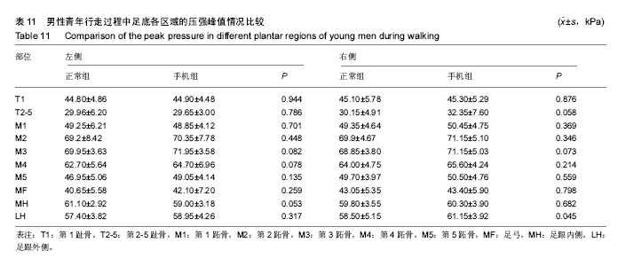
总之,男性青年凝视于手机站立20 s过程中,足底重心轨迹的波动速度、位移以及轨迹面积明显大于正常站立时,并且足底区域中的M1和M2区域的接触面积、压力峰值以及平均压强的大小也明显大于正常站立时,而HL区域的参数明显低于正常站立时。 2.3 男性青年行走过程中时空参数和各区域参数情况 2.3.1 男性青年行走过程中步态周期中时间参数情况 由表6可知,男性青年在行走过程,凝视于手机的左脚支撑所用时间非常明显高于正常组(P < 0.01),凝视于手机的右脚支撑所用时间非常明显高于正常行走(P < 0.01),凝视于手机的双脚同时支撑所用时间明显高于正常行走(P < 0.05)。 2.3.2 男性青年行走过程中的步态周期中空间参数情况由表7可知,男性青年在行走过程中,凝视于手机时左右脚步长均明显短于正常行走(P < 0.01),凝视于手机的左右脚的夹角均明显大于正常组行走(P < 0.01)。 2.3.3 男性青年行走的足底压力区域参数情况 由表8可知,男性青年行走过程中,凝视于手机时左右侧足底M4、M5和LH的接触面积明显大于正常行走时(P < 0.05),其他区域的接触面积与正常行走时差异无显著性意义。 由表9可知,男性青年行走过程中,凝视于手机时左右侧足底M4、M5和LH的压力峰值明显大于正常行走时(P < 0.05),其他区域的接触面积与正常行走时差异无显著性意义。 由表10可知,男性青年行走过程中,凝视于手机的左侧足底M3、M4和LH的平均压强明显大于正常行走时(P < 0.05);右侧足底T2-5、M3、M4和LH的平均压强明显高于正常行走时(P < 0.05),其他区域的接触面积与正常行走时差异无显著性意义。 由表11可知,男性青年行走过程中,凝视于手机时右侧足底LH的压强峰值明显大于正常行走时(P < 0.05),其他区域的接触面积与正常行走时差异无显著性意义。 总之,男性青年凝视于手机行走过程中行走步长和步速明显小于正常行走,双脚支撑的时间和左右脚行走的夹角明显大于正常行走,足底区域中的M4、M5和LH的接触面积、压力峰值以及平均压强大小明显大于正常行走时。"
"
| [1] Jenaro C, Flores N, Gómez-Vela M, et al. Problematic internet and cell-phone use: Psychological, behavioral, and health correlates. Add Res Theor. 2007;15(3):309-320.[2] Gates DH, Wilken JM, Scott SJ, et al. Kinematic strategies for walking across a destabilising rock surface. Gait Posture. 2012;35(1):36-42.[3] Victor H,Nordin FM. 骨骼系统的生物力学基础[M].上海: 学林出版社,1985: 117-182.[4] Caselli A, Pham H,Giurini JM, et al. The forefoot-to-rearfoot plantar pressure ratio is increased in severe diabetic neuropathy and can predict foot ulceration. Diabetes Care. 2002;25(6):1066-1071.[5] 李伟, 汪宗保,李国平,等.膝关节骨性关节炎患者步态运动学参数的研究[J].中国康复医学杂志,2008,23(1):11-13.[6] 李峰,李沂,文静,等.基于步态分析的击剑运动员膝损伤原因探讨[J].中国康复医学杂志,2008,23(3):254-255.[7] 江晓峰,胡雪艳.偏瘫步态膝关节角度分析[J].中国康复医学杂志, 2007, 22(10): 918-920.[8] 李海,周安艳, 黄东锋,等.痉挛型脑瘫儿童步行时的动态足底压力特征[J].中国康复医学杂志,2007,22(1):44-47.[9] 袁刚,张木勋,王中琴,等. 正常人足底压力分布及其影响因素分析[J]. 中华物理医学与康复杂志,2004,26(3):156-159.[10] Yan SH, Zhang K, Tan GQ, et al. Effects of obesity on dynamic plantar pressure distribution in Chinese prepubescent children during walking. Gait Posture. 2013;7(1): 37-42.[11] 朱奕,朱晓军,孟殿怀,等.三维运动分析系统在健康人平衡功能质心检测中三种方法一致性的研究[J].中国康复医学杂志,2012, 27(3):216-222.[12] 金德闻,张济川,王人成,等.康复工程与生物机械学[M]. 北京:清华大学出版社,2011.[13] 霍洪峰,吴艳霞,高峰. 男性老年人健步走足底压力分布与步态特征[J]. 中国康复医学杂志,2009,24(12):1119-1121.[14] Akiyo T, Tomoyuki M. Characteristics of stability limits and foot pressure distributions during reaching movements by stroke patients. Rigakuryoho Kagaku. 2015;30(4): 635-640.[15] Pasma JH, Engelhart D, Maier AB, et al. Changes in sensory reweighting of proprioceptive information during standing balance with age and disease. J Neurophysiol. 2015;114(6): 3220-3233.[16] Ruhe A, Fejer R, Walker B. Center of pressure excursion as a measure of balance performance in patients with non-specific low back pain compared to healthy controls: a systematic review of the literature. Euro Spine J. 2011;20: 358-368. [17] Park H, Branson D, Kim S, et al. Effect of armor and carrying load on body balance and leg muscle function. Gait Posture. 2014;39(1): 430-435. [18] Bastien GJ, Willems PA, Schepens B, et al. Effect of load andspeed on the energetic cost of human walking. Eur J Appl Physiol.2005;94(1-2):76-83. [19] 实淑霞. 痉挛型脑瘫患儿躯干控制能力的评估及疗效观察[D].安徽医科大学, 2013. [20] Emfery CA, Cassidy JD, Klassen TP, et al. Development of a clinical static and dynamic standing balance measurement tool appropriate for use in adolescents. Phys Ther. 2005;85: 502-514.[21] Clark RA, Bell SW, Feller JA, et al. Standing balance and inter-limb balance asymmetry at one year post primary anterior cruciate ligament reconstruction: Sex differences in a cohort study of 414 patients. Gait Posture. 2016;52: 318-324.[22] McIntosh PT, Case LE, Chan JM, et al. Characterization of gait in late onset Pompe disease. Mol Genet Metab. 2015; 116(3):152-156.[23] Cronin NJ. The effects of high heeled shoes on female gait: A review.J Electromyogr Kinesiol. 2014 Apr;24(2):258-263. [24] Andriacci TP,Ogle JA,Galonte JO.Walking speed as a basis for normal and abnormal gait measurement. J Biomech. 1978;10 (4) : 261-268.[25] Sofuwa O, Nieuwboer A, Desloovere K, et al.Quantitative gait analysis in Parkinson's disease: comparison with a healthy control group. Arch Phys Med Rehabil. 2005;86(5): 1007-1013.[26] Rajachandrakumar R, Fraser JE, Schinkel-Ivy A, et al. Atypical anticipatory postural adjustments during gait initiation among individuals with sub-acute stroke. Gait Posture. 2016; 52:325-331.[27] Clark DJ. Automaticity of walking: functional significance, mechanisms, measurement and rehabilitation strategies. Front Hum Neurosci. 2015;9: 246-259.[28] 赵焕彬,李建设. 运动生物力学[M].北京:高等教育出版社,2008: 165-166.[29] Boudarham J, Hameau S, Zory R, et al. Coactivation of lower limb muscles during gait in patients with multiple sclerosis. PLoS One. 2016;11(6):1-13.[30] Fasano A,Bloem BR. Gait disorders. Continuum( Minneap Minn). 2013;19(5): 1344-1382.[31] 徐晴岩,周大伟,李立峰,等.使用硅胶足垫分解足底压力的研究[J].中国康复医学杂志,2007,22(8):736-738.[32] Englan SA, Granata KP.The influence of gait speed on local dynamic stability of walking. Gait Posture. 2007;25(2): 172-178.[33] 赵焕彬,霍洪峰,张静,等.老年人健身背向走的足底压力与步态特征[J].中国康复医学杂志,2010, 25(5): 435-438.[34] 霍洪峰,吴艳霞,高峰,等.男性老年人健步走足底压力分布与步态特征[J].中国康复医学杂志,2009,24(12): 1119-1121.[35] Mohd Said A, Justine M, Manaf H. Plantar Pressure Distribution among Older Persons with Different Types of Foot and Its Correlation with Functional Reach Distance. Scientifica. 2016;2016:1-7.[36] Imaizumi K, Iwakami Y, Yamashita K. Analysis of foot pressure distribution data for the evaluation of foot arch type. Conf Proc IEEE Eng Med Biol Soc. 2011;2011:7388-7392.[37] Schepers T, Van der Stoep A, Van der Avert H, et al. Plantar pressure analysis after percutaneous repair of displaced intra-articular calcaneal fractures. Foot Ankle Int. 2008;29(2): 128-135.[38] Cattaneo D, Rabuffetti M, Bovi G, et al. Assessment of postural stabilization in three task oriented movements in people with multiple sclerosis. Disabil Rehabil. 2014; 36(26): 2237-2243. [39] Frank R, Larimore J. Yoga as a method of symptom management in multiple sclerosis. Front Neurosci. 2015; 9:133.[40] Michailidou I, De Vries HE, Hol EM, et al. Activation of endogenous neural stem cells for multiple sclerosis therapy. Front Neurosci. 2015;8:454. |
| [1] | Zhang Jichao, Dong Yuefu, Mou Zhifang, Zhang Zhen, Li Bingyan, Xu Xiangjun, Li Jiayi, Ren Meng, Dong Wanpeng. Finite element analysis of biomechanical changes in the osteoarthritis knee joint in different gait flexion angles [J]. Chinese Journal of Tissue Engineering Research, 2022, 26(9): 1357-1361. |
| [2] | Bai Zixing, Cao Xuhan, Sun Chengyi, Yang Yanjun, Chen Si, Wen Jianmin, Lin Xinxiao, Sun Weidong. Construction and biomechanical analysis of ankle joint finite element model in gait cycle [J]. Chinese Journal of Tissue Engineering Research, 2022, 26(9): 1362-1366. |
| [3] | Liu Feng, Feng Yi. Finite element analysis of different Kirschner wire tension bands on transverse patella fractures during gait cycle [J]. Chinese Journal of Tissue Engineering Research, 2022, 26(9): 1367-1371. |
| [4] | Yao Xiaoling, Peng Jiancheng, Xu Yuerong, Yang Zhidong, Zhang Shuncong. Variable-angle zero-notch anterior interbody fusion system in the treatment of cervical spondylotic myelopathy: 30-month follow-up [J]. Chinese Journal of Tissue Engineering Research, 2022, 26(9): 1377-1382. |
| [5] | Liu Gang, Ma Chao, Wang Le, Zeng Jie, Jiao Yong, Zhao Yi, Ren Jingpei, Hu Chuanyu, Xu Lin, Mu Xiaohong. Ankle-foot orthoses improve motor function of children with cerebral palsy: a Meta-analysis based on 12 randomized controlled trials [J]. Chinese Journal of Tissue Engineering Research, 2022, 26(8): 1299-1304. |
| [6] | Zhang Jinglin, Leng Min, Zhu Boheng, Wang Hong. Mechanism and application of stem cell-derived exosomes in promoting diabetic wound healing [J]. Chinese Journal of Tissue Engineering Research, 2022, 26(7): 1113-1118. |
| [7] | An Weizheng, He Xiao, Ren Shuai, Liu Jianyu. Potential of muscle-derived stem cells in peripheral nerve regeneration [J]. Chinese Journal of Tissue Engineering Research, 2022, 26(7): 1130-1136. |
| [8] | Shao Yangyang, Zhang Junxia, Jiang Meijiao, Liu Zelong, Gao Kun, Yu Shuhan. Kinematics characteristics of lower limb joints of young men running wearing knee pads [J]. Chinese Journal of Tissue Engineering Research, 2022, 26(6): 832-837. |
| [9] | Huang Hao, Hong Song, Wa Qingde. Finite element analysis of the effect of femoral component rotation on patellofemoral joint contact pressure in total knee arthroplasty [J]. Chinese Journal of Tissue Engineering Research, 2022, 26(6): 848-852. |
| [10] | Shan Sharui, Huang Xuming, Zhang Mingxing, Wang Xiukun, Zheng Xiang, Bao Sairong, Hong Feng. Three-dimensional gait analysis of low-frequency repetitive transcranial magnetic stimulation for post-stroke hemiplegia [J]. Chinese Journal of Tissue Engineering Research, 2022, 26(5): 762-767. |
| [11] | Chen Xiaoxu, Luo Yaxin, Bi Haoran, Yang Kun. Preparation and application of acellular scaffold in tissue engineering and regenerative medicine [J]. Chinese Journal of Tissue Engineering Research, 2022, 26(4): 591-596. |
| [12] | Kang Kunlong, Wang Xintao. Research hotspot of biological scaffold materials promoting osteogenic differentiation of bone marrow mesenchymal stem cells [J]. Chinese Journal of Tissue Engineering Research, 2022, 26(4): 597-603. |
| [13] | Shen Jiahua, Fu Yong. Application of graphene-based nanomaterials in stem cells [J]. Chinese Journal of Tissue Engineering Research, 2022, 26(4): 604-609. |
| [14] | Zhang Tong, Cai Jinchi, Yuan Zhifa, Zhao Haiyan, Han Xingwen, Wang Wenji. Hyaluronic acid-based composite hydrogel in cartilage injury caused by osteoarthritis: application and mechanism [J]. Chinese Journal of Tissue Engineering Research, 2022, 26(4): 617-625. |
| [15] | Li Hui, Chen Lianglong. Application and characteristics of bone graft materials in the treatment of spinal tuberculosis [J]. Chinese Journal of Tissue Engineering Research, 2022, 26(4): 626-630. |
| Viewed | ||||||
|
Full text |
|
|||||
|
Abstract |
|
|||||