Chinese Journal of Tissue Engineering Research ›› 2017, Vol. 21 ›› Issue (26): 4258-4264.doi: 10.3969/j.issn.2095-4344.2017.26.027
Advances and clinical application in digital complete denture
- Department of Prosthodontics, College of Stomatology, Guangxi Medical University, Nanning 530021, Guangxi Zhuang Autonomous Region, China
-
Received:2017-06-06Online:2017-09-18Published:2017-09-28 -
Contact:Li Xiao-jie, M.D., Associate professor, Master's supervisor, Department of Prosthodontics, College of Stomatology, Guangxi Medical University, Nanning 530021, Guangxi Zhuang Autonomous Region, China -
About author:Xue Wen-li, Master, Department of Prosthodontics, College of Stomatology, Guangxi Medical University, Nanning 530021, Guangxi Zhuang Autonomous Region, China -
Supported by:the Natural Science Foundation of Guangxi Zhuang Autonomous Region, No. 2013GXNSFBA019162: Ability Improving Program of Young and Middle-aged Teachers of Colleges and Universities in Guangxi Zhuang Autonomous Region, No. 2017KY0092
CLC Number:
Cite this article
Xue Wen-li, Li Xiao-jie .
share this article
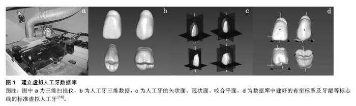
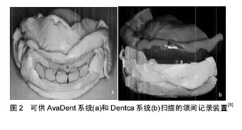
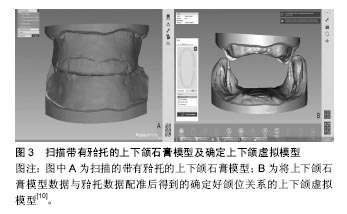
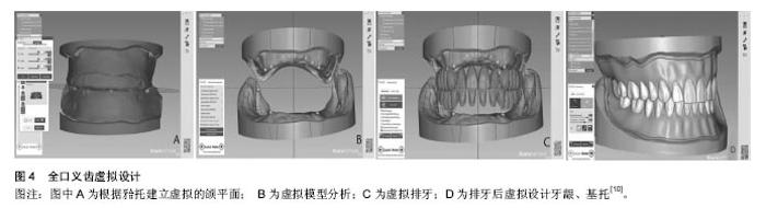
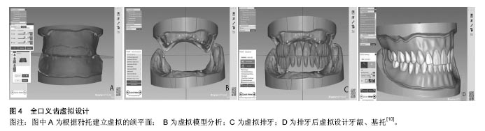
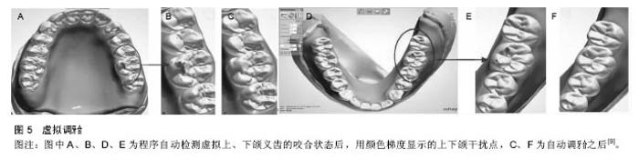
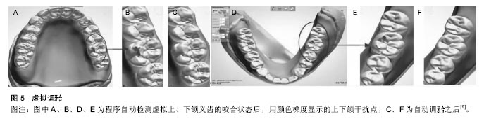
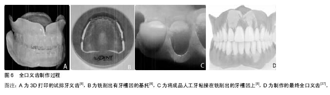
2.1 数字化全口义齿主要技术 数字化全口义齿是用三维扫描技术获取人工牙、无牙颌模型或印模、颌位关系,面部信息等数据,用专用软件进行数据参数化、模型重建、配准融合等,形成虚拟的确定好颌位关系的无牙颌,并虚拟确定牙合平面、中线等,然后虚拟排牙、设计基托、牙龈等全口义齿的三维形态,之后用虚拟牙合架调牙合并保存数据,将数据传输到CAM设备按照设计进行自动加工成型,获得最终全口义齿。 2.1.1 数据采集 数字化全口义齿数据采集分为2部分:第1部分需要采集人工牙数据,用CAD软件建立人工牙三维数据库并储存(图1)[16],这是建立全口义齿数字化系统的前提,为后续CAD排牙做准备;第2部分相当于传统全口义齿获取初印模、终印模、颌位关系、面部丰满度等信息的过程,传统方式中,这个过程患者需要多次就诊,较繁琐,对医师的技术要求较高且有一定主观性。数字化采集信息患者第1次就诊就可完成,效率高,且可减小对医师技术和主观性的依赖程度。数字化印模采集主要有直接法和间接法。直接法是用三维扫描数字传感器直接在患者口内扫描牙槽嵴、黏膜,获取所需数据[17]。目前此技术虽可减少患者的不适感,但还存在较大误差[18],因黏膜的可移动性等特点,获取的无牙颌黏膜表面数据还不够准确,扫描耗时较长,不能获得肌功能整塑的边界且口内扫描设备成本较高[11]。因此口内直接三维扫描获取数字印模的技术应用仍受限。目前主要采用扫描牙颌印模或模型的间接方式。 全口义齿数据采集的核心问题在于颌位的选择和确定。由于直接扫描处于下颌姿势位的上下颌牙槽嵴时,不能确保上下颌的稳定性,还不能采用。目前可间接扫描在口内确定好颌位关系的颌间记录装置[8, 19](如可确定下颌正中关系位与垂直距离的解剖测量装置,见图2)或者扫描带有牙合托的上下颌石膏模型[20]。扫描后可得到高精度的无牙颌数字模型正中关系和垂直距离数据[21-22]。还应采集患者下颌的个性化运动轨迹信息,以便与后续设计好的全口义齿数字化模型及牙合堤的三维数据配准形成虚拟牙合架。目前可使用电子面弓、下颌咬合运动轨迹描记仪等进行下颌运动轨迹数据采集[11,23]。但由于目前记录下颌运动轨迹的设备及软件应用较复杂,还未推广应用。修复后的面部容貌也是重要的一方面,用数字技术获取患者修复前后面部信息,然后用软件进行定量分析,可更精确地研究患者面部信息变化,甚至可预测模拟修复后面容[24-25],提高义齿制作效果。用三维扫描技术获取患者修复前面部信息、颌位关系数据,可用于在CAD排牙完成后用软件进行虚拟试戴牙、虚拟临床评估排牙情况等,减少就诊次数[19]。在完成临床上的上述信息采集工作后,将获取的数据或制取的印模、颌间记录装置等送至义齿加工中心,进行扫描、整合、配准等(图3),用于后续CAD。 数据采集涉及到的技术主要有:①接触式三坐标测量技术[6,26]:是通过探头与物体(如人工牙、无牙颌模型或印模)表面点接触获取其表面三维数据。此法可能会对物体表面产生划痕,且探头不能接触到的表面就无法获取其数据,扫描速率低,操作时间久,对环境要求高,成本高,可获取印模、无牙颌模型或人工牙的数据,现已较少使用;②自动切层扫描技术[16,27]:是采用数控铣床或层析机将被测物体切成层,然后用数据采集系统收集断层的二维信息数据,用图像处理软件再现被测物体三维图形。此技术可扫描不容易测量的物体,精度高,成本低,速度快,基本无测量盲区,还能得到物体内部数据,但会破坏被测物体,现应用较少,可用于获取人工石膏牙或无牙颌石膏模型的数据[16,27];③三维激光扫描[6,9,16,24]:是一种非接触测量技术,通过激光(点激光、线激光)扫描获取被测物体表面的三维坐标点云数据,快速重构物体三维虚拟模型,其测量精度高,分辨率高,操作简便,成本较低。但由于模型存在倒凹,会有扫描盲区[16]。由于目前口内直接扫描的使用仍受限,其主要用于人工牙、无牙颌模型、在口内确定好颌位关系的颌间记录装置、带有牙合托的上下无牙颌模型、患者面部信息等数据的获取;④计算机体层摄影技术[7,24,28]:即锥形束CT扫描技术,是利用X射线围绕物体特定部位旋转扫描获得二维图像,通过计算机处理获取物体的三维重建图形,其适用性好,但空间分辨率不够高,重建图像的速度较低,成本较高且属介入性方式,可用于获取人工牙、无牙颌模型等数据;⑤三维立体摄影测量技术[25,29-30]:是通过2台以上的摄像机从不同角度获取患者面部的二维图像,通过计算机分析整合成能显示出面部质地的三维图像[31],属于非介入性,安全、快速[32-33],主要用于患者修复前后面部软组织信息的获取。 2.1.2 CAD技术 CAD是数字化全口义齿系统的关键技术之一,不同的系统有各自专用的CAD软件,如Ceramill Mind软件、3Shape Dental System软件等,均结合了计算机数据库中储存的与全口义齿设计相关的原理、数据等,用CAD软件将获取的无牙颌终印模或模型扫描数据、颌关系记录扫描数据、面部扫描数据等进行参数化、上下颌模型三维重建、配准融合等,用软件检测排牙重要参考标志,自动确定牙合平面、中线、横牙合曲线、纵牙合曲线及腭后缘封闭区等,并创建空间三维坐标系,排牙曲线,在曲线上标出牙齿的三维定位标志点,从人工牙三维数据库中自动选牙并按照排牙原则进行三维虚拟排列,设计基托、牙龈形态,最后在计算机屏幕呈现完成的全口义齿虚拟三维图像[9,16,34](图4),到此全口义齿数字模型完成。接下来是虚拟牙合架的建立并运用。将数字模型的三维坐标与采集的个性化下颌运动轨迹坐标,基于扫描的颌堤三维数据进行配准融合,形成个性化虚拟牙合架进行虚拟调牙合。虚拟牙合架可根据采集的数据进行三维空间观察,模拟患者开闭口、前伸、后退、侧方等功能性运动,自动识别并检测咬合接触关系(早接触、牙合干扰等),用颜色梯度显示接触区位置、接触范围、上下虚拟人工牙之间非接触区的距离,并在接收计算机指令后自动进行精准的虚拟选磨调牙合,瞬时去除咬颌高点[35-36](图5)。上述完成之后,还可将设计好的3D图像发送给医生检查,确定好后再开始义齿的CAM[37]。虽然目前各系统的CAD软件都已相对成熟,但仍有需要继续改进的方面,比如采集的三维数据转换配准融合精度有待继续提高;人工牙与铣削的基托粘接后原本的位置存在偏离[38-39],需要在设计时校正;虚拟牙合架还不能完全模拟人体口颌系统的生理运动等[40]。 2.1.3 CAM技术 将全口义齿三维虚拟模型通过CAD软件设计好后,三维数据传输到CAM设备自动加工进行实物制造。目前全口义齿的CAM技术主要有以下几种:加法加工技术、减法加工技术、机器人辅助排牙技术。加法加工技术[2,13],也称增量加工技术,即快速成型或3D打印技术,是将设计好的物体三维图形用软件分割成连续多层叠加的二维图像,并在加工程序控制下使快速成型设备通过不断堆放材料形成连续层,然后逐层堆积形成物体原型,最后再进行表面处理[13],主要包括立体光刻技术、选择性激光烧结技术、分层实体制造技术等。加法加工技术目前用于4种类型:打印出物理石膏阴模,插入人工牙,充胶,然后按照传统方式制作出最终义齿;整体3D打印出牙列和基托,由于材料性能的限制,这种方式目前主要用树脂材料或者蜡来打印试排牙阶段义齿[8,41](图6A),且精度能达到临床要求;激光快速成形技术打印钛基托[42];3D打印全口义齿个别托盘[43]。加法加工直接、快速,不受加工物体形状的复杂性制约,但此种加工类型会使最终模型因为逐层堆积后固化产生收缩而出现差异,且其美观性有待提高[2,7]。加法加工技术能打印的聚合物和金属还有限,且不能打印牙科陶瓷材料[2]。目前用于全口义齿3D打印的材料主要有石膏、树脂类、聚乳酸类、钛和蜡。减法加工技术[2,13],也称去除式加工技术,如计算机数控制造是将物体三维CAD数据传入由计算机程序控制的电动铣削设备、将成型的大块材料铣削得到设计的物体原形、按铣削环境分为湿铣削和干铣削;按轴的数量分3轴、4轴、5轴、4轴和5轴可通过不同的XYZ轴线性上下移动。铣削钻直径越小切割精度就越高[44],因此小于铣削钻直径的一些表面特征铣削精度就不足。目前减法制作全口义齿主要有以下方式:铣削有牙槽凹刻的基托,然后将成品人工牙粘接在铣削基托的凹刻处,完成全口义齿[28、45] (图6B-D);分别铣削出牙列和有牙槽凹刻的基托[9,34],将两部分粘接后得到最终全口义齿;用将牙齿和基托材料交联为整体的材料,将基托和人工牙整体铣削作为最终全口义齿[2,37]。减法加工生产效率高[28],整体铣销材料人工牙部分可由牙本质和牙釉质类材料多层组成,相比传统成品人工牙更自然美观、更耐磨、义齿整体强度更高[37]。目前用于全口义齿的切割材料主要限于蜡块和树脂类材料[2]。由于存在倒凹和切削误差,切削的牙列和基托在对位粘接时会有不能直接完全吻合的情况[34,38-39]。国内创建的机器人排牙技术是在完成CAD设计后,以数字化控制机器人方式实现机器人辅助全口义齿排牙制作牙列,相比传统方式更加快速精确,但此技术只能排牙,不能制造基托,仅处于试验阶段[5]。 2.2 数字化全口义齿的临床应用情况 目前数字化全口义齿应用到临床的报道主要有以下几方面:①数字化全口义齿临床具体操作过程:2013年开始陆续有学者报道了AvaDent系统[8,46]、Dentca系统[8]、Ceramill系统的具体临床过程[10],均可二次就诊即完成义齿制作;②与传统全口义齿相比,数字化全口义齿的临床效果: Inokoshi[7]、Kattadiyil[47]、Saponaro[48]和Bidra等[49]将数字化与传统全口义齿从美观、适合性、整体满意度等方面进行横向或者纵向比较,表明CAD/CAM全口义齿的临床效果良好,无论从医生还是患者角度,满意度均 显著高于传统全口义齿,特别是在椅旁操作时间、加工密度、可操作性方面。其中Bidra等为期1年的纵向研究表明医生、患者均较满意,且患者的整体满意度高于医生;③与传统全口义齿相比,数字化全口义齿的优越性:McLaughlin等[50]用AvaDent系统铣削的树脂基托作为颌位记录及试排牙时的暂基托,并将该暂基托作为终基托,为无牙颌患者制作全口义齿,相比传统基托适合性更好,义齿基托收缩小,咬合误差更少。Kurahashi等[14]用3D打印技术复制患者原有义齿后,作为个别托盘,制取压力印模转关系后制作最终义齿,相比传统个别托盘制作过程更简便,节约了材料、椅旁时间,患者就诊次数少,适用于需要更换义齿而旧义齿可直接复制或稍作修改可复制的病例。另外,应用数字化全口义齿技术可更好地完成临床上传统技术不易解决的问题,并取得了更好的临床效果。关于牙槽嵴吸收严重、颏孔离牙槽嵴很近甚至位于牙槽嵴顶的病例,Ohkubo等[15]报道了用Dentca系统对一病例使用数字化双CT扫描定位颏孔位置,然后精确缓冲基托,戴上最终义齿后咀嚼无异常感觉,取得了良好效果,显示了独特优势。数字化全口义齿重要的优势是减少了医生椅旁操作时间和患者的复诊时间,但缺少长期临床数据。虽然国内外已出现了多个数字化全口义齿系统且有其独特优势,但仍有需要改进的方面[48]。如由于RP义齿的美观性不足,义齿的尺寸稳定性不足,亟需开发使其更美观的材料,用3D激光扫描或CAD软件运算数据弥补扫描过程中的差异,提高义齿的尺寸稳定性[7]。 "


"

"
"
"

"
| [1]Miyazaki T,Hotta Y,Kunii J,et al.A review of dental CAD/CAM: current status and future perspectives from 20 years of experience.Dent Mater J.2009;28(1):44-56. [2]Alghazzawi TF.Advancements in CAD/CAM technology: Options for practical implementation. J Prosthodont Res. 2016;60(2):72-84. [3]吕培军,李国珍,谭京.计算机辅助设计总义齿排牙方案及可调式排牙器的研究[J].现代口腔医学杂志,1988,2(3):166-167. [4]吕培军,李国珍.计算机辅助设计在全口义齿排牙的应用[J].中华口腔医学杂志,1992,27(3):134-6. [5]吕培军,王勇,李国珍,等.机器人辅助全口义齿排牙系统的初步研究[J].中华口腔医学杂志, 2001,36(2):139-42.[6]Maeda Y,Minoura M,Tsutsumi S,et al.A CAD/CAM system for removable denture. Part I: Fabrication of complete dentures. Int J Prosthodont.1994;7(1):17-21. [7]Inokoshi M,Kanazawa M,Minakuchi S.Evaluation of a complete denture trial method applying rapid prototyping.Dent Mater J.2012;31(1):40-46. [8]Kattadiyil MT,Goodacre CJ,Baba NZ.CAD/CAM complete dentures: a review of two commercial fabrication systems.J Calif Dent Assoc.2013;41(6):407-416. [9]Han WL,Li YF,Zhang Y,et al.Design and fabrication of complete dentures using CAD/CAM technology. Medicine (Baltimore).2017;96(1):e5435.[10]Wimmer T,Gallus K,Eichberger M,et al.Complete denture fabrication supported by CAD/CAM.J Prosthet Dent. 2016; 115(5):541-546.[11]张鹏,李伟伟,王勇,等.多源数据获取技术在全口义齿数字修复中的应用进展[J].中华口腔医学杂志. 2016,51(2):124-8.[12]Bidra AS,Taylor TD,Agar JR.Computer-aided technology for fabricating complete dentures: systematic review of historical background, current status, and future perspectives.J Prosthet Dent. 2013;109(6):361-366. [13]Bilgin MS,Baytaroglu EN,Erdem A,et al.A review of computer-aided design/computer-aided manufacture techniques for removable denture fabrication.Eur J Dent.2016;10(2):286-291. [14]Kurahashi K,Matsuda T,Goto T,et al.Duplication of complete dentures using general-purpose handheld optical scanner and 3-dimensional printer: Introduction and clinical considerations. J Prosthodont Res.2017;61(1):81-86.[15]Ohkubo C,Park EJ,Kim TH,et al.Digital Relief of the Mental Foramen for a CAD/CAM-Fabricated Mandibular Denture.J Prosthodont.2016.[Epub ahead of print][16]Sun Y,Lu P,Wang Y.Study on CAD&RP for removable complete denture.Comput Methods Programs Biomed. 2009;93(3):266-272. [17]Patzelt SB,Bishti S,Stampf S,et al.Accuracy of computer-aided design/computer-aided manufacturing- generated dental casts based on intraoral scanner data.J Am Dent Assoc. 2014;145(11):1133-1140. [18]Patzelt SB,Emmanouilidi A,Stampf S,et al.Accuracy of full-arch scans using intraoral scanners. Clin Oral Investig. 2014;18(6):1687-1694. [19]Schweiger J,Guth JF,Edelhoff D,et al.Virtual evaluation for CAD-CAM-fabricated complete dentures.J Prosthet Dent. 2017;117(1):28-33. [20]Sun Y,Yuan F,Li H,et al.Evaluation of the accuracy of a common regional registration method for three-dimensional reconstruction of edentulous jaw relation by a 7-axis three-dimensional measuring system.Biomed Mater Eng. 2014;24(1):1275-1287. [21]Yuan FS,Sun YC,Wang Y,et al.Accuracy evaluation of a new three-dimensional reproduction method of edentulous dental casts, and wax occlusion rims with jaw relation.Int J Oral Sci. 2013;5(3):155-161. [22]Li W,Yuan F,Lv P,et al.Evaluation of the quantitative accuracy of 3D reconstruction of edentulous jaw models with jaw relation based on reference point system alignment.PLoS One.2015;10(2):e0117320. [23]Hayashi K,Hayashi M,Reich B,et al.Functional data analysis of mandibular movement using third-degree b-spline basis functions and self-modeling regression.Orthodontic Waves. 2012; 71(1):17-25. [24]Katase H,Kanazawa M,Inokoshi M,et al.Face simulation system for complete dentures by applying rapid prototyping.J Prosthet Dent.2013;109(6):353-360. [25]Iordanova MV,Iordanova SV,Chaprashikian OG.Changes of the facial soft tissue profile in complete denture prosthetic treatment.Folia Med(Plovdiv).2006;48(3-4):74-78. [26]王勇,刘明丽,霍平,等.人工牙解剖形态三维坐标系的建立[J].中华口腔医学杂志,2006,41(11):684-686.[27]王晓波,高勃,姚月玲,等.上颌半口义齿金属基托的计算机辅助设计[J].实用口腔医学杂志,2005,21(1):5-7. [28]Kanazawa M,Inokoshi M,Minakuchi S,et al.Trial of a CAD/CAM system for fabricating complete dentures.Dent Mater J.2011;30(1):93-96. [29]Ushijima M,Kamashita Y,Nishi Y,et al.Changes in lip forms on three-dimensional images with alteration of lip support and/or occlusal vertical dimension in complete denture wearers.J Prosthodont Res.2013;57(2):113-121. [30]Sakar O,Sulun T,Kurt H,et al.Reliability and comparison of two facial measurements to detect changes of occlusal vertical dimension in complete denture wearers.Gerodontology. 2011;28(3):205-208. [31]Lee S.Three-dimensional photography and its application to facial plastic surgery.Arch Facial Plast Surg.2004;6(6):410-414. [32]van Loon B,Maal TJ,Plooij JM,et al.3D Stereophotogrammetric assessment of pre- and postoperative volumetric changes in the cleft lip and palate nose.Int J Oral Maxillofac Surg.2010;39(6):534-540. [33]Riphagen JM,van Neck JW,van Adrichem LN.3D surface imaging in medicine: a review of working principles and implications for imaging the unsedated child.J Craniofac Surg. 2008;19(2):517-524. [34]李岩峰,郝文君,韩卫丽,等.计算机辅助设计及制作技术在全口义齿设计制作中应用[J].山东医药,2015 55(35):64-6. [35]Koralakunte PR,Aljanakh M.The role of virtual articulator in prosthetic and restorative dentistry.J Clin Diagn Res.2014; 8(7):ZE25-28. [36]孙玉春,吕培军,王勇.用于全口义齿计算机辅助设计的虚拟半可调架[J].北京大学学报(医学版),2008,40(1):92-6. [37]AvaDent Digital Dentures.Global Dental Science LLC. Available at: http://www.avadent.com[38]Yamamoto S,Kanazawa M,Hirayama D,et al.In vitro evaluation of basal shapes and offset values of artificial teeth for CAD/CAM complete dentures.Comput Biol Med.2016;68: 84-89. [39]Yamamoto S,Kanazawa M,Iwaki M,et al.Effects of offset values for artificial teeth positions in CAD/CAM complete denture. Comput Biol Med.2014;52:1-7. [40]吴冰,吴国锋.口腔修复CAD/CAM系统中的虚拟牙合架[J].实用口腔医学杂志,2016,32(2):293-297. [41]Chen H,Wang H,Lv P,et al.Quantitative Evaluation of Tissue Surface Adaption of CAD-Designed and 3D Printed Wax Pattern of Maxillary Complete Denture.Biomed Res Int. 2015;2015:453968. [42]吴江,赵湘辉,沈丽娟,等.应用激光扫描法测量激光快速成形技术制作全口义齿钛基托的适合性研究[J]. 实用口腔医学杂志,2011, 27(3):293-297. [43]魏菱,陈虎,周永胜,等.数字化全口义齿个别托盘制作与临床应用时间评价[J].北京大学学报(医学版),2017,49(1):86-91. [44]Bosch G,Ender A,Mehl A.A 3-dimensional accuracy analysis of chairside CAD/CAM milling processes. J Prosthet Dent. 2014;112(6):1425-1431. [45]Goodacre CJ,Garbacea A,Naylor WP,et al.CAD/CAM fabricated complete dentures: concepts and clinical methods of obtaining required morphological data.J Prosthet Dent. 2012;107(1):34-46.[46]Infante L,Yilmaz B,McGlumphy E,et al.Fabricating complete dentures with CAD/CAM technology. J Prosthet Dent.2014; 111(5):351-355. [47]Kattadiyil MT,Jekki R,Goodacre CJ,et al.Comparison of treatment outcomes in digital and conventional complete removable dental prosthesis fabrications in a predoctoral setting.J Prosthet Dent.2015;114(6):818-825. [48]Saponaro PC,Yilmaz B,Heshmati RH,et al.Clinical performance of CAD-CAM-fabricated complete dentures: A cross-sectional study.J Prosthet Dent.2016;116(3):431-435. [49]Bidra AS,Farrell K,Burnham D,et al.Prospective cohort pilot study of 2-visit CAD/CAM monolithic complete dentures and implant-retained overdentures: Clinical and patient-centered outcomes.J Prosthet Dent.2016;115(5):578-586 e1. [50]McLaughlin JB,Ramos V Jr.Complete denture fabrication with CAD/CAM record bases.J Prosthet Dent. 2015;114(4): 493-497. [51]Fernandez MA,Nimmo A,Behar-Horenstein LS.Digital Denture Fabrication in Pre- and Postdoctoral Education: A Survey of U.S. Dental Schools.J Prosthodont.2016;25(1):83-90. |
| [1] | Yao Xiaoling, Peng Jiancheng, Xu Yuerong, Yang Zhidong, Zhang Shuncong. Variable-angle zero-notch anterior interbody fusion system in the treatment of cervical spondylotic myelopathy: 30-month follow-up [J]. Chinese Journal of Tissue Engineering Research, 2022, 26(9): 1377-1382. |
| [2] | Zhang Jinglin, Leng Min, Zhu Boheng, Wang Hong. Mechanism and application of stem cell-derived exosomes in promoting diabetic wound healing [J]. Chinese Journal of Tissue Engineering Research, 2022, 26(7): 1113-1118. |
| [3] | An Weizheng, He Xiao, Ren Shuai, Liu Jianyu. Potential of muscle-derived stem cells in peripheral nerve regeneration [J]. Chinese Journal of Tissue Engineering Research, 2022, 26(7): 1130-1136. |
| [4] | He Yunying, Li Lingjie, Zhang Shuqi, Li Yuzhou, Yang Sheng, Ji Ping. Method of constructing cell spheroids based on agarose and polyacrylic molds [J]. Chinese Journal of Tissue Engineering Research, 2022, 26(4): 553-559. |
| [5] | He Guanyu, Xu Baoshan, Du Lilong, Zhang Tongxing, Huo Zhenxin, Shen Li. Biomimetic orientated microchannel annulus fibrosus scaffold constructed by silk fibroin [J]. Chinese Journal of Tissue Engineering Research, 2022, 26(4): 560-566. |
| [6] | Chen Xiaoxu, Luo Yaxin, Bi Haoran, Yang Kun. Preparation and application of acellular scaffold in tissue engineering and regenerative medicine [J]. Chinese Journal of Tissue Engineering Research, 2022, 26(4): 591-596. |
| [7] | Kang Kunlong, Wang Xintao. Research hotspot of biological scaffold materials promoting osteogenic differentiation of bone marrow mesenchymal stem cells [J]. Chinese Journal of Tissue Engineering Research, 2022, 26(4): 597-603. |
| [8] | Shen Jiahua, Fu Yong. Application of graphene-based nanomaterials in stem cells [J]. Chinese Journal of Tissue Engineering Research, 2022, 26(4): 604-609. |
| [9] | Zhang Tong, Cai Jinchi, Yuan Zhifa, Zhao Haiyan, Han Xingwen, Wang Wenji. Hyaluronic acid-based composite hydrogel in cartilage injury caused by osteoarthritis: application and mechanism [J]. Chinese Journal of Tissue Engineering Research, 2022, 26(4): 617-625. |
| [10] | Li Hui, Chen Lianglong. Application and characteristics of bone graft materials in the treatment of spinal tuberculosis [J]. Chinese Journal of Tissue Engineering Research, 2022, 26(4): 626-630. |
| [11] | Gao Cangjian, Yang Zhen, Liu Shuyun, Li Hao, Fu Liwei, Zhao Tianyuan, Chen Wei, Liao Zhiyao, Li Pinxue, Sui Xiang, Guo Quanyi. Electrospinning for rotator cuff repair [J]. Chinese Journal of Tissue Engineering Research, 2022, 26(4): 637-642. |
| [12] | Guan Jian, Jia Yanfei, Zhang Baoxin , Zhao Guozhong. Application of 4D bioprinting in tissue engineering [J]. Chinese Journal of Tissue Engineering Research, 2022, 26(3): 446-455. |
| [13] | Liu Jiali, Suo Hairui, Yang Han, Wang Ling, Xu Mingen. Influence of lay-down angles on mechanical properties of three-dimensional printed polycaprolactone scaffolds [J]. Chinese Journal of Tissue Engineering Research, 2022, 10(16): 2612-2617. |
| [14] | Huang Bo, Chen Mingxue, Peng Liqing, Luo Xujiang, Li Huo, Wang Hao, Tian Qinyu, Lu Xiaobo, Liu Shuyun, Guo Quanyi . Fabrication and biocompatibility of injectable gelatin-methacryloyl/cartilage-derived matrix particles composite hydrogel scaffold [J]. Chinese Journal of Tissue Engineering Research, 2022, 10(16): 2600-2606. |
| [15] | Li Xuan, Sun Yimin, Li Longbiao, Wang Zhenming, Yang Jing, Wang Chenglin, Ye Ling. Manufacturing of nano-modified polycaprolactone microspheres and its biological effects in dental pulp cells [J]. Chinese Journal of Tissue Engineering Research, 2022, 26(10): 1530-1536. |
| Viewed | ||||||
|
Full text |
|
|||||
|
Abstract |
|
|||||