Chinese Journal of Tissue Engineering Research ›› 2022, Vol. 26 ›› Issue (10): 1530-1536.doi: 10.12307/2022.199
Previous Articles Next Articles
Manufacturing of nano-modified polycaprolactone microspheres and its biological effects in dental pulp cells
Li Xuan, Sun Yimin, Li Longbiao, Wang Zhenming, Yang Jing, Wang Chenglin, Ye Ling
- West China School of Stomatology, Sichuan University, Chengdu 610041, Sichuan Province, China
-
Received:2020-10-12Revised:2020-10-14Accepted:2020-11-28Online:2022-04-08Published:2021-10-25 -
Contact:Ye Ling, MD, Professor, West China School of Stomatology, Sichuan University, Chengdu 610041, Sichuan Province, China -
About author:Li Xuan, Doctoral candidate, West China School of Stomatology, Sichuan University, Chengdu 610041, Sichuan Province, China -
Supported by:the Foundation of Basic Research for Application from the Department of Science and Technology, Sichuan Province, No.2019YFS0035 (to YL); the National Science Foundation for Distinguished Young Scholars of China, No.81825005 (to YL)
CLC Number:
Cite this article
Li Xuan, Sun Yimin, Li Longbiao, Wang Zhenming, Yang Jing, Wang Chenglin, Ye Ling. Manufacturing of nano-modified polycaprolactone microspheres and its biological effects in dental pulp cells[J]. Chinese Journal of Tissue Engineering Research, 2022, 26(10): 1530-1536.
share this article
Add to citation manager EndNote|Reference Manager|ProCite|BibTeX|RefWorks

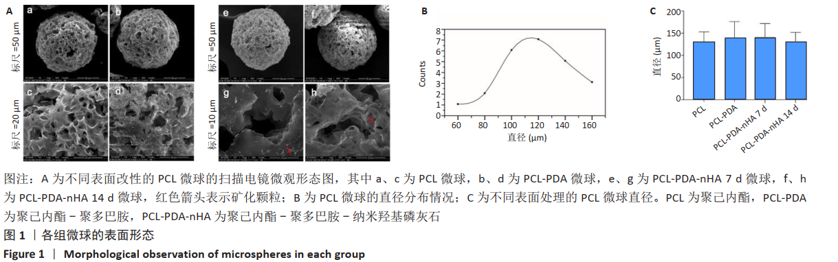
2.1 各组微球的表面形态 如图1A所示,3种微球表面均可见均匀疏松的多孔结构,PCL微球在多巴胺溶液中浸泡 24 h,清洗干燥后可见PDA颗粒覆盖。PCL-PDA-nHA 7 d组和PCL-PDA-nHA 14 d组的尺寸和孔隙无明显差异,但PCL-PDA-nHA 14 d组的微球表面结构更加粗糙,可见更加明显的矿化颗粒形成。由扫描电镜结果计算出PCL组、PCL-PDA组、PCL-PDA-nHA 7 d组和PCL-PDA-nHA 14 d组的直径分布呈正态分布,平均直径分别为(127.82±25.13),(138.93±37.07),(141.50±30.39),(131.50±20.02) μm,4组材料之间的粒径分布无明显差异(P > 0.05),见图1B、C。压汞仪检测得到PCL微球的孔隙率为87.4%,表面孔隙直径在20-50 μm之间。 "
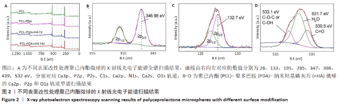
2.2 各组微球X射线光电子能谱分析结果 X射线光电子能谱分析结果如图2A所示,全谱扫描结果显示,PCL组在532,285 eV处有O1s、C1s轨道波峰,这主要来源于PCL的C-O、C=O、C-C等键;PCL-PDA组在398 eV处出现了N1s轨道吸收峰,其来源于PDA的氨基及其形成的化学键;PCL-PDA-nHA组在347 eV处和133 eV处中出现了Ca2p、P2p吸收峰,这可能来源于羟基磷灰石的钙和磷酸根。为了进一步确认化学键的种类,对PCL-PDA-nHA微球的Ca2p、P2p和O1s进行了窄谱扫描。图2B中Ca 2p 3/2轨道的吸收峰在346.98eV处出现,与之前文献报道中HA的吸收峰(347.4,347.2 eV)接 近[17-18],表明钙离子与磷酸基团结合。图2C中P2p吸收峰可以分为2p1/2和2p3/2两个峰,其中2p3/2轨道的吸收峰出现在132.7eV,这与文献报道中羟基磷灰石的P2p3/2轨道的吸收峰(133.4eV)较为相近[18]。在羟基磷灰石中,磷主要与氧形成磷酸根离子。图2D显示O1s的3个吸收峰分别来自O-C、O-H和O=C,这主要来源于PCL自身的C-O-C、C=O化学键和羟基磷灰石中的O-H化学键。结果表明PDA和nHA成功修饰于PCL微球表面。"
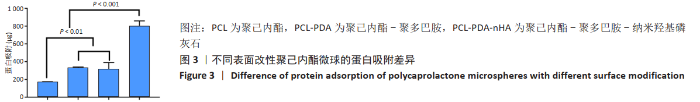
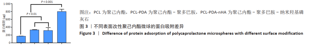
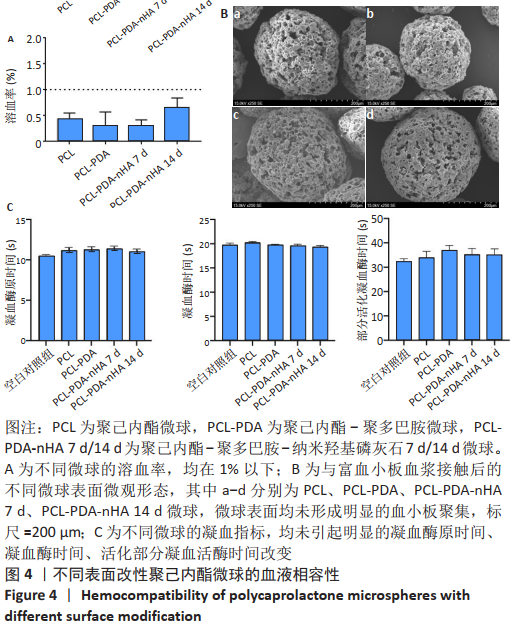
2.3 各组微球的蛋白吸附能力 如图3显示,与未修饰的PCL组相比,PCL-PDA组、PCL-PDA-nHA 7 d组和PCL-PDA-nHA 14 d组的蛋白吸附能力明显增强(P < 0.01);PCL-PDA组与PCL-PDA-nHA 7 d组的蛋白吸附能力相似(P > 0.05),PCL-PDA-nHA 14 d组的蛋白吸附能力高于PCL-PDA-nHA 7 d组(P < 0.001)。 2.4 各组微球血液相容性检测结果 如图4A所示,各组微球的溶血率均在1%以下,符合ISO 10993-4:2017《医疗器械生物学评价第4部分:与血液相互作用试验选择》中标准溶血试验的相关要求(<5%),结果显示修饰了PDA和nHA的PCL微球不会引起明显的溶血。nHA修饰的PCL微球植入体内后不会造成红细胞破裂,避免了因为血红蛋白释放而带来的细胞毒性。"
扫描电镜观察不同微球表面的血小板黏附情况,如图4B所示,各组微球表面均未形成明显的血小板聚集。各组微球的凝血实验结果如图4C所示,与空白对照组相比,PCL-PDA-nHA微球未引起凝血酶原时间、凝血酶时间、活化部分凝血活酶时间的明显改变(P > 0.05),这说明内源性凝血途径和外源性凝血途径均未被激活,不会发生凝血反应。"
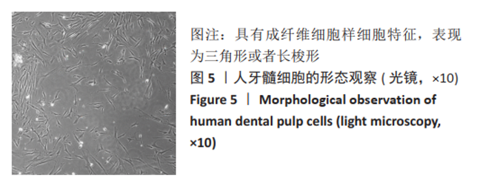
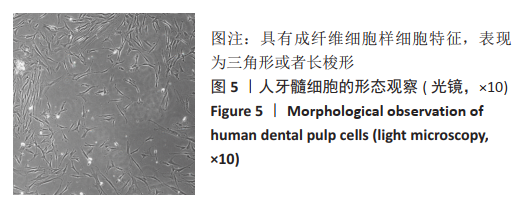
2.5 人牙髓细胞形态观察 从人牙髓组织中分离获得的原代人牙髓细胞具有成纤维细胞样细胞特征,表现为三角形或者长梭形,见图5。"
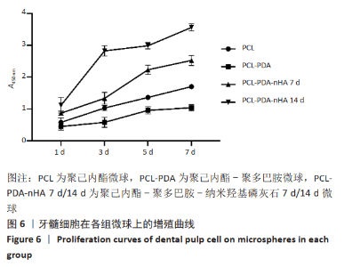
2.6 与微球三维共培养后牙髓细胞的增殖 CCK-8法评价微球的细胞毒性结果如图6所示。细胞与微球共培养1 d后,PCL-PDA组/PCL-PDA-nHA 7 d组的细胞数量与PCL组相比差异无显著性意义(P > 0.05),PCL-PDA-nHA 14 d组的细胞数量多于PCL组(P < 0.05)。随着培养时间的增加,各组微球中的细胞数量均呈上升趋势,PCL-PDA组、PCL-PDA-nHA 7 d组培养5,7 d的细胞数量少于PCL组(P < 0.05),PCL-PDA-nHA 14 d组培养3,5,7 d的细胞数量多于PCL组(P < 0.05),PCL-PDA-nHA 7 d组培养3,5,7 d的细胞数量少于PCL-PDA-nHA 14 d组(P < 0.05)。结果可以看出,经羟基磷灰石修饰后材料的生物相容性明显增强,且羟基磷灰石修饰量增加后,材料的细胞相容性得到了进一步提高。"
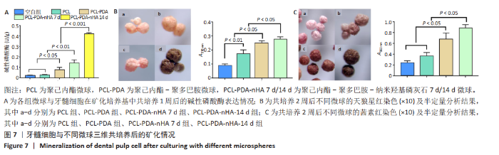
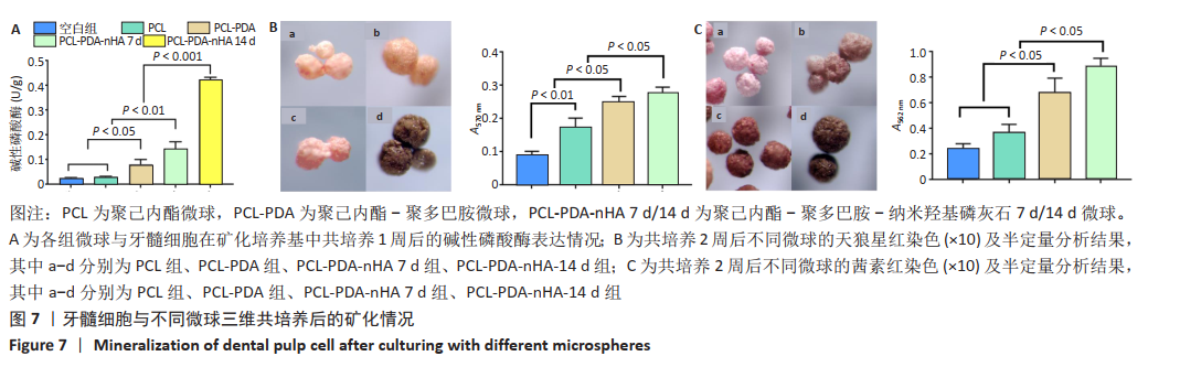
2.7 不同微球对牙髓细胞的矿化诱导作用 碱性磷酸酶检测结果显示,PCL组与空白组的碱性磷酸酶表达水平比较差异无显著性意义(P > 0.05),PCL-PDA组高于空白组、PCL组 (P < 0.05),PCL-PDA-nHA 7 d组高于空白组、PCL微球组、PCL-PDA组(P < 0.01),PCL-PDA-nHA 14 d组高于空白组、PCL微球组、PCL-PDA组、PCL-PDA-nHA 7 d组(P < 0.001),见图7A。 采用天狼星红染色及半定量分析检测胶原分泌水平,见图7B。半定量分析结果显示,PCL-PDA组的细胞胶原分泌水平高于PCL组(P < 0.01),PCL-PDA-nHA 7 d组高于PCL微球组、PCL-PDA组(P < 0.05),PCL-PDA-nHA 14 d组高于PCL组、PCL-PDA组、PCL-PDA-nHA 7 d组(P < 0.05)。 采用茜素红染色及半定量分析检测细胞外基质的矿化水平,见图7C。半定量分析结果显示,PCL-PDA组与PCL组矿化水平比较差异无显著性意义(P > 0.05),PCL-PDA-nHA 7 d组高于PCL-PDA组、PCL组(P < 0.05),PCL-PDA-nHA 14 d组高于PCL-PDA-nHA 7 d组、PCL-PDA组、PCL组(P < 0.05),说明PDA和nHA在PCL表面的共同修饰促进了骨样表型的建立。"
| [1] 樊明文.牙体牙髓病学[M]. 4版.人民卫生出版社,2012. [2] SPIELMAN H, SCHAFFER SB, COHEN MG, et al. Restorative outcomes for endodontically treated teeth in the Practitioners Engaged in Applied Research and Learning Network. J Am Dent Assoc. 2012;143(7):746-755. [3] PETRINO JA, BODA KK, SHAMBARGER S, et al. Challenges in regenerative endodontics: a case series. J Endod. 2010;36(3):536-541. [4] DOYON GE, DUMSHA T, FRAUNHOFER JAV. Fracture Resistance of Human Root Dentin Exposed to Intracanal Calcium Hydroxide. J Endod. 2005;31(12):895-897. [5] Yao Q, Wei B, Liu N, et al. Chondrogenic regeneration using bone marrow clots and a porous polycaprolactone-hydroxyapatite scaffold by three-dimensional printing. Tissue Eng Part A. 2015;21(7-8):1388-1397. [6] 朱惠光,计剑,高长有,等.聚乳酸组织工程支架材料[J].功能高分子学报,2001,14(4):488-92. [7] 戴炜枫,茹敏良,何月英,等.新型官能团化聚己内酯的研究进展[J].高分子通报,2008(9):10-19. [8] LEE H, LEE BP, MESSERSMITH PB. A reversible wet/dry adhesive inspired by mussels and geckos. Nature. 2007;448(7151):338-341. [9] HO CC, DING SJ. Structure, properties and applications of mussel-inspired polydopamine. J Biomed Nanotechnol. 2014;10(10):3063-3084. [10] SONG Y, JIANG H, WANG B, et al. Silver-Incorporated Mussel-Inspired Polydopamine Coatings on Mesoporous Silica as an Efficient Nanocatalyst and Antimicrobial Agent. Acs Appl Mater Interfaces. 2018;10(2):1792-1801. [11] QIAN Y, ZHOU X, ZHANG F, et al. Triple PLGA/PCL Scaffold Modification Including Silver Impregnation, Collagen Coating, and Electrospinning Significantly Improve Biocompatibility, Antimicrobial, and Osteogenic Properties for Orofacial Tissue Regeneration. ACS Appl Mater Interfaces. 2019;11(41):37381-37396. [12] PAN H, ZHENG Q, YANG S, et al. Effects of functionalization of PLGA-[Asp-PEG]n copolymer surfaces with Arg-Gly-Asp peptides, hydroxyapatite nanoparticles, and BMP-2-derived peptides on cell behavior in vitro. J Biomed Mater Res A. 2014;102(12):4526-4535. [13] 谭帼馨,欧阳孔友,周蕾,等.聚多巴胺螯合钙离子对钛表面的修饰及修饰后的细胞相容性[J].无机材料学报,2015(10):1075-1080. [14] GAO X, SONG J, JI P, et al. Polydopamine-Templated Hydroxyapatite Reinforced Polycaprolactone Composite Nanofibers with Enhanced Cytocompatibility and Osteogenesis for Bone Tissue Engineering. ACS Appl Mater Interfaces. 2016;8(5): 3499-3515. [15] LIN B, ZHONG M, ZHENG C, et al. Preparation and characterization of dopamine-induced biomimetic hydroxyapatite coatings on the AZ31 magnesium alloy. Surf Coat Technol. 2015;281:82-88. [16] RYU J, KU SH, LEE H, et al. Mussel‐Inspired Polydopamine Coating as a Universal Route to Hydroxyapatite Crystallization. Adv Funct Mater. 2010; 20(13):2132-2139. [17] NEGRILA CC, PREDOI MV, ICONARU SL, et al. Development of Zinc-Doped Hydroxyapatite by Sol-Gel Method for Medical Applications. Molecules. 2018; 23(11):2986. [18] STOICA TF, MOROSANU C, SLAV A, et al. Hydroxyapatite films obtained by sol–gel and sputtering. Thin Solid Films. 2008;516(22):8112-8116. [19] NAKAO S, TSUKAMOTO T, UEYAMA T, et al. STAT3 for Cardiac Regenerative Medicine: Involvement in Stem Cell Biology, Pathophysiology, and Bioengineering. Int J Mol Sci. 2020;21(6):1937. [20] RAHAMAN MN, MAO JJ. Stem cell-based composite tissue constructs for regenerative medicine. Biotechnol Bioeng. 2010;91(3):261-284. [21] WILLIAMS JM, ADEWUNMI A, SCHEK RM, et al. Bone tissue engineering using polycaprolactone scaffolds fabricated via selective laser sintering. Biomaterials. 2005;26(23):4817-4827. [22] YANG X, YANG F, WALBOOMERS XF, et al. The performance of dental pulp stem cells on nanofibrous PCL/gelatin/nHA scaffolds. J Biomed Mater Res A. 2010; 93(1):247-257. [23] LOUVRIER A, EUVRARD E, NICOD L, et al. Odontoblastic differentiation of dental pulp stem cells from healthy and carious teeth on an original PCL-based 3D scaffold. Int Endod J. 2018;51 Suppl 4:e252-e263. [24] 张一.聚己内酯的表面改性及其对细胞行为的影响[D].上海:华东理工大学, 2012. [25] Yoon H, Kim GH, Koh YH. A micro-scale surface-structured PCL scaffold fabricated by a 3D plotter and a chemical blowing agent. J Biomater Sci Polym Ed. 2010;21(2):159-70. [26] 李俊达,陈美霖,韦晓英,等.覆盖富血小板血浆3D打印聚己内酯支架对牙髓细胞体外生物学行为的影响[J].中华口腔医学研究杂志(电子版),2017, 11(3):149-156. [27] 刘宗光,屈树新,翁杰.聚多巴胺在生物材料表面改性中的应用[J].化学进展, 2015,27(2):212-219. [28] 张嘉敏,汪涛,汤春波.颅骨修复钛网表面PDA/CPP涂层的制备及性能研究[J].材料科学,2017,7(1):134-142. [29] ZHAO H, WAITE JH. Linking adhesive and structural proteins in the attachment plaque of Mytilus californianus. J Biol Chem. 2006;281(36): 26150-26158. [30] 叶玲.再生性牙髓治疗方法的前景[J].口腔医学,2016,36(11):961-967. [31] 侯丽,乔春霞,赵增琳.解读ISO 10993-4:2017《医疗器械生物学评价第4部分:与血液相互作用试验选择》[J].中国医疗设备,2018,33(11):1-6. [32] NAKASHIMA M, IOHARA K, MURAKAMI M, et al. Pulp regeneration by transplantation of dental pulp stem cells in pulpitis: a pilot clinical study. Stem Cell Res Ther. 2017;8(1):61. [33] GRONTHOS S, MANKANI M, BRAHIM J, et al. Postnatal human dental pulp stem cells (DPSCs) in vitro and invivo. Proc Natl Acad Sci U S A. 2000;97(25): 13625-13630. [34] MIURA M, GRONTHOS S, ZHAO M, et al. SHED: stem cells from human exfoliated deciduous teeth . Proc Natl Acad Sci U S A. 2003;100(10):5807-5812. [35] D’ANTÒ V, RAUCCI MG, GUARINO V, et al. Behaviour of human mesenchymal stem cells on chemically synthesized HA-PCL scaffolds for hard tissue regeneration. J Tissue Eng Regen Med. 2016;10(2):E147-154. [36] GUO T, CAO G, LI Y, et al. Signals in Stem Cell Differentiation on Fluorapatite-Modified Scaffolds. J Dent Res. 2018;97(12):1331-1338. [37] ALIPOUR M, AGHAZADEH M, AKBARZADEH A, et al. Towards osteogenic differentiation of human dental pulp stem cells on PCL-PEG-PCL/zeolite nanofibrous scaffolds. Artif Cells Nanomed Biotechnol. 2019;47(1):3431-3437. [38] 梁倩.PCOS/bFGF调控人牙槽骨成骨细胞增殖矿化的作用研究[D].广州:暨南大学,2018. |
| [1] | Dai Xianglin, Zhang Wenfeng, Yao Xijun, Shang Jiaqi, Huang Qiujin, Ren Yifan, Deng Jiupeng. Barium titanate/polylactic acid piezoelectric composite film affects adhesion, proliferation, and osteogenic differentiation of MC3T3-E1 cells [J]. Chinese Journal of Tissue Engineering Research, 2023, 27(3): 367-373. |
| [2] | Liu Huan, Li Han, Ma Yunhao, Zhong Weijian, Ma Guowu. Osteogenic capacity of partially demineralized dentin particles in the maxillary sinus lift [J]. Chinese Journal of Tissue Engineering Research, 2023, 27(3): 354-359. |
| [3] | Wu Lihao, Shao Anliang, Xu Lin, Ren Kang, Wang Hongjian, Chen Liang, Xu Ling. Evaluation of immunotoxicity of the absorbable macroporous polysaccharides composite hemostatic material [J]. Chinese Journal of Tissue Engineering Research, 2023, 27(3): 329-334. |
| [4] | Bi Jintong, Hu Xin, Liu Jinshu. Wear properties of dental ceramics [J]. Chinese Journal of Tissue Engineering Research, 2023, 27(3): 406-412. |
| [5] | Jiang Haifang, Liu Rong, Hu Peng, Chen Wei, Wei Zairong, Yang Chenglan, Nie Kaiyu. Application of 3D printing technology in the precise and personalized treatment of cleft lip and palate [J]. Chinese Journal of Tissue Engineering Research, 2023, 27(3): 413-419. |
| [6] | Zhang Jinglin, Leng Min, Zhu Boheng, Wang Hong. Mechanism and application of stem cell-derived exosomes in promoting diabetic wound healing [J]. Chinese Journal of Tissue Engineering Research, 2022, 26(7): 1113-1118. |
| [7] | Shen Song, Xu Bin. Diffuse distribution of bone cement in percutaneous vertebroplasty reduces the incidence of refracture of adjacent vertebral bodies [J]. Chinese Journal of Tissue Engineering Research, 2022, 26(4): 499-503. |
| [8] | Hou Wanxing, Li Hongwei, Zheng Xin, Zhu Xianren. Correlation between preoperative magnetic resonance imaging findings and bone cement leakage after percutaneous vertebral augmentation [J]. Chinese Journal of Tissue Engineering Research, 2022, 26(4): 504-509. |
| [9] | Yang Sidi, Wang Qian, Xu Nuo, Wang Ronghan, Jin Chuanqi, Lu Ying, Dong Ming. Biodentine enhances the proliferation and differentiation of osteoblasts through upregulating bone morphogenetic protein-2 [J]. Chinese Journal of Tissue Engineering Research, 2022, 26(4): 516-520. |
| [10] | Le Guoping, Zhang Ming, Xi Licheng, Luo Hanwen. Preparation and in vitro evaluation of vancomycin hydrochloride@polylactic acid-glycolic acid copolymer-chitosan-hyaluronic acid composite sustained-release microspheres [J]. Chinese Journal of Tissue Engineering Research, 2022, 26(4): 528-534. |
| [11] | Liu Tongbin, Lin Peng, Zhang Xiaoming, Dong Xiling, Cao Fei, Wang Le, Guo Xinxing. Optimization of preparation method of atorvastatin calcium sustained-release microspheres [J]. Chinese Journal of Tissue Engineering Research, 2022, 26(4): 535-539. |
| [12] | Qiu Peng, Fu Qilin, Liu Min, Lan Yuyan, Wang Pin. Comparison of oral micro-adhesion on polyetheretherketone, zirconium dioxide, and pure titanium abutment [J]. Chinese Journal of Tissue Engineering Research, 2022, 26(4): 540-545. |
| [13] | He Guanyu, Xu Baoshan, Du Lilong, Zhang Tongxing, Huo Zhenxin, Shen Li. Biomimetic orientated microchannel annulus fibrosus scaffold constructed by silk fibroin [J]. Chinese Journal of Tissue Engineering Research, 2022, 26(4): 560-566. |
| [14] | Wang Can, Gu Weiping, Jiang Yubin, Zhu Lin, Chen Gang. Finite element analysis of the influence of different implant designs on the stress of mandibular edentulous jaw [J]. Chinese Journal of Tissue Engineering Research, 2022, 26(4): 573-578. |
| [15] | Yang Feng, Zhao Qian, Zhang Shixuan, Zhao Tienan, Feng Bo. Effectiveness and safety of rapamycin combined with CD133 antibody stent in preventing vascular restenosis [J]. Chinese Journal of Tissue Engineering Research, 2022, 26(4): 579-584. |
| Viewed | ||||||
|
Full text |
|
|||||
|
Abstract |
|
|||||