Chinese Journal of Tissue Engineering Research ›› 2017, Vol. 21 ›› Issue (25): 3956-3963.doi: 10.3969/j.issn.2095-4344.2017.25.004
Previous Articles Next Articles
Mutant hypoxia inducible factor 1alpha transfection promotes proliferation of bone marrow mesenchymal stem cells
Zhang Wei-wei1, Wang Jue2
- 1Department of Plastic Surgery, 2Center for Clinical Biological Samples, First Affiliated Hospital of Jinzhou Medical University, Jinzhou 121000, Liaoning Province, China
-
Revised:2017-03-24Online:2017-09-08Published:2017-10-09 -
Contact:Wang Jue, Attending physician, Center for Clinical Biological Samples, First Affiliated Hospital of Jinzhou Medical University, Jinzhou 121000, Liaoning Province, China -
About author:Zhang Wei-wei, Master, Physician, Department of Plastic Surgery, First Affiliated Hospital of Jinzhou Medical University, Jinzhou 121000, Liaoning Province, China -
Supported by:the Principal’s Fund of Liaoning Medical University – Special Fund for Clinical Medical Construction, No. XZJJ20140215
CLC Number:
Cite this article
Zhang Wei-wei, Wang Jue. Mutant hypoxia inducible factor 1alpha transfection promotes proliferation of bone marrow mesenchymal stem cells[J]. Chinese Journal of Tissue Engineering Research, 2017, 21(25): 3956-3963.
share this article
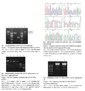
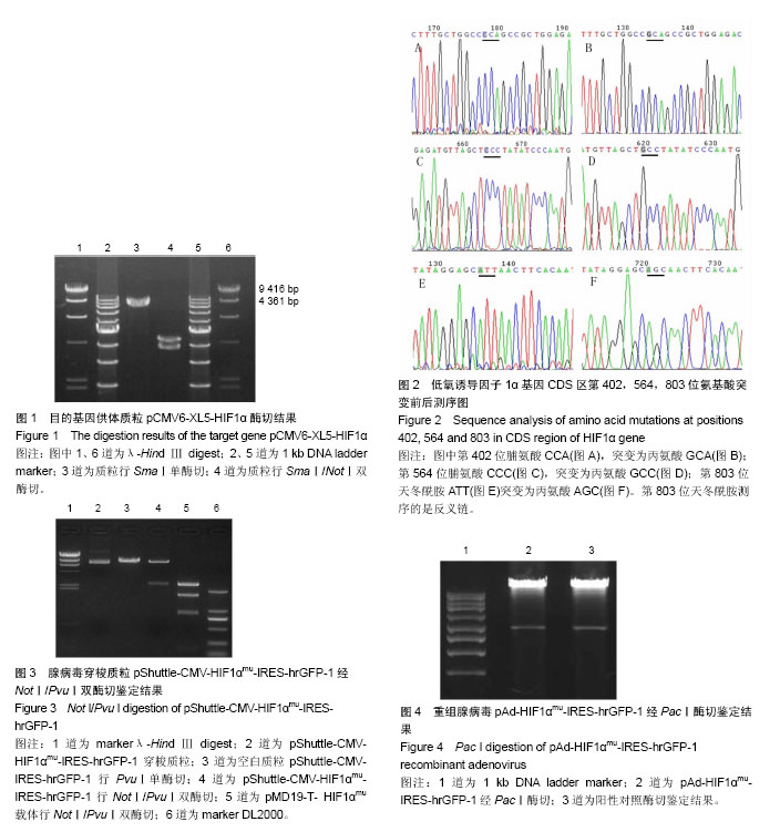
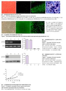
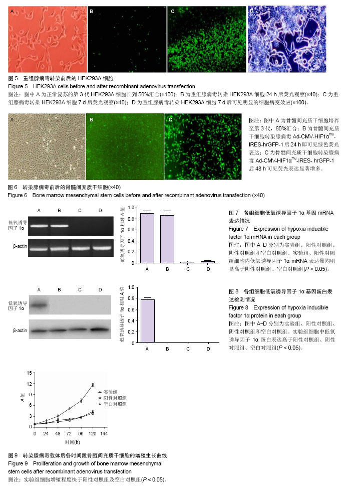
2.1 HIF1α基因测序结果 测序显示,供体质粒携带的HIF1α基因与GenBank上登记的HIF1α mRNA序列(NM_001530)完全一致,序列全长为4 082 bp,CDS区序列长度为2 481 bp(405-2 885 bp),共编码了826个氨基酸。对供体质粒行SmaⅠ单酶切及SmaⅠ/NotⅠ双酶切,与预期结果相符(图1)。对HIF1α基因序列内部的限制性内切酶识别位点分析结果显示,序列内部不含有PacⅠ、PvuⅠ、XhoⅠ及NotⅠ、PmeⅠ、SamⅠ等关键酶切位点。 2.2 HIF1α突变后基因测序结果 测序显示,供体质粒中所携带的HIF1α基因CDS区402位氨基酸由CCA突变成GCA,564位氨基酸由CCC突变成GCC,803位氨基酸由ATT突变成AGC,均为丙氨酸(图2)。 2.3 双基因载体构建结果 电泳显示,腺病毒穿梭质粒pShuttle-CMV-HIF1αmu-IRES-hrGFP-1及重组腺病毒突变型真核表达载体pAd-HIF1αmu-IRES-hrGFP-1均构建成功(图3,4)。 2.4 HEK293A细胞培养情况 HEK293A细胞从50%汇合到70%汇合约需2 d;显微镜下观察细胞密度大时呈铺路石状并有伪足伸展(图5A),腺病毒经Lipofectamine 2000转染HEK293A细胞后24 h可见绿色荧光表达,随着时间延长,绿色荧光表达越来越强,可见明显的细胞脱落、变圆等病变效应(图5B-D)。 2.5 骨髓间充质干细胞培养情况 骨髓间充质干细胞原代培养5 d首次换液后,在倒置显微镜下见有长条形细胞贴壁生长,培养至第3代80%汇合,细胞纯度较高、状态饱满、贴壁良好(图6A);最佳MOI值经计算为100,以MOI=100转染腺病毒后24 h即可见绿色荧光表达(图6B),48 h后荧光细胞显著增多(图6C)。 2.6 腺病毒滴度测定结果 重组腺病毒Ad-CMV- HIF1αmu-IRES-hrGFP-1按终点稀释法测得病毒滴度为1.6×10⁸ pfu/mL,阳性及阴性对照的病毒滴度分别为1.3× 108 pfu/mL和2.0×108 pfu/mL,滴度满足后续实验要求。 2.7 RT-PCR检测结果 实验组和阳性对照组细胞内HIF1α mRNA表达比较差异无显著性意义(P > 0.05);阴性对照组和空白对照组细胞内HIF1α mRNA表达量非常低,组间比较差异无显著性意义(P > 0.05);实验组、阳性对照组细胞内HIF1α mRNA表达量均明显高于阴性对照组、空白对照组(P < 0.05),见图7。 2.8 Western blot检测结果 实验组细胞中HIF1α蛋白表达高于阳性对照组、阴性对照组、空白对照组(P < 0.05),后3组间比较差异无显著性意义(P > 0.05),见图8。提示三点突变型HIF1α可在常氧条件下正常且高效表达,有利于促进种子细胞移植后的持续增殖及新血管再生。 2.9 细胞增殖结果测定 阳性对照组及空白对照组的骨髓间充质干细胞生长速度均较慢,两组之间相比较差异无显著性意义(P > 0.05);实验组细胞增殖程度快于阳性对照组及空白对照组(P < 0.05),见图9,说明三点突变型HIF1α可显著提高骨髓间充质干细胞的增殖。"
"
| [1] Verseijden F,Posthumus-van Sluijs SJ,van Neck JW,et al. Comparing scaffold-free and fibrin-based adipose-derived stromal cell constructs for adipose tissue engineering: an in vitro and in vivo study. Cell Transplant.2012;21(10):2283-2297.[2] 伞光,宋佳.血管内皮生长因子165转染可促进脂肪间充质干细胞增殖[J].中国组织工程研究, 2015,19(36):5782-5788.[3] Joo HH,Jo HJ,Jung TD,et al.Adipose-derived stem cells on the healing of ischemic colitis: a therapeutic effect by angiogenesis.Int J Colorectal Dis.2012;27(11): 1437-1443.[4] Chua KH,Raduan F,Wan Safwani WK,et al.Effects of serum reduction and VEGF supplementation on angiogenic potential of human adipose stromal cells in vitro.Cell Prolif.2013;46(3): 300-311.[5] 潘玮敏,刘民,杨建昌,等. LIM矿化蛋白1/低氧诱导因子1α慢病毒载体转染脂肪源性干细胞的成骨分化[J].中国组织工程研究, 2015,19(32):5140-5147. [6] Ide C,Nakano N,Kanekiyo K.Cell transplantation for the treatment of spinal cord injury - bone marrow stromal cells and choroid plexus epithelial cells.Neural Regen Res.2016; 11(9):1385-1388.[7] 李沙,李树仁,张倩辉.缺氧诱导因子1a基因转染心脏干细胞修复坏死心肌:应用前景[J].中国组织工程研究,2015,19(23):3750- 3754. [8] Fiorenzo P,Mongiardi MP,Dimitri D,et al. HIF1-positive and HIF1-negative glioblastoma cells compete in vitro but cooperate in tumor growth in vivo.Int J Oncol. 2010;36(4): 785-791.[9] Hao C,Wang Y,Shao L,et al.Local Injection of Bone Mesenchymal Stem Cells and Fibrin Glue Promotes the Repair of Bone Atrophic Nonunion In Vivo.Adv Ther.2016; 33(5):824-833.[10] Zhang W,Chang Q,Xu L,et al.Graphene Oxide-Copper Nanocomposite-Coated Porous CaP Scaffold for Vascularized Bone Regeneration via Activation of Hif-1α.Adv Healthc Mater. 2016;5(11):1299-1309.[11] 王钰,朱志图,陈峻江.血管内皮生长因子165基因促进人脂肪间充质干细胞的增殖[J].中国组织工程研究,2015,19(28):4485-4492. [12] 张馨,周琳,张晓雷.重组腺病毒介导突变型低氧诱导因子1α基因转染脂肪间充质干细胞并促进其增殖[J].中国组织工程研究, 2016,20(23):3386-3393.[13] Ou M,Sun X,Liang J,et al.A polysaccharide from Sargassum thunbergii inhibits angiogenesis via downregulating MMP-2 activity and VEGF/HIF-1α signaling.Int J Biol Macromol.2016; 94(Pt A):451-458.[14] Song B,Zhang Q,Yu M,et al.Ursolic acid sensitizes radioresistant NSCLC cells expressing HIF-1α through reducing endogenous GSH and inhibiting HIF-1α.Oncol Lett. 2017;13(2):754-762.[15] Zhang G,Zhao C,Wang Q,et al.Identification of HIF-1 signaling pathway in Pelteobagrus vachelli using RNA-Seq: effects of acute hypoxia and reoxygenation on oxygen sensors, respiratory metabolism, and hematology indices.J Comp Physiol B. 2017.doi: 10.1007/s00360-017-1083-8.[Epub ahead of print][16] Liu H,Zhang Z,Xiong W,et al.HIF-1α promotes cells migration and invasion by upregulating autophagy in endometriosis. Reproduction. 2017. pii: REP-16-0643. doi: 10.1530/REP-16-0643.[Epub ahead of print][17] Xiang GL,Zhu XH,Lin CZ,et al.125I seed irradiation induces apoptosis and inhibits angiogenesis by decreasing HIF-1α and VEGF expression in lung carcinoma xenografts.Oncol Rep.2017.doi:10.3892/or.2017.5521.[Epub ahead of print][18] Zhao H,Jiang H,Li Z,et al.2-Methoxyestradiol enhances radiosensitivity in radioresistant melanoma MDA-MB-435R cells by regulating glycolysis via HIF-1α/PDK1 axis.Int J Oncol.2017.doi: 10.3892/ijo.2017.3924.[Epub ahead of print][19] Tanaka Y,Hosoyama T,Mikamo A,et al.Hypoxic preconditioning of human cardiosphere-derived cell sheets enhances cellular functions via activation of the PI3K/Akt/mTOR/HIF-1α pathway.Am J Transl Res.2017; 9(2):664-673.[20] Wu W,Hu Q,Nie E,et al.Hypoxia induces H19 expression through direct and indirect Hif-1α activity, promoting oncogenic effects in glioblastoma.Sci Rep.2017;7:45029.[21] Xue L,Chen H,Lu K,et al.Clinical significance of changes in serum neuroglobin and HIF-1α concentrations during the early-phase of acute ischemic stroke.J Neurol Sci. 2017;375: 52-57.[22] He F,Qi Q,Li X,et al.Association of Indoor Air Pollution, Single Nucleotide Polymorphism of HIF-1α Gene with Susceptibility to Lung Cancer in Han Population in Fujian Province. Zhongguo Fei Ai Za Zhi.2017;20(3):149-156.[23] Yang SJ,Park YS,Cho JH,et al.Regulation of hypoxia responses by flavin adenine dinucleotide-dependent modulation of HIF-1α protein stability.EMBO J. 2017. pii: e201694408. doi: 10.15252/embj.201694408.[Epub ahead of print][24] Alexander-Shani R,Mreisat A,Smeir E,et al.Long term HIF-1α transcriptional activation is essential for heat-acclimation mediated cross-tolerance: Mitochondrial target genes.Am J Physiol Regul Integr Comp Physiol. 2017:ajpregu.00461.2016. doi: 10.1152/ ajpregu.00461.2016. [Epub ahead of print][25] Luo QQ,Qian ZM,Zhou YF,et al.Expression of Iron Regulatory Protein 1 Is Regulated not only by HIF-1 but also pCREB under Hypoxia.Int J Biol Sci.2016;12(10):1191-1202.[26] 孟祥超,刘卓超,王君,等. MicroRNAs在缺氧诱导因子1α缺失椎间盘组织中的差异性表达[J].中国组织工程研究,2016,20(7): 940-946.[27] Choi SH,Park JY,Kang W,et al.Knockdown of HIF-1α and IL-8 induced apoptosis of hepatocellular carcinoma triggers apoptosis of vascular endothelial cells.Apoptosis. 2016; 21(1):85-95.[28] Jiang YZ,Li Y,Wang K,et al.Distinct roles of HIF1A in endothelial adaptations to physiological and ambient oxygen.Mol Cell Endocrinol.2014;391(1-2):60-67.[29] 刘竹影,陈颖,刘倩,等.血管内皮祖细胞改善骨质疏松大鼠骨髓间充质干细胞的增殖及凋亡[J].中国组织工程研究,2016,20(14): 1999-2006. [30] Christoph M,Ibrahim K,Hesse K,et al.Local inhibition of hypoxia-inducible factor reduces neointima formation after arterial injury in ApoE-/- mice.Atherosclerosis.2014; 233(2): 641-647.[31] 杨超,张雷,周利武,等.低氧诱导因子2α调控骨关节炎:机制研究与转化应用[J].中国组织工程研究,2016,20(24):3634-3641. |
| [1] | Yao Xiaoling, Peng Jiancheng, Xu Yuerong, Yang Zhidong, Zhang Shuncong. Variable-angle zero-notch anterior interbody fusion system in the treatment of cervical spondylotic myelopathy: 30-month follow-up [J]. Chinese Journal of Tissue Engineering Research, 2022, 26(9): 1377-1382. |
| [2] | Wang Jing, Xiong Shan, Cao Jin, Feng Linwei, Wang Xin. Role and mechanism of interleukin-3 in bone metabolism [J]. Chinese Journal of Tissue Engineering Research, 2022, 26(8): 1260-1265. |
| [3] | Xiao Hao, Liu Jing, Zhou Jun. Research progress of pulsed electromagnetic field in the treatment of postmenopausal osteoporosis [J]. Chinese Journal of Tissue Engineering Research, 2022, 26(8): 1266-1271. |
| [4] | Tian Chuan, Zhu Xiangqing, Yang Zailing, Yan Donghai, Li Ye, Wang Yanying, Yang Yukun, He Jie, Lü Guanke, Cai Xuemin, Shu Liping, He Zhixu, Pan Xinghua. Bone marrow mesenchymal stem cells regulate ovarian aging in macaques [J]. Chinese Journal of Tissue Engineering Research, 2022, 26(7): 985-991. |
| [5] | Hou Jingying, Guo Tianzhu, Yu Menglei, Long Huibao, Wu Hao. Hypoxia preconditioning targets and downregulates miR-195 and promotes bone marrow mesenchymal stem cell survival and pro-angiogenic potential by activating MALAT1 [J]. Chinese Journal of Tissue Engineering Research, 2022, 26(7): 1005-1011. |
| [6] | Zhou Ying, Zhang Huan, Liao Song, Hu Fanqi, Yi Jing, Liu Yubin, Jin Jide. Immunomodulatory effects of deferoxamine and interferon gamma on human dental pulp stem cells [J]. Chinese Journal of Tissue Engineering Research, 2022, 26(7): 1012-1019. |
| [7] | Liang Xuezhen, Yang Xi, Li Jiacheng, Luo Di, Xu Bo, Li Gang. Bushen Huoxue capsule regulates osteogenic and adipogenic differentiation of rat bone marrow mesenchymal stem cells via Hedgehog signaling pathway [J]. Chinese Journal of Tissue Engineering Research, 2022, 26(7): 1020-1026. |
| [8] | Wang Jifang, Bao Zhen, Qiao Yahong. miR-206 regulates EVI1 gene expression and cell biological behavior in stem cells of small cell lung cancer [J]. Chinese Journal of Tissue Engineering Research, 2022, 26(7): 1027-1031. |
| [9] | Liu Feng, Peng Yuhuan, Luo Liangping, Wu Benqing. Plant-derived basic fibroblast growth factor maintains the growth and differentiation of human embryonic stem cells [J]. Chinese Journal of Tissue Engineering Research, 2022, 26(7): 1032-1037. |
| [10] | An Weizheng, He Xiao, Ren Shuai, Liu Jianyu. Potential of muscle-derived stem cells in peripheral nerve regeneration [J]. Chinese Journal of Tissue Engineering Research, 2022, 26(7): 1130-1136. |
| [11] | Fan Yiming, Liu Fangyu, Zhang Hongyu, Li Shuai, Wang Yansong. Serial questions about endogenous neural stem cell response in the ependymal zone after spinal cord injury [J]. Chinese Journal of Tissue Engineering Research, 2022, 26(7): 1137-1142. |
| [12] | Wen Dandan, Li Qiang, Shen Caiqi, Ji Zhe, Jin Peisheng. Nocardia rubra cell wall skeleton for extemal use improves the viability of adipogenic mesenchymal stem cells and promotes diabetes wound repair [J]. Chinese Journal of Tissue Engineering Research, 2022, 26(7): 1038-1044. |
| [13] | Zhu Bingbing, Deng Jianghua, Chen Jingjing, Mu Xiaoling. Interleukin-8 receptor enhances the migration and adhesion of umbilical cord mesenchymal stem cells to injured endothelium [J]. Chinese Journal of Tissue Engineering Research, 2022, 26(7): 1045-1050. |
| [14] | Luo Xiaoling, Zhang Li, Yang Maohua, Xu Jie, Xu Xiaomei. Effect of naringenin on osteogenic differentiation of human periodontal ligament stem cells [J]. Chinese Journal of Tissue Engineering Research, 2022, 26(7): 1051-1056. |
| [15] | Wang Xinmin, Liu Fei, Xu Jie, Bai Yuxi, Lü Jian. Core decompression combined with dental pulp stem cells in the treatment of steroid-associated femoral head necrosis in rabbits [J]. Chinese Journal of Tissue Engineering Research, 2022, 26(7): 1074-1079. |
| Viewed | ||||||
|
Full text |
|
|||||
|
Abstract |
|
|||||