Chinese Journal of Tissue Engineering Research ›› 2017, Vol. 21 ›› Issue (18): 2906-2912.doi: 10.3969/j.issn.2095-4344.2017.18.019
Previous Articles Next Articles
Treatment of osteosarcoma by targeting transportation of pHSP70-shPLK1/DOX compounds with bacterial magnetosomes
- Department of Orthopaedics, the Second Affiliated Hospital of Anhui Medical University, Hefei 230601, Anhui Province, China
-
Received:2017-04-26Online:2017-06-28Published:2017-07-07 -
Contact:Jing Jue-hua, Chief physician, Master’s supervisor, Department of Orthopaedics, the Second Affiliated Hospital of Anhui Medical University, Hefei 230601, Anhui Province, China -
About author:Yu Shui-sheng, Master, Department of Orthopaedics, the Second Affiliated Hospital of Anhui Medical University, Hefei 230601, Anhui Province, China -
Supported by:the National Natural Science Foundation of China, No. 81441068
CLC Number:
Cite this article
Yu Shui-sheng, Cheng Li, Xu Xin-zhong, Ke You-qun, Jing Jue-hua.
Treatment of osteosarcoma by targeting transportation of pHSP70-shPLK1/DOX compounds with bacterial magnetosomes [J]. Chinese Journal of Tissue Engineering Research, 2017, 21(18): 2906-2912.
share this article

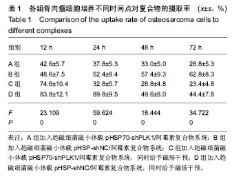
2.1 各组细胞对复合物的摄取率及形态学变化 对复合物的摄取率:B组摄取率随时间延长逐渐增加;D组24 h达峰值,48 h逐渐回落;A组和C组12 h达峰值,24 h逐渐回落。培养12 h时,D组摄取率最高,其次为C组,B组和A组最少;培养24 h时,D组最高,其次为B组,A组和C组最少;培养48,72 h时,B组最高,其次为D组,A组和C组最少,见表1。"
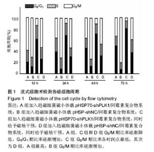
细胞形态变化:①A组、C组:培养12 h时,细胞形态不完整,细胞质浓缩,染色质凝集,细胞膜表面微绒毛消失,胞质内许多空泡样变,凋亡小体形成,凋亡较多,荧光较强;培养24,48,72 h时,细胞形态不完整,凋亡逐渐减少,荧光逐渐减弱;②B组:培养12 h时,细胞形态相对较完整,内可见多个细胞核,核仁大明显,微绒毛和内质网丰富,染色质分布较均匀,凋亡相对较少,荧光较强;培养24,48,72 h时,细胞形态较完整,荧光逐渐增强;③D组:培养12,24 h时,细胞形态不完整,凋亡较多,荧光较强;培养48,72 h时,细胞形态不完整,凋亡逐渐减少,荧光逐渐减弱。 2.2 各组细胞周期的比较 A组、C组和D组G2/M期比率逐渐降低,G0/G1期比率逐渐增加;C组G2/M期比率各时间点最低,其次为D组,A组最高;B组G2/M期比率逐渐增加,见图1。培养12 h时,G2/M期比率除了A组与D组、B组与D组比较无差异外,其余组间两两比较差异均有显著性意义(P < 0.05);G0/G1期比率除了A组与D组比较无差异外,其余组间两两比较差异均有显著性意义(P < 0.05)。培养24,48,72 h时,G2/M期、G0/G1期比率组间两两比较差异均有显著性意义(P < 0.05)。"


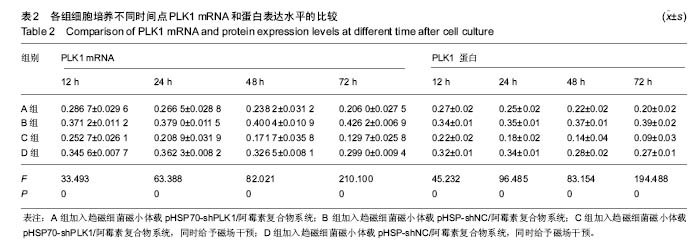
2.3 各组细胞中PLK1 mRNA和蛋白表达水平的比较 B组PLK1 mRNA和蛋白表达水平随时间延长逐渐增加;D组24 h达峰值,48 h逐渐回落;A组和C组12 h达峰值,24 h逐渐回落。培养12 h时,B组、D组PLK1 mRNA和蛋白表达高于A组、C组(P < 0.05),A组高于C组(P < 0.05),B组与D组比较无差异。培养24 h时,B组、D组PLK1 mRNA和蛋白表达高于A组、C组(P < 0.05),B组与D组比较无差异,A组高于C组(P < 0.05)。培养48,72 h时,B组PLK1 mRNA和蛋白表达高于D组、A组、C组(P < 0.05),D组高于A组、C组(P < 0.05),A组高于C组(P < 0.05),见表2。"

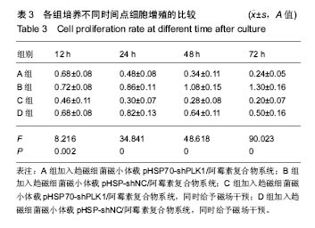
2.4 各组细胞增殖的比较 B组增殖随时间延长逐渐增加;D组24 h达峰值,48 h逐渐回落;A组和C组12 h达峰值,24 h逐渐回落。培养12 h时,A组、B组、D组细胞增殖高于C组(P < 0.05),A组、B组、D组间比较无差异。培养24,48,72 h时,B组细胞增殖高于D组、A组、C组(P < 0.05),D组高于A组、C组(P<0.05),A组高于C组(P< 0.05),见表3。"

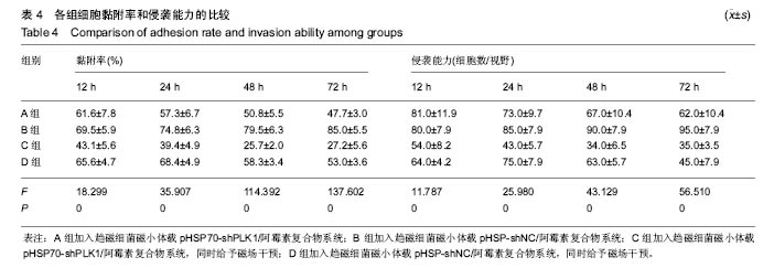
2.5 各组细胞黏附率和侵袭能力的比较 B组细胞黏附率和侵袭能力随时间延长逐渐增加;D组黏附率和侵袭能力24 h达峰值,48 h逐渐回落;A组和C组黏附率和侵袭能力12 h达峰值,24 h逐渐回落,见表4。"

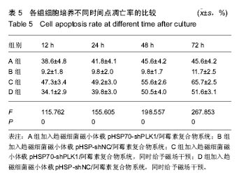
2.6 各组细胞凋亡率的比较 B组凋亡率无改变且始终最低,其余3组随时间延长逐渐增加;C组各时间点凋亡率均高于A组、D组(P < 0.05);A组和D组各时间点凋亡率比较均无差异,见表5。"
| [1]Luetke A,Meyers PA,Lewis I,et al.Osteosarcoma treatment-Where do we stand? A state of the art review. Cancer Treatt Rev.2013;40(4):523-532.[2]Tacar O,Sriamornsak P,Dass CR.Doxorubicin: an update on anticancer molecular action, toxicity and novel drug delivery systems.J Pharm Pharmacol.2013;65(2):157-170.[3]Seow HF,Yip WK,Fifis T.Advances in targeted and immunobased therapies for colorectal cancer in the genomic era.Onco Targets Ther.2016;9:1899-1920.[4]Melero I,Berman DM,Aznar MA,et al.Evolving synergistic combinations of targeted immunotherapies to combat cancer. Nat Rev Cancer.2015;15(8):457-472.[5]Galluzzi L,Buque A,Kepp O,et al.Immunological Effects of Conventional Chemotherapy and Targeted Anticancer Agents. Cancer Cell.2015;28(6):690-714.[6]Chou YS,Yen CC,Chen WM,et al.Cytotoxic mechanism of PLK1 inhibitor GSK461364 against osteosarcoma: Mitotic arrest,apoptosis,cellular senescence,and synergistic effect with paclitaxel.Int J Oncol.2016;48(3):1187-1194.[7]Ma H,He C,Cheng Y,et al.PLK1shRNA and doxorubicin co-loaded thermosensitive PLGA-PEG-PLGA hydrogels for osteosarcoma treatment.Biomaterials. 2014;35(30): 8723-8734.[8]Gjertsen BT,Schoffski P.Discovery and development of the Polo-like kinase inhibitor volasertib in cancer therapy. Leukemia.2015;29(1):11-19.[9]Cheng L,Wang C,Jing J. Polo-like kinase 1 as a potential therapeutic target for osteosarcoma.Curr Pharm Des.2015; 21(10):1347-1350.[10]Zhao L,Jiang BO,Wang D,et al.Triptolide reduces the viability of osteosarcoma cells by reducing MKP-1 and Hsp70 expression.Exp Ther Med.2016;11(5):2005-2010. [11]姚琴,赵慧毅,谢柏臻.Ezrin shRNA联合HSP70对骨肉瘤细胞凋亡和增殖的影响[J].中华骨科杂志,2015, 35(2):165-173.[12]Mathuriya AS.Magnetotactic bacteria: nanodrivers of the future.Crit RevBiotechnol. 2015;22(6):1-15.[13]Goldhawk DE,Gelman N,Thompson RT,et al.Forming Magnetosome-Like Nanoparticles in Mammalian Cells for Molecular MRI[M]//Design and Applications of Nanoparticles in Biomedical Imaging.Springer International Publishing, 2017:187-203.[14]张薇薇,许乙凯,刘瑞源.基于趋磁细菌磁小体的新型主动靶向线粒体的MR双功能探针制备[J].中华放射学杂志, 2014,48(6): 509-511.[15]Chen CY,Chen CF,Yong Y,et al.Construction of a microrobot system using magnetotactic bacteria for the separation of Staphylococcus aureus.Biomed Microdevices. 2014;16(5): 761-770. [16]Alphandéry E,Guyot F,Chebbi I.Preparation of chains of magnetosomes, isolated from Magnetospirillum magneticum, strain AMB-1 magnetotactic bacteria, yielding efficient treatment of tumors using magnetic hyperthermia.Int J Pharm. 2012;434(1-2):444-452.[17]Yuan-Gang L,Qing-Lei D,Shi-Bin W,et al.Preparation and in vitro antitumor effects of cytosine arabinoside-loaded genipin-poly-l-glutamic acid-modified bacterial magnetosomes. Int J Nanomedicine. 2015;10:1387-1397.[18]Isakoff MS,Bielack SS,Meltzer P,et al.Osteosarcoma: Current Treatment and a Collaborative Pathway to Success.J Clin Oncol.2015;33(27):3029-3035.[19]Gill J,Ahluwalia MK,Geller D,et al.New targets and approaches in osteosarcoma.Pharmacol Ther. 2013;137(1): 89-99.[20]Teagle AR,Birchall JC,Hargest R.Gene Therapy for Pyoderma Gangrenosum: Optimal Transfection Conditions and Effect of Drugs on Gene Delivery in the HaCaT Cell Line Using Cationic Liposomes.Skin Pharmacol Physiol. 2016;29(3): 119-129.[21]Lohße A,Ullrich S,Katzmann E,et al.Functional Analysis of the Magnetosome Island in Magnetospirillum gryphiswaldense: The mamAB Operon Is Sufficient for Magnetite Biomineralization. Plos One.2011;6(10):e25561.[22]Alphandéry E,Chebbi I,Guyot F,et al.Use of bacterial magnetosomes in the magnetic hyperthermia treatment of tumours: A review.Int J Hyperthermia.2013;29(8):801.[23]Hervault A,Dunn AE,Lim M,et al.Doxorubicin loaded dual pH-and thermo-responsive magnetic nanocarrier for combined magnetic hyperthermia and targeted controlled drug delivery applications. Nanoscale.2016;8(24):12152.[24]Sun J,Li Y,Liang XJ,et al.Bacterial magnetosome: a novel biogenetic magnetic targeted drug carrier with potential multifunctions.J Nanomater. 2011;2011(2011):469031- 469043.[25]王熙,耿圆圆,侯蔷,等.细菌磁小体作为新型非病毒载体用于肿瘤基因治疗的研究进展[J].肿瘤学杂志, 2016,22(4):322-326.[26]尹燕鹰,顾宁,洪敏,等.靶向磁性热疗对小鼠结肠癌皮下移植模型作用的观察[J].中华肿瘤防治杂志,2013, 20(4):249-253.[27]Li C,Ke Y,Yu S,et al.Co-delivery of doxorubicin and recombinant plasmid pHSP70-Plk1-shRNA by bacterial magnetosomes for osteosarcoma therapy.Int J Nanomedicine. 2016;11:5277-5286.[28]Sero V,Tavanti E,Vella S,et al.Targeting polo-like kinase 1 by NMS-P937 in osteosarcoma cell lines inhibits tumor cell growth and partially overcomes drug resistance.Invest New Drugs.2014; 32(6):1167-1180.[29]Otsu H,Iimori M,Ando K,et al.Gastric Cancer Patients with High PLK1 Expression and DNA Aneuploidy Correlate with Poor Prognosis.Oncology.2016;2(6):15-16.[30]Fischer M,Quaas M,Nickel A,et al.Indirect p53-dependent transcriptional repression of Survivin, CDC25C, and PLK1 genes requires the cyclin-dependent kinase inhibitor p21/CDKN1A and CDE/CHR promoter sites binding the DREAM complex.Oncotarget.2015;6(39):41402-41417.[31]Ogawa R,Morii A,Watanabe A,et al.Regulation of gene expression in human prostate cancer cells with artificially constructed promoters that are activated in response to ultrasound stimulation.Ultrason Sonochem.2012;20(1): 460-467.[32]Bhayani KR,Rajwade JM,Paknikar KM.Radio frequency induced hyperthermia mediated by dextran stabilized LSMO nanoparticles: in vitro evaluation of heat shock protein response. Nanotechnology.2013;24(1):015102.[33]Yamaguchi M,Ito A,Okamoto N,et al.Heat-inducible transgene expression system incorporating a positive feedback loop of transcriptional amplification for hyperthermia-induced gene therapy.J Biosci Bioeng.2012;114(4):460-465.[34]Yamaguchi M,Ito A,Ono A,et al.Heat-inducible gene expression system by applying alternating magnetic field to magnetic nanoparticles.ACS Synth Biol 2014;3(5):273-279.[35]Tantos A,Kalmar L,Tompa P.The role of structural disorder in cell cycle regulation, related clinical proteomics, disease development and drug targeting.Expert Rev Proteomics.2015; 12(3):221-233.[36]Liu Y,Bi T,Dai W,et al.Lupeol Induces Apoptosis and Cell Cycle Arrest of Human Osteosarcoma Cells Through PI3K/AKT/mTOR Pathway.TechnolCancer Res Treat.2016; 15(6):NP16. |
| [1] | Yao Xiaoling, Peng Jiancheng, Xu Yuerong, Yang Zhidong, Zhang Shuncong. Variable-angle zero-notch anterior interbody fusion system in the treatment of cervical spondylotic myelopathy: 30-month follow-up [J]. Chinese Journal of Tissue Engineering Research, 2022, 26(9): 1377-1382. |
| [2] | Wen Dandan, Li Qiang, Shen Caiqi, Ji Zhe, Jin Peisheng. Nocardia rubra cell wall skeleton for extemal use improves the viability of adipogenic mesenchymal stem cells and promotes diabetes wound repair [J]. Chinese Journal of Tissue Engineering Research, 2022, 26(7): 1038-1044. |
| [3] | Zhang Yujie, Yang Jiandong, Cai Jun, Zhu Shoulei, Tian Yuan. Mechanism by which allicin inhibits proliferation and promotes apoptosis of rat vascular endothelial cells [J]. Chinese Journal of Tissue Engineering Research, 2022, 26(7): 1080-1084. |
| [4] | Zhang Jinglin, Leng Min, Zhu Boheng, Wang Hong. Mechanism and application of stem cell-derived exosomes in promoting diabetic wound healing [J]. Chinese Journal of Tissue Engineering Research, 2022, 26(7): 1113-1118. |
| [5] | An Weizheng, He Xiao, Ren Shuai, Liu Jianyu. Potential of muscle-derived stem cells in peripheral nerve regeneration [J]. Chinese Journal of Tissue Engineering Research, 2022, 26(7): 1130-1136. |
| [6] | Deng Shuang, Pu Rui, Chen Ziyang, Zhang Jianchao, Yuan Lingyan . Effects of exercise preconditioning on myocardial protection and apoptosis in a mouse model of myocardial remodeling due to early stress overload [J]. Chinese Journal of Tissue Engineering Research, 2022, 26(5): 717-723. |
| [7] | Yang Sidi, Wang Qian, Xu Nuo, Wang Ronghan, Jin Chuanqi, Lu Ying, Dong Ming. Biodentine enhances the proliferation and differentiation of osteoblasts through upregulating bone morphogenetic protein-2 [J]. Chinese Journal of Tissue Engineering Research, 2022, 26(4): 516-520. |
| [8] | He Yunying, Li Lingjie, Zhang Shuqi, Li Yuzhou, Yang Sheng, Ji Ping. Method of constructing cell spheroids based on agarose and polyacrylic molds [J]. Chinese Journal of Tissue Engineering Research, 2022, 26(4): 553-559. |
| [9] | He Guanyu, Xu Baoshan, Du Lilong, Zhang Tongxing, Huo Zhenxin, Shen Li. Biomimetic orientated microchannel annulus fibrosus scaffold constructed by silk fibroin [J]. Chinese Journal of Tissue Engineering Research, 2022, 26(4): 560-566. |
| [10] | Chen Xiaoxu, Luo Yaxin, Bi Haoran, Yang Kun. Preparation and application of acellular scaffold in tissue engineering and regenerative medicine [J]. Chinese Journal of Tissue Engineering Research, 2022, 26(4): 591-596. |
| [11] | Kang Kunlong, Wang Xintao. Research hotspot of biological scaffold materials promoting osteogenic differentiation of bone marrow mesenchymal stem cells [J]. Chinese Journal of Tissue Engineering Research, 2022, 26(4): 597-603. |
| [12] | Shen Jiahua, Fu Yong. Application of graphene-based nanomaterials in stem cells [J]. Chinese Journal of Tissue Engineering Research, 2022, 26(4): 604-609. |
| [13] | Zhang Tong, Cai Jinchi, Yuan Zhifa, Zhao Haiyan, Han Xingwen, Wang Wenji. Hyaluronic acid-based composite hydrogel in cartilage injury caused by osteoarthritis: application and mechanism [J]. Chinese Journal of Tissue Engineering Research, 2022, 26(4): 617-625. |
| [14] | Li Hui, Chen Lianglong. Application and characteristics of bone graft materials in the treatment of spinal tuberculosis [J]. Chinese Journal of Tissue Engineering Research, 2022, 26(4): 626-630. |
| [15] | Gao Cangjian, Yang Zhen, Liu Shuyun, Li Hao, Fu Liwei, Zhao Tianyuan, Chen Wei, Liao Zhiyao, Li Pinxue, Sui Xiang, Guo Quanyi. Electrospinning for rotator cuff repair [J]. Chinese Journal of Tissue Engineering Research, 2022, 26(4): 637-642. |
| Viewed | ||||||
|
Full text |
|
|||||
|
Abstract |
|
|||||