Chinese Journal of Tissue Engineering Research ›› 2017, Vol. 21 ›› Issue (17): 2708-2713.doi: 10.3969/j.issn.2095-4344.2017.17.014
Previous Articles Next Articles
Cultivation, identification and differentiation of neural stem cells
Zhu Qiong1, Hao Yue-juan2, Gao Shun-ji1, Chen Zhong1, Liu Zheng1, Xu Ya-li1
- 1Department of Ultrasound, Second Affiliate Hospital of Third Military Medical University, Chongqing 400037, China; 2Department of Ultrasound, the 302nd Hospital of PLA, Beijing 100039, China
-
Revised:2017-01-10Online:2017-06-18Published:2017-06-29 -
Contact:Xu Ya-li, M.D., Associate chief physician, Department of Ultrasound, Second Affiliate Hospital of Third Military Medical University, Chongqing 400037, China -
About author:Zhu Qiong, Studying for master’s degree, Department of Ultrasound, Second Affiliate Hospital of Third Military Medical University, Chongqing 400037, China -
Supported by:the National Natural Science Foundation of China, No. 81471795; the Military Medical Incubation Plan for the Youth, No. 16QNP102
CLC Number:
Cite this article
Zhu Qiong, Hao Yue-juan, Gao Shun-ji, Chen Zhong, Liu Zheng, Xu Ya-li. Cultivation, identification and differentiation of neural stem cells[J]. Chinese Journal of Tissue Engineering Research, 2017, 21(17): 2708-2713.
share this article
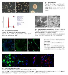
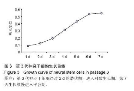
2.1 神经干细胞的形态 在解剖显微镜下剥离脑膜及血管,得到大脑半球。采用机械分离和酶消化法得到大量单细胞。原代培养第1天光镜下见部分细胞贴壁,部分细胞悬浮呈单细胞,折光性好,第2天悬浮细胞部分聚集呈细胞团,大小不均,较大者约有十几个细胞,克隆球直径逐渐增大,呈桑葚状,随着直径的增大,部分克隆球中心变暗。将克隆球消化传代后,克隆球大小更加均匀,形态更加规整,未见明显突起,折光性好(图2)。 2.2 神经干细胞的增殖能力 由细胞生长曲线(图3)可见,前2 d细胞生长缓慢,之后细胞增殖较快,进入对数生长期,从第6天开始,细胞增殖缓慢,进入平台期。细胞周期结果显示(图4),53.23%的神经干细胞处于G0+G1期,G2期占12.58%,S期占34.19%,G2/G1=1.99。根据增殖公式计算得增殖指数为87.86%,说明绝大部分细胞处于增殖状态。 2.3 神经干细胞的超微结构 透射电镜观察神经干细胞体积较小,胞核大且明显,核内染色质丰富,以常染色质为主,核仁较大,胞浆少,核浆比大,胞浆中含有大量的核糖体和线粒体,其余细胞器很少,表明该细胞分化程度低。大量的核糖体和线粒体表明细胞合成代谢能力旺盛,生命活动力强。细胞间可见细胞连接(图5)。 2.4 神经干细胞标志蛋白Nestin的表达 免疫荧光结果显示(图6),无论是接种的克隆球,还是单个细胞,细胞核均较小,被DAPI染成蓝色,胞浆呈绿色,说明Nestin呈阳性表达。克隆球向外呈放射状,越靠近中心,细胞密度越大,细胞越紧密,不易被染色,故中心未见染色细胞。 2.5 神经干细胞的诱导分化 免疫荧光结果显示,大多数细胞GFAP反应阳性,胞浆丰富呈绿色,有多个短突起,贴壁牢固,说明大部分神经干细胞分化为星形胶质细胞(图7A);少量细胞MBP反应阳性,呈绿色,说明少数神经干细胞分化为少突胶质细胞(图7B);部分细胞βtubulin Ⅲ反应阳性,呈红色,突起较少但较长,说明部分神经干细胞分化为神经元(图7C,D)。其中,含体积分数为1%胎牛血清的培养基能诱导分化生成更多的神经元(图7D),而含体积分数为10%胎牛血清的培养基能诱导分化生成更多的星形胶质细胞,说明低体积分数的血清有助于神经干细胞向神经元细胞分化,而高体积分数血清促使神经干细胞向胶质细胞分化。该分化结果进一步证实了分离培养得到的细胞为神经干细胞,且该细胞具有多向分化能力。"

"

| [1] Gage FH. Mammalian neural stem cells. Science. 2000;287 (5457):14331438. [2] Ma W, Fitzgerald W, Liu QY, et al. CNS stem and progenitor cell differentiation into functional neuronal circuits in three-dimensional collagen gels. Exp Neurol. 2004;190(2): 276-288.[3] Swistowski A, Peng J, Liu Q, et al. Efficient generation of functional dopaminergic neurons from human induced pluripotent stem cells under defined conditions. Stem Cells. 2010;28(10):1893-1904.[4] Li Z, Zeng Y, Chen X, et al. Neural stem cells transplanted to the subretinal space of rd1 mice delay retinal degeneration by suppressing microglia activation. Cytotherapy. 2016;18(6): 771-784. [5] Haus DL, López-Velázquez L, Gold EM, et al. Transplantation of human neural stem cells restores cognition in an immunodeficient rodent model of traumatic brain injury. Exp Neurol. 2016;281:1-16.[6] Curtis E, Gabel BC, Marsala M, et al. 172 A Phase I, Open-Label, Single-Site, Safety Study of Human Spinal Cord-Derived Neural Stem Cell Transplantation for the Treatment of Chronic Spinal Cord Injury. Neurosurgery. 2016;63 Suppl 1:168-169. [7] Mazzini L, Gelati M, Profico DC, et al. Human neural stem cell transplantation in ALS: initial results from a phase I trial. J Transl Med. 2015;13:17. [8] Zhang W, Wang PJ, Sha HY, et al. Neural stem cell transplants improve cognitive function without altering amyloid pathology in an APP/PS1 double transgenic model of Alzheimer's disease. Mol Neurobiol. 2014;50(2):423-437.[9] George PM, Steinberg GK. Novel Stroke Therapeutics: Unraveling Stroke Pathophysiology and Its Impact on Clinical Treatments. Neuron. 2015;87(2):297-309.[10] Merkle FT, Alvarez-Buylla A. Neural stem cells in mammalian development. 2006;18(6):704-709.[11] Gritti A, Parati EA, Cova L, et al. Multipotential stem cells from the adult mouse brain proliferate and self-renew in response to basic fibroblast growth factor. J Neurosci. 1996;16(3): 1091-1100.[12] Gage FH. Neurogenesis in the adult brain. J Neurosci. 2002; 22(3):612-613.[13] Arvidsson A, Collin T, Kirik D, et al. Neuronal replacement from endogenous precursors in the adult brain after stroke. Nat Med. 2002;8(9):963-970.[14] Yamashita T, Ninomiya M, Hernández Acosta P, et al. Subventricular zone-derived neuroblasts migrate and differentiate into mature neurons in the post-stroke adult striatum. J Neurosci. 2006;26(24):6627-6636.[15] Qu T, Brannen CL, Kim HM, et al. Human neural stem cells improve cognitive function of aged brain. Neuroreport. 2001; 12(6):1127-1132.[16] Zhao R, Xuan Y, Li X, et al. Age-related changes of germline stem cell activity, niche signaling activity and egg production in Drosophila. Aging Cell. 2008;7(3):344-354. [17] Kirik OV, Vlasov TD, Korzhevski? DÉ. Nestin and Musashi1 as the markers of neural stem cells in rat telencephalon following transitory focal ischemia. Morfologiia. 2012;142(4):19-24.[18] Eng LF. Glial fibrillary acidic protein (GFAP): the major protein of glial intermediate filaments in differentiated astrocytes. J Neuroimmunol. 1985;8(4-6):203-214.[19] Sun L, Lee J, Fine HA. Neuronally expressed stem cell factor induces neural stem cell migration to areas of brain injury. J Clin Invest. 2004;113(9):1364-1374.[20] Warrington AE, Barbarese E, Pfeiffer SE. Stage specific, (O4+GalC-) isolated oligodendrocyte progenitors produce MBP+ myelin in vivo. Dev Neurosci. 1992;14(2):93-97.[21] Ciccolini F. Identification of two distinct types of multipotent neural precursors that appear sequentially during CNS development. Mol Cell Neurosci. 2001;17(5):895-907.[22] Han J, Calvo CF, Kang TH, et al. Vascular endothelial growth factor receptor 3 controls neural stem cell activation in mice and humans. Cell Rep. 2015;10(7):1158-1172.[23] Yang JW, Ma W, Luo T, et al. BDNF promotes human neural stem cell growth via GSK-3β-mediated crosstalk with the wnt/β-catenin signaling pathway. Growth Factors. 2016; 34(1-2):19-32. [24] Huat TJ, Khan AA, Pati S, et al. IGF-1 enhances cell proliferation and survival during early differentiation of mesenchymal stem cells to neural progenitor-like cells BMC Neurosci. 2014;15:91.[25] Xiong LL, Chen ZW, Wang TH. Nerve growth factor promotes in vitro proliferation of neural stem cells from tree shrews. Neural Regen Res. 2016;11(4):591-596.[26] Zhao H, Steiger A, Nohner M, et al. Specific Intensity Direct Current (DC) Electric Field Improves Neural Stem Cell Migration and Enhances Differentiation towards βIII-Tubulin+ Neurons. PLoS One. 2015;10(6):e0129625.[27] Chen X, Liu Y, Zhang Z, et al. Hypoxia stimulates the proliferation of rat neural stem cells by regulating the expression of metabotropic glutamate receptors: an in vitro study. Cell Mol Biol (Noisy-le-grand). 2016;62(3):105-114.[28] Jiang XF, Yang K, Yang XQ, et al. Elastic modulus affects the growth and differentiation of neural stem cells. Neural Regen Res. 2015;10(9):1523-1527.[29] Keirstead HS. Stem cell transplantation into the central nervous system and the control of differentiation. J Neurosci Res. 2001;63(3):233-236.[30] Hung CH, Young TH. Differences in the effect on neural stem cells of fetal bovine serum in substrate-coated and soluble form. Biomaterials. 2006;27(35):5901-5908. [31] Chen WW, Blurton-Jones M. Concise review: Can stem cells be used to treat or model Alzheimer's disease. Stem Cells. 2012;30(12):2612-2618.[32] Obernier K, Tong CK, Alvarez-Buylla A. Restricted nature of adult neural stem cells: re-evaluation of their potential for brain repair. Front Neurosci. 2014;8:162.[33] Crawford DC, Jiang X, Taylor A, et al. Astrocyte-derived thrombospondins mediate the development of hippocampal presynaptic plasticity in vitro. J Neurosci. 2012;32(38): 13100-13110.[34] Bernardinelli Y, Muller D, Nikonenko I. Astrocyte-synapse structural plasticity. Neural Plast. 2014;2014:232105. [35] Blanco-Suárez E, Caldwell AL, Allen NJ. Role of astrocyte-synapse interactions in CNS disorders. J Physiol. 2016 Jul 5. [Epub ahead of print][36] Hynes SR, Rauch MF, Bertram JP, et al. A library of tunable poly(ethylene glycol)/poly(L-lysine) hydrogels to investigate the material cues that influence neural stem cell differentiation. J Biomed Mater Res A. 2009;89(2):499-509.[37] Lee IC, Wu YC, Cheng EM, et al. Biomimetic niche for neural stem cell differentiation using poly-L-lysine/hyaluronic acid multilayer films. J Biomater Appl. 2015;29(10):1418-1427. |
| [1] | Yao Xiaoling, Peng Jiancheng, Xu Yuerong, Yang Zhidong, Zhang Shuncong. Variable-angle zero-notch anterior interbody fusion system in the treatment of cervical spondylotic myelopathy: 30-month follow-up [J]. Chinese Journal of Tissue Engineering Research, 2022, 26(9): 1377-1382. |
| [2] | Zhang Jinglin, Leng Min, Zhu Boheng, Wang Hong. Mechanism and application of stem cell-derived exosomes in promoting diabetic wound healing [J]. Chinese Journal of Tissue Engineering Research, 2022, 26(7): 1113-1118. |
| [3] | Huang Chenwei, Fei Yankang, Zhu Mengmei, Li Penghao, Yu Bing. Important role of glutathione in stemness and regulation of stem cells [J]. Chinese Journal of Tissue Engineering Research, 2022, 26(7): 1119-1124. |
| [4] | An Weizheng, He Xiao, Ren Shuai, Liu Jianyu. Potential of muscle-derived stem cells in peripheral nerve regeneration [J]. Chinese Journal of Tissue Engineering Research, 2022, 26(7): 1130-1136. |
| [5] | Fan Yiming, Liu Fangyu, Zhang Hongyu, Li Shuai, Wang Yansong. Serial questions about endogenous neural stem cell response in the ependymal zone after spinal cord injury [J]. Chinese Journal of Tissue Engineering Research, 2022, 26(7): 1137-1142. |
| [6] | Chen Xiaoxu, Luo Yaxin, Bi Haoran, Yang Kun. Preparation and application of acellular scaffold in tissue engineering and regenerative medicine [J]. Chinese Journal of Tissue Engineering Research, 2022, 26(4): 591-596. |
| [7] | Kang Kunlong, Wang Xintao. Research hotspot of biological scaffold materials promoting osteogenic differentiation of bone marrow mesenchymal stem cells [J]. Chinese Journal of Tissue Engineering Research, 2022, 26(4): 597-603. |
| [8] | Shen Jiahua, Fu Yong. Application of graphene-based nanomaterials in stem cells [J]. Chinese Journal of Tissue Engineering Research, 2022, 26(4): 604-609. |
| [9] | Zhang Tong, Cai Jinchi, Yuan Zhifa, Zhao Haiyan, Han Xingwen, Wang Wenji. Hyaluronic acid-based composite hydrogel in cartilage injury caused by osteoarthritis: application and mechanism [J]. Chinese Journal of Tissue Engineering Research, 2022, 26(4): 617-625. |
| [10] | Li Hui, Chen Lianglong. Application and characteristics of bone graft materials in the treatment of spinal tuberculosis [J]. Chinese Journal of Tissue Engineering Research, 2022, 26(4): 626-630. |
| [11] | Gao Cangjian, Yang Zhen, Liu Shuyun, Li Hao, Fu Liwei, Zhao Tianyuan, Chen Wei, Liao Zhiyao, Li Pinxue, Sui Xiang, Guo Quanyi. Electrospinning for rotator cuff repair [J]. Chinese Journal of Tissue Engineering Research, 2022, 26(4): 637-642. |
| [12] | Yang Sidi, Wang Qian, Xu Nuo, Wang Ronghan, Jin Chuanqi, Lu Ying, Dong Ming. Biodentine enhances the proliferation and differentiation of osteoblasts through upregulating bone morphogenetic protein-2 [J]. Chinese Journal of Tissue Engineering Research, 2022, 26(4): 516-520. |
| [13] | Liu Tongbin, Lin Peng, Zhang Xiaoming, Dong Xiling, Cao Fei, Wang Le, Guo Xinxing. Optimization of preparation method of atorvastatin calcium sustained-release microspheres [J]. Chinese Journal of Tissue Engineering Research, 2022, 26(4): 535-539. |
| [14] | He Yunying, Li Lingjie, Zhang Shuqi, Li Yuzhou, Yang Sheng, Ji Ping. Method of constructing cell spheroids based on agarose and polyacrylic molds [J]. Chinese Journal of Tissue Engineering Research, 2022, 26(4): 553-559. |
| [15] | He Guanyu, Xu Baoshan, Du Lilong, Zhang Tongxing, Huo Zhenxin, Shen Li. Biomimetic orientated microchannel annulus fibrosus scaffold constructed by silk fibroin [J]. Chinese Journal of Tissue Engineering Research, 2022, 26(4): 560-566. |
| Viewed | ||||||
|
Full text |
|
|||||
|
Abstract |
|
|||||