Chinese Journal of Tissue Engineering Research ›› 2017, Vol. 21 ›› Issue (16): 2558-2564.doi: 10.3969/j.issn.2095-4344.2017.16.017
Previous Articles Next Articles
Dynamic pH measurement in the skeletal muscle during ischemic postconditioning and simulated infusion with acidic perfusate to attenuate ischemia/reperfusion injury
Ruan Si-jie, Yang Fu-chun, Yang Mao-chun, Liu Jun-ting, Hu Feng, Wang Jing-wei
- Department of Trauma Orthopedics & Hand Surgery, the First Affiliated Hospital of Guangxi Medical University, Nanning 530021, Guangxi Zhuang Autonomous Region, China
-
Revised:2017-04-20Online:2017-06-08Published:2017-07-06 -
Contact:Yang Fu-chun, M.D., Professor, Department of Trauma Orthopedics & Hand Surgery, the First Affiliated Hospital of Guangxi Medical University, Nanning 530021, Guangxi Zhuang Autonomous Region, China -
About author:Ruan Si-jie, Studying for master’s degree, Department of Trauma Orthopedics & Hand Surgery, the First Affiliated Hospital of Guangxi Medical University, Nanning 530021, Guangxi Zhuang Autonomous Region, China -
Supported by:the National Natural Science Foundation of China, No. 81260276
CLC Number:
Cite this article
Ruan Si-jie, Yang Fu-chun, Yang Mao-chun, Liu Jun-ting, Hu Feng, Wang Jing-wei. Dynamic pH measurement in the skeletal muscle during ischemic postconditioning and simulated infusion with acidic perfusate to attenuate ischemia/reperfusion injury[J]. Chinese Journal of Tissue Engineering Research, 2017, 21(16): 2558-2564.
share this article

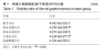
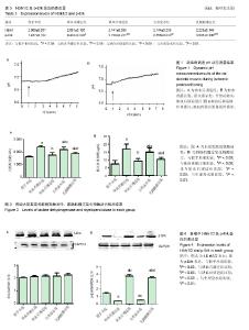
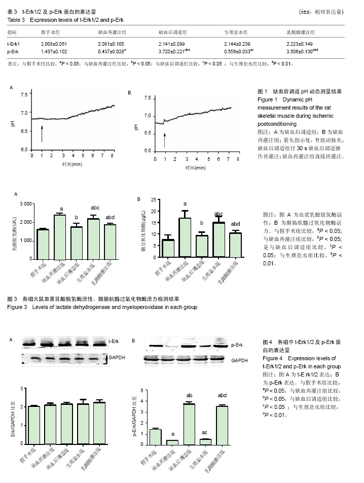
2.1 实验动物数量分析 实验选用大鼠25只,分为5组,实验过程未有脱失。 2.2 缺血后调适pH动态测定结果 大鼠股动脉手术显露后,将玻纤pH探头植入腓肠肌,选取主缺血期酸中毒至稳定时段、缺血再灌注组缺血后再灌注pH值回复至正常稳定时段及缺血后调适组缺血后调适完成至pH回复正常时段进行连续测量,对比缺血再灌注组缺血后调适组会出现一个酸中毒延长稳定平台,pH为6.810±0.133,时长为2 min 40 s。见图1。 2.3 腓肠肌湿/干质量(W/D)比值 见表1,生理盐水组和缺血再灌注组相较假手术组的湿/干质量比值显著增高(P < 0.05),说明骨骼肌前期缺血导致组织水肿明显;乳酸酸灌注组与缺血后调适组较假手术组差异无显著性意义(P < 0.05),说明酸性灌注有效模拟缺血后调适作用,可有效减轻组织水肿。"
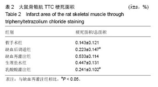
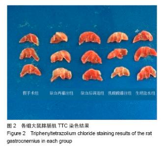
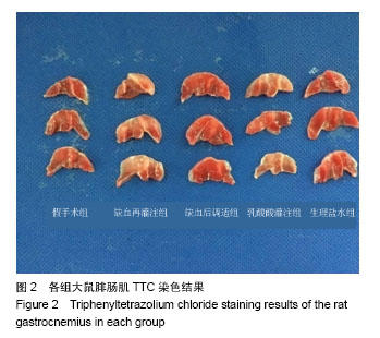
2.4 骨骼肌TTC染色梗死面积结果 与缺血再灌注组相比,缺血后调适组及乳酸酸灌注组的梗死面积较缺血再灌注组明显减小,差异有显著性意义(P < 0.05);而缺血后调适组和乳酸酸灌注组与假手术组相比,梗死面积虽然有所增大,但其差异均无显著性意义(P > 0.05),见图2,表2。"
2.5 血浆乳酸脱氢酶检测 各组血浆乳酸脱氢酶检测结果见图3A。缺血后调适组及乳酸酸灌注组血浆乳酸脱氢酶活性与假手术组比较差异不明显(P > 0.05),相较于缺血再灌注组显著降低(P < 0.05),表明缺血后调适组及乳酸酸灌注组均可降低骨骼肌缺血再灌注所致的乳酸脱氢酶活性反应性升高,乳酸酸灌注组可以有效模拟缺血后调适对骨骼肌的保护作用。 2.6 腓肠肌髓过氧化物酶检测 各组血浆髓过氧化物酶结果见图3B。缺血后调适组及乳酸酸灌注组腓肠肌髓过氧化物酶活性与假手术组比较差异不明显(P > 0.05),相较于缺血再灌注组显著降低(P < 0.05),表明缺血后调适组及乳酸酸灌注组均可降低骨骼肌缺血再灌注所致的腓肠肌髓过氧化物酶活性反应性升高,乳酸酸灌注组可以有效模拟缺血后调适对骨骼肌的保护作用。 2.7 Werstern blot检测t-Erk1/2及p-Erk结果 与假手术组对比,其余各组中t-Erk1/2的表达量增加,但表达量差异无显著性意义(P > 0.05),见图4A。 p-Erk检测结果显示,与缺血后调适组及乳酸酸灌注组比较缺血再灌注组表达量增高明显(P < 0.05),差异有显著性意义;而缺血后调适组与乳酸酸灌注组之间比较,缺血后调适组较乳酸酸灌注组表达增加,但差异无显著性意义(P > 0.05),见图4B。 故得出,乳酸灌注能有效模拟缺血后调适作用,通过激活p-Erk表达,激活MAPK通路,激活RISK信号通路,对缺血再灌注的骨骼肌起到保护作用。结果见图4,表3。"
"
| [1] Jennings RB,Sommers HM,Smyth GA, et al. Myocardial necrosis induced by temporary occlusion of a coronary artery in the dog. Arch Pathol. 1960;70:68-78.[2] Orsini F,Chrysanthou E, Dudler T, et al. Mannan binding lectin-associated serine protease-2 (MASP-2) critically contributes to post-ischemic brain injury independent of MASP-1. J Neuroinflammation. 2016;13(1):213.[3] Ferchichi H, Bacha S, Kourda N, et al. Animal model of liver ischemia reperfusion: biochemical and histological evaluation.La Tunisie Me?dicale.2016;94(3):235.[4] Na KR,Choi H,Jeong JY, et al.Nafamostat Mesilate Attenuates Ischemia-Reperfusion-Induced Renal Injury. Transplant Proc. 2016;48(6):2192-2199.[5] Zu G,Yao J,Ji A,et al.Nurr1 promotes intestinal regeneration after ischemia/reperfusion injury by inhibiting the expression of p21 (Waf1/Cip1). J Mol Med (Berl). 2017; 95(1):83-95.[6] Eckert P, Schnackerz K. Ischemic tolerance of human skeletal muscle. Ann Plast Surg. 1991;26(1):77-84.[7] Blaisdell FW.The pathophysiology of skeletal muscle ischemia and the reperfusion syndrome: a review. Cardiovasc Surg. 2002;10(6):620-630. [8] Appell HJ,Glöser S,Duarte JA,et al.Skeletal muscle damage during tourniquet-induced ischaemia. The initial step towards atrophy after orthopaedic surgery?. Eur J Appl Physiol Occup Physiol. 1993;67(4):342-347.[9] Gillani S,Cao J,Suzuki T,et al.The effect of ischemia reperfusion injury on skeletal muscle. Injury.2012;43(6): 670-675.[10] Murry CE,Jennings RB,Reimer KA. Preconditioning with ischemia: a delay of lethal cell injury in ischemic myocardium. Circulation.1986; 74(5):1124-1136.[11] Zaman J,Jeddi S,Daneshpour MS,et al.Ischemic postconditioning provides cardioprotective and antiapoptotic effects against ischemia-reperfusion injury through iNOS inhibition in hyperthyroid rats. Gene.2015; 570(2):185.[12] Cao QF,Qu MJ,Yang WQ,et al.Ischemia postconditioning preventing lung ischemia-reperfusion injury.Gene.2015; 554(1):120-124.[13] Hahn JY, Yu CW, Park HS, et al.Long-term effects of ischemic postconditioning on clinical outcomes: 1-year follow-up of the POST randomized trial. Am Heart J. 2015;169(5):639-646.[14] Duanmu WS,Cao L,Chen JY,et al.Ischemic postconditioning protects against ischemic brain injury by up-regulation of acid-sensing ion channel 2a. Neural Regen Res. 2016;11(4): 641-645.[15] Zhao ZQ,Corvera JS,Halkos ME,et al. Inhibition of myocardial injury by ischemic postconditioning during reperfusion: comparison with ischemic preconditioning. Am J Physiol Heart Circ Physiol. 2003;285(2):H579-88..[16] Park JW, Kang JW, Jeon WJ, et al. Postconditioning protects skeletal muscle from ischemia-reperfusion injury.Microsurgery.2010;30(3):223-229.[17] Cohen MV,Yang XM,Downey JM.The pH hypothesis of postconditioning: staccato reperfusion reintroduces oxygen and perpetuates myocardial acidosis. Circulation.2007; 115(14):1895.[18] 彭龙龙, 阳富春, 薄占东,等. 不同缺血后调适方案对大鼠模型肢体骨骼肌缺血再灌注损伤的影响[J]. 中国组织工程研究, 2015,19(5):739-744.[19] Jäger R, Roberts MD, Lowery RP, et al.Oral adenosine-5’-triphosphate (ATP) administration increases blood flow following exercise in animals and humans. J Int Soc Sports Nutr. 2014;11:28[20] Cohen MV,Yang XM,Downey JM.Acidosis, oxygen, and interference with mitochondrial permeability transition pore formation in the early minutes of reperfusion are critical to postconditioning's success. Basic Res Cardiol. 2008;103(5): 464-471.[21] Baines CP, Song CX, Zheng YT,et al.Protein Kinase Cε Interacts With and Inhibits the Permeability Transition Pore in Cardiac Mitochondria. Circ Res. 2003;92(8):873-880.[22] Juel C.Skeletal muscle Na+/H+ exchange in rats: pH dependency and the effect of training. Acta Physiol Scand. 1998;164(2):135-140.[23] Murphy RM,Dutka TL,Horvath D,et al.Ca2+-dependent proteolysis of junctophilin-1 and junctophilin-2 in skeletal and cardiac muscle. J Physiol. 2013;591(3):719-729.[24] Lejay A,Meyer A,Schlagowski AI,et al.Mitochondria: mitochondrial participation in ischemia-reperfusion injury in skeletal muscle. Int J Biochem Cell Biol. 2014;50:101-105.[25] Inserte J,Barba I,Hernando V,et al. Effect of acidic reperfusion on prolongation of intracellular acidosis and myocardial salvage. Cardiovasc Res. 2008;77(4):782-790. [26] Schulman D,Latchman DS,Yellon DM. Urocortin protects the heart from reperfusion injury via upregulation of p42/p44 MAPK signaling pathway. Am J Physiol Heart Circ Physiol. 2002;283(4):H1481-488.[27] Hausenloy DJ, Yellon DM. Reperfusion injury salvage kinase signalling: taking a RISK for cardioprotection. Heart Failure Reviews.2007;12(3):217-234.[28] Yang XM, Proctor JB, Cui L,et al.Multiple, brief coronary occlusions during early reperfusion protect rabbit hearts by targeting cell signaling pathways. J Am Coll Cardiol. 2004; 44(5):1103-1110.[29] Hausenloy DJ,Yellon DM.New directions for protecting the heart against ischaemia-reperfusion injury: targeting the Reperfusion Injury Salvage Kinase (RISK)-pathway. Cardiovasc Res.2004;61(3):448-460.[30] Chiu WC,Chiou TJ,Chung MJ,et al.β2-Glycoprotein I Inhibits Vascular Endothelial Growth Factor-Induced Angiogenesis by Suppressing the Phosphorylation of Extracellular Signal-Regulated Kinase 1/2, Akt, and Endothelial Nitric Oxide Synthase. PLoS One.2016 ;11(8):e0161950. [31] Wang L,Shan Y,Chen L,et al.Colchicine protects rat skeletal muscle from ischemia/reperfusion injury by suppressing oxidative stress and inflammation. Iran J Basic Med Sci.2016; 19(6):670-675. |
| [1] | Yao Xiaoling, Peng Jiancheng, Xu Yuerong, Yang Zhidong, Zhang Shuncong. Variable-angle zero-notch anterior interbody fusion system in the treatment of cervical spondylotic myelopathy: 30-month follow-up [J]. Chinese Journal of Tissue Engineering Research, 2022, 26(9): 1377-1382. |
| [2] | Wang Shuo, Liu Wenying, Lü Chaofan, Li Jiacong, Geng Yi, Zhao Yungang. Cardioprotective effect of 3-nitro-N-methyl salicylamide on the isolated rat heart under cold ischemia preservation [J]. Chinese Journal of Tissue Engineering Research, 2022, 26(8): 1194-1201. |
| [3] | Li Zhiyi, He Pengcheng, Bian Tianyue, Xiao Yuxia, Gao Lu, Liu Huasheng. Bibliometric and visualized analysis of ferroptosis mechanism research [J]. Chinese Journal of Tissue Engineering Research, 2022, 26(8): 1202-1209. |
| [4] | Yang Shenglin, Pu Xingwei, Luo Chunshan, Yang Jianwen. Neuroprotective effects of tetrandrine preconditioning in rabbits with spinal cord ischemia-reperfusion injury [J]. Chinese Journal of Tissue Engineering Research, 2022, 26(8): 1223-1227. |
| [5] | An Weizheng, He Xiao, Ren Shuai, Liu Jianyu. Potential of muscle-derived stem cells in peripheral nerve regeneration [J]. Chinese Journal of Tissue Engineering Research, 2022, 26(7): 1130-1136. |
| [6] | Zhang Jinglin, Leng Min, Zhu Boheng, Wang Hong. Mechanism and application of stem cell-derived exosomes in promoting diabetic wound healing [J]. Chinese Journal of Tissue Engineering Research, 2022, 26(7): 1113-1118. |
| [7] | Dong Miaomiao, Lai Han, Li Manling, Xu Xiuhong, Luo Meng, Wang Wenhao, Zhou Guoping. Effect of electroacupuncture on expression of nucleotide binding oligomerization domain-like receptor protein 3/cysteinyl aspartate specific proteinase 1 in rats with cerebral ischemia/reperfusion injury [J]. Chinese Journal of Tissue Engineering Research, 2022, 26(5): 749-755. |
| [8] | He Yunying, Li Lingjie, Zhang Shuqi, Li Yuzhou, Yang Sheng, Ji Ping. Method of constructing cell spheroids based on agarose and polyacrylic molds [J]. Chinese Journal of Tissue Engineering Research, 2022, 26(4): 553-559. |
| [9] | He Guanyu, Xu Baoshan, Du Lilong, Zhang Tongxing, Huo Zhenxin, Shen Li. Biomimetic orientated microchannel annulus fibrosus scaffold constructed by silk fibroin [J]. Chinese Journal of Tissue Engineering Research, 2022, 26(4): 560-566. |
| [10] | Chen Xiaoxu, Luo Yaxin, Bi Haoran, Yang Kun. Preparation and application of acellular scaffold in tissue engineering and regenerative medicine [J]. Chinese Journal of Tissue Engineering Research, 2022, 26(4): 591-596. |
| [11] | Kang Kunlong, Wang Xintao. Research hotspot of biological scaffold materials promoting osteogenic differentiation of bone marrow mesenchymal stem cells [J]. Chinese Journal of Tissue Engineering Research, 2022, 26(4): 597-603. |
| [12] | Shen Jiahua, Fu Yong. Application of graphene-based nanomaterials in stem cells [J]. Chinese Journal of Tissue Engineering Research, 2022, 26(4): 604-609. |
| [13] | Zhang Tong, Cai Jinchi, Yuan Zhifa, Zhao Haiyan, Han Xingwen, Wang Wenji. Hyaluronic acid-based composite hydrogel in cartilage injury caused by osteoarthritis: application and mechanism [J]. Chinese Journal of Tissue Engineering Research, 2022, 26(4): 617-625. |
| [14] | Li Hui, Chen Lianglong. Application and characteristics of bone graft materials in the treatment of spinal tuberculosis [J]. Chinese Journal of Tissue Engineering Research, 2022, 26(4): 626-630. |
| [15] | Gao Cangjian, Yang Zhen, Liu Shuyun, Li Hao, Fu Liwei, Zhao Tianyuan, Chen Wei, Liao Zhiyao, Li Pinxue, Sui Xiang, Guo Quanyi. Electrospinning for rotator cuff repair [J]. Chinese Journal of Tissue Engineering Research, 2022, 26(4): 637-642. |
| Viewed | ||||||
|
Full text |
|
|||||
|
Abstract |
|
|||||