Chinese Journal of Tissue Engineering Research ›› 2017, Vol. 21 ›› Issue (14): 2215-2221.doi: 10.3969/j.issn.2095-4344.2017.14.014
Previous Articles Next Articles
Comparative study on cytocompatibility of Pluronic copolymer nanoparticles modified with folic acid and polylactic acid
Huang Han, Hu Qing-juan, Xiong Xiang-yuan, Ge Xiang-jun, Chen Chong, Li Yu-ping
-
Received:2016-12-16Online:2017-05-18Published:2017-06-10 -
Contact:Yu-ping, M.D., Professor, School of Life Science, Jiangxi Science & Technology Normal University, Nanchang 330013, Jiangxi Province, China -
About author:Huang Han, Studying for master’s degree, School of Life Science, Jiangxi Science & Technology Normal University, Nanchang 330013, Jiangxi Province, China -
Supported by:the National Natural Science Foundation of China, No. 31360376, 21264009; the Cultivation Project for Academic and Technical Leaders of Major Disciplines of Jiangxi Province, No. 20153BCB22009; the Scientific and Technological Landing Project of Higher Education of Jiangxi Province, No. KJLD13071
CLC Number:
Cite this article
Huang Han, Hu Qing-juan, Xiong Xiang-yuan, Ge Xiang-jun, Chen Chong, Li Yu-ping. Comparative study on cytocompatibility of Pluronic copolymer nanoparticles modified with folic acid and polylactic acid[J]. Chinese Journal of Tissue Engineering Research, 2017, 21(14): 2215-2221.
share this article
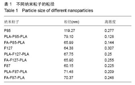
2.1 纳米粒子的粒度 采用动态激光光散射仪对制备的各种纳米粒子的粒度进行分析,结果如表1所示,聚合物在水中自组装形成的纳米粒子胶束的粒径在60-120 nm之间,粒度比较集中。"


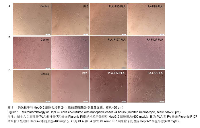
2.2 细胞形态学评价结果 将不同质量浓度的纳米粒子与HepG-2细胞共同培养24 h后,选取每种材料的最高质量浓度的细胞形态进行比较观察(图1),各实验组与空白对照组无明显差异,细胞形态正常,生长良好。但是高质量浓度(400 mg/L)F87纳米粒子处理后,HepG-2细胞有一定的圆缩漂浮,细胞数量与空白组相比有所减少。"
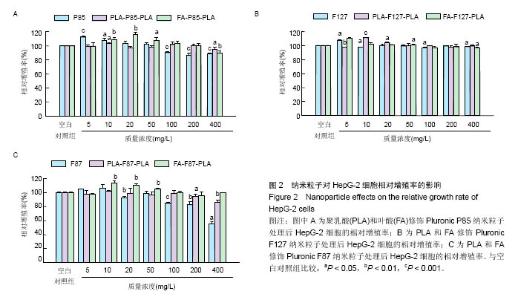
2.3 细胞代谢活性评价结果 用P85、PLA-P85-PLA和FA-P85-PLA纳米粒子处理24 h后,对细胞生存活力的影响如图2A所示,低质量浓度P85纳米粒子(5,10,20,50 mg/L)对HepG-2细胞的增殖有促进作用,高质量浓度的P85纳米粒子(100,200,400 mg/L)对HepG-2细胞的增殖有抑制作用(P < 0.01),对应的细胞相对增殖率分别为90.4%,86.4%,88.2%;而经PLA和FA修饰的P85聚合物纳米粒子(PLA-P85-PLA NPs和FA-P85-PLA NPs)在100,200,400 mg/L条件下对应的细胞相对增殖率分别为101.9%,100.7%,94.9%和103%,100.6%,89.5%,与对照组相比,细胞增殖率差异无显著性意义 (P > 0.05)。 由图2B可知,F127、PLA-F127-PLA和FA-F127- PLA纳米粒子与HepG-2共同培养后,与对照组相比较,细胞相对活力差异无显著性意义(P > 0.05),低质量浓度的纳米粒子可促进HepG-2细胞的生长,表现出良好的生物相容性。"
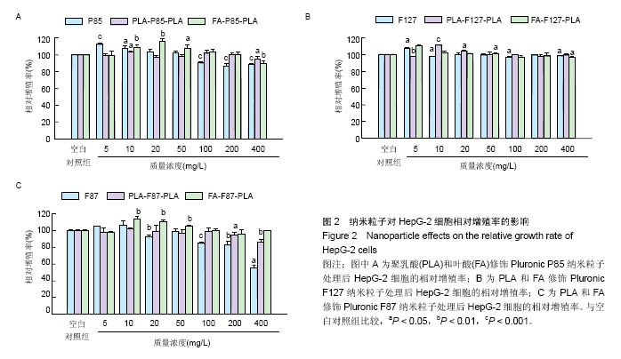
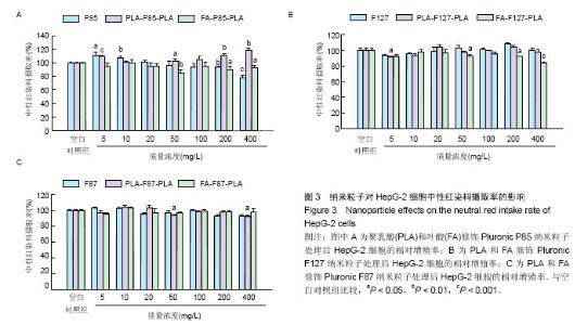
由图2C可知,低质量浓度F87纳米粒子(5,10,20,50 mg/L)促进细胞生长,高质量浓度F87纳米粒子(100,200,400 mg/L)对HepG-2细胞生长有一定的抑制作用,其对应的细胞相对增殖率分别为84.8%,82.8%,55.4%,与细胞形态观察结果相一致(图1);PLA-F87-PLA纳米粒子实验组与对照组相比较,细胞相对活力差异无显著性意义(P > 0.05),但高质量浓度PLA-F87-PLA纳米粒子(400 mg/L)对细胞生长表现出一定的抑制作用(P < 0.01);FA-F87-PLA纳米粒子可促进HepG-2细胞的生长。 2.4 中性红摄取实验结果 由图3A可知,低质量浓度P85纳米粒子对细胞生长有促进作用,高质量浓度P85纳米粒子(400 mg/L)抑制细胞生长,其细胞相对增殖率为77.9%,与对照组相比差异有极显著性意义(P < 0.001);PLA-P85- PLA纳米粒子表现促进生长的作用,在质量浓度为200 mg/L和400 mg/L时明显促进细胞增长,与空白对照组相比差异有非常显著性意义(P < 0.01);FA-P85-PLA 纳米粒子对细胞增殖的影响与对照组相比差异无显著性意义(P > 0.05),高质量浓度时对HepG-2细胞的增殖有抑制作用(P < 0.05)。 由图3B和3C可知,F127、PLA-F127-PLA、FA-F127-PLA、F87、PLA-F87-PLA、FA-F87-PLA 纳米粒子处理组和对照组相比,HepG-2细胞对中性红染料的摄入量无显著性变化(P > 0.05),高质量浓度FA-F127-PLA 纳米粒子条件下细胞对中性红染料的摄入量表现极显著性变化(P < 0.001)。 "

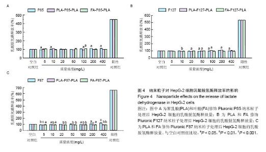
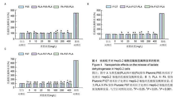
2.5 乳酸脱氢酶实验结果 由图4可知,不同质量浓度的PLA-P85-PLA、FA-P85-PLA、F127、PLA-F127-PLA和FA-F127-PLA纳米粒子与HepG-2细胞共培养24 h后,乳酸脱氢酶的释放率与空白组相比较,差异无显著性意义 (P > 0.05)。 P85纳米粒子在质量浓度为200和400 mg/L时,与空白组相比,乳酸脱氢酶的释放量增加(P < 0.05)。PLA-F87-PLA和FA-F87-PLA纳米粒子处理HepG-2细胞24 h后,明显抑制乳酸脱氢酶的释放(P < 0.01);用 400 mg/L的F87纳米粒子处理时,乳酸脱氢酶的释放量增加非常明显(P < 0.001)。"
| [1]Fahmy TM, Fong PM, Goyal A, et al. Targeted for drug delievey. Materials Today. 2005; 8(8): 18-26.[2]Sun Y, Yu B, Wang G, et al. Enhanced antitumor efficacy of vitamin E TPGS-emulsified PLGA nanoparticles for delivery of paclitaxel. Colloids Surf B Biointerfaces. 2014;123:716-723.[3]Zhong Y, Zhang J, Cheng R, et al. Reversibly crosslinked hyaluronic acid nanoparticles for active targeting and intelligent delivery of doxorubicin to drug resistant CD44+ human breast tumor xenografts. J Control Release. 2015; 205:144-154.[4]Oberdörster G, Ferin J, Gelein R, et al. Role of the alveolar macrophage in lung injury: studies with ultrafine particles. Environ Health Perspect. 1992;97:193-199.[5]Srivastav AK, Kumar M, Ansari NG, et al. A comprehensive toxicity study of zinc oxide nanoparticles versus their bulk in Wistar rats: Toxicity study of zinc oxide nanoparticles. Hum Exp Toxicol. 2016;35(12):1286-1304.[6]Curtis J, Greenberg M, Kester J, et al. Nanotechnology and nanotoxicology: a primer for clinicians. Toxicol Rev. 2006; 25(4): 245-260.[7]Hanot-Roy M, Tubeuf E, Guilbert A, et al. Oxidative stress pathways involved in cytotoxicity and genotoxicity of titanium dioxide (TiO2) nanoparticles on cells constitutive of alveolo-capillary barrier in vitro. Toxicol In Vitro. 2016;33: 125-135.[8]Alexandridis P, Hatton TA. Poly(ethylene oxide)-poly(propylene oxide)-poly(ethylene oxide) block copolymer surfactants in aqueous solutions and at interfaces: thermodynamics, structure, dynamics, and modeling. Colloids and Surfaces A: Physicochemical and Engineering Aspects. 1995; 96(1–2): 1-46.[9]Alakhov VYu, Moskaleva EYu, Batrakova EV, et al. Hypersensitization of multidrug resistant human ovarian carcinoma cells by pluronic P85 block copolymer. Bioconjug Chem. 1996;7(2):209-216.[10]Xia W, Low PS. Folate-targeted therapies for cancer. J Med Chem. 2010;53(19):6811-6824.[11]Hilgenbrink AR, Low PS. Folate receptor-mediated drug targeting: from therapeutics to diagnostics .J Pharm Sci. 2005;94(10):2135-2146.[12]Xiong XY, Qin X, Li ZL, et al. Synthesis, drug release and targeting behaviors of Novel Folated Pluronic F87/poly(lactic acid) block copolymer. European Polymer Journal. 2015; 68:233-242.[13]秦翔.新型含叶酸靶向Pluronic/PLA高分子药物制剂的研究[D]. 南昌:江西科技师范大学,2015.[14]Xiong XY, Li QH, Li YP, et al. Pluronic P85/poly(lactic acid) vesicles as novel carrier for oral insulin delivery. Colloids Surf B Biointerfaces. 2013;111:282-288.[15]李资玲,熊向源,龚妍春,等.包埋紫杉醇的Pluronic P85 /聚乳酸纳米粒子制备及体外释放行为考察[J].中国实验方剂学杂志, 2014,20(1):1-4.[16]Kabanov AV, Batrakova EV, Alakhov VY. Pluronic block copolymers as novel polymer therapeutics for drug and gene delivery. J Control Release. 2002;82(2-3):189-212.[17]Mosmann T. Rapid colorimetric assay for cellular growth and survival: application to proliferation and cytotoxicity assays. J Immunol Methods. 1983;65(1-2):55-63.[18]孙利珍,李玉萍,邱维曌,等. 纳米级PLA-P85-PLA两亲性共聚物的细胞相容性研究[J]. 功能材料, 2013, 44(21): 3135-3142.[19]Gomes-Cornélio AL, Rodrigues EM, Mestieri LB, et al. Cytotoxicity and genotoxicity of calcium silicate-based cements on an osteoblast lineage. Braz Oral Res. 2016;30(1): e48.[20]Saberi EA, Karkehabadi H, Mollashahi NF. Cytotoxicity of Various Endodontic Materials on Stem Cells of Human Apical Papilla. Iran Endod J. 2016;11(1):17-22.[21]Ta?l? PN, Yalvaç ME, Sofiev N, et al. Effect of F68, F127, and P85 pluronic block copolymers on odontogenic differentiation of human tooth germ stem cells. J Endod. 2013;39(10): 1265-1271.[22]Borenfreund E, Puerner JA. Cytotoxicity of metals, metal-metal and metal-chelator combinations assayed in vitro. Toxicology. 1986;39(2):121-134.[23]Babich H, Sinensky MC. Indirect cytotoxicity of dental materials: a study with Transwell inserts and the neutral red uptake assay. Altern Lab Anim. 2001;29(1):9-13.[24]Risbud MV, Hambir S, Jog J, et al. Biocompatibility assessment of polytetrafluoroethylene/wollastonite composites using endothelial cells and macrophages. J Biomater Sci Polym Ed. 2001;12(11):1177-1189.[25]Ivask A, Titma T, Visnapuu M, et al. Toxicity of 11 Metal Oxide Nanoparticles to Three Mammalian Cell Types In Vitro. Curr Top Med Chem. 2015;15(18):1914-1929.[26]Weyermann J, Lochmann D, Zimmer A. A practical note on the use of cytotoxicity assays. Int J Pharm. 2005;288(2): 369-376. |
| [1] | Yao Xiaoling, Peng Jiancheng, Xu Yuerong, Yang Zhidong, Zhang Shuncong. Variable-angle zero-notch anterior interbody fusion system in the treatment of cervical spondylotic myelopathy: 30-month follow-up [J]. Chinese Journal of Tissue Engineering Research, 2022, 26(9): 1377-1382. |
| [2] | Zhang Jinglin, Leng Min, Zhu Boheng, Wang Hong. Mechanism and application of stem cell-derived exosomes in promoting diabetic wound healing [J]. Chinese Journal of Tissue Engineering Research, 2022, 26(7): 1113-1118. |
| [3] | An Weizheng, He Xiao, Ren Shuai, Liu Jianyu. Potential of muscle-derived stem cells in peripheral nerve regeneration [J]. Chinese Journal of Tissue Engineering Research, 2022, 26(7): 1130-1136. |
| [4] | Chen Xiaoxu, Luo Yaxin, Bi Haoran, Yang Kun. Preparation and application of acellular scaffold in tissue engineering and regenerative medicine [J]. Chinese Journal of Tissue Engineering Research, 2022, 26(4): 591-596. |
| [5] | Kang Kunlong, Wang Xintao. Research hotspot of biological scaffold materials promoting osteogenic differentiation of bone marrow mesenchymal stem cells [J]. Chinese Journal of Tissue Engineering Research, 2022, 26(4): 597-603. |
| [6] | Shen Jiahua, Fu Yong. Application of graphene-based nanomaterials in stem cells [J]. Chinese Journal of Tissue Engineering Research, 2022, 26(4): 604-609. |
| [7] | Zhang Tong, Cai Jinchi, Yuan Zhifa, Zhao Haiyan, Han Xingwen, Wang Wenji. Hyaluronic acid-based composite hydrogel in cartilage injury caused by osteoarthritis: application and mechanism [J]. Chinese Journal of Tissue Engineering Research, 2022, 26(4): 617-625. |
| [8] | Li Hui, Chen Lianglong. Application and characteristics of bone graft materials in the treatment of spinal tuberculosis [J]. Chinese Journal of Tissue Engineering Research, 2022, 26(4): 626-630. |
| [9] | Gao Cangjian, Yang Zhen, Liu Shuyun, Li Hao, Fu Liwei, Zhao Tianyuan, Chen Wei, Liao Zhiyao, Li Pinxue, Sui Xiang, Guo Quanyi. Electrospinning for rotator cuff repair [J]. Chinese Journal of Tissue Engineering Research, 2022, 26(4): 637-642. |
| [10] | Le Guoping, Zhang Ming, Xi Licheng, Luo Hanwen. Preparation and in vitro evaluation of vancomycin hydrochloride@polylactic acid-glycolic acid copolymer-chitosan-hyaluronic acid composite sustained-release microspheres [J]. Chinese Journal of Tissue Engineering Research, 2022, 26(4): 528-534. |
| [11] | He Yunying, Li Lingjie, Zhang Shuqi, Li Yuzhou, Yang Sheng, Ji Ping. Method of constructing cell spheroids based on agarose and polyacrylic molds [J]. Chinese Journal of Tissue Engineering Research, 2022, 26(4): 553-559. |
| [12] | He Guanyu, Xu Baoshan, Du Lilong, Zhang Tongxing, Huo Zhenxin, Shen Li. Biomimetic orientated microchannel annulus fibrosus scaffold constructed by silk fibroin [J]. Chinese Journal of Tissue Engineering Research, 2022, 26(4): 560-566. |
| [13] | Guan Jian, Jia Yanfei, Zhang Baoxin , Zhao Guozhong. Application of 4D bioprinting in tissue engineering [J]. Chinese Journal of Tissue Engineering Research, 2022, 26(3): 446-455. |
| [14] | Liu Jiali, Suo Hairui, Yang Han, Wang Ling, Xu Mingen. Influence of lay-down angles on mechanical properties of three-dimensional printed polycaprolactone scaffolds [J]. Chinese Journal of Tissue Engineering Research, 2022, 10(16): 2612-2617. |
| [15] | Huang Bo, Chen Mingxue, Peng Liqing, Luo Xujiang, Li Huo, Wang Hao, Tian Qinyu, Lu Xiaobo, Liu Shuyun, Guo Quanyi . Fabrication and biocompatibility of injectable gelatin-methacryloyl/cartilage-derived matrix particles composite hydrogel scaffold [J]. Chinese Journal of Tissue Engineering Research, 2022, 10(16): 2600-2606. |
| Viewed | ||||||
|
Full text |
|
|||||
|
Abstract |
|
|||||