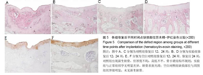
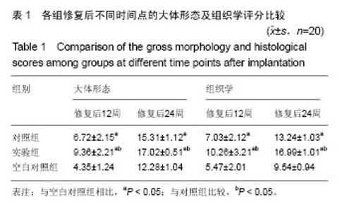
| [1]雷鸣,肖德明,熊建义,等.藻酸钠微球三维立体培养恢复去分化软骨细胞表型的实验研究[J].中国矫形外科杂志, 2011,19(9): 779-782. [2]Hansen OM,Foldager CB,Christensen BB,et al.Increased chondrocyte seeding density has no positive effect on cartilage repair in an MPEG-PLGA scaffold.Knee Surg Sports Traumatol Arthrosc. 2013;21(2):485-493. [3]陈加荣,张余,黄华扬,等. 磁力靶向传递SPIO标记的BMSC修复关节软骨缺损的研究进展[J]. 中国骨科临床与基础研究杂志, 2014,6(2):118-122.[4]刘拴,杨洪平,张卫国.关节镜下关节清理术治疗膝关节骨性关节炎的疗效分析[J].实用临床医学杂志,2014,18(1):58-60.[5]Ito S,Sato M,Yamato M,et al.Repair of articular cartilage defect with layered chondrocyte sheets and cultured synovial cells.Biomaterials.2012;33(21):5278-5286. [6]Yi L,Lin X.Tissue engineering technology for repair of articular cartilage injury. Chin J Tissue Eng Res. 2013;17(41): 7310-7316. [7]张海峰,杜子婧,姜闻博,等.3D打印PLA-HA复合材料与骨髓基质细胞的相容性研究[J].组织工程与重建外科杂志,2015,11(6): 349-353. [8]Xue Z,Niu LY,An G,et al.Repairing rabbit’s radial bone defects using injectable nano-hydroxyapatite composite scaffold co-cultured with bone marrow mesenchymal stem cells. Chin J Tissue Eng Res.2015;3:378-383. [9]王虔,马云胜,李德华.不同来源蚕丝蛋白修复骨软骨组织缺损的效果比较[J].中国组织工程研究,2015,19(52):8412-8417.[10]Wray LS,Rnjak-Kovacina J,Mandal BB,et al.A silk-based scaffold platform with tunable architecture for engineering critically-sized tissue constructs. Biomaterials. 2012;33: 9214-9224.[11]Stenhamre H,Nannmark U,Lindahl A,et al.Brittberg M Influence of pore size on the redifferentiation potential of human articular chondrocytes in poly(urethane urea) scaffolds.J Tissue Eng Regen Med.2011;5(7):578-588.[12]Im GI,Ko JY,Lee JH.Chondrogenesis of adipose stem cells in a porous polymer scaffold: influence of the pore size.Cell Transplant.2012;21(11):2397-2405.[13]高晓珺,解骏,肖涟波,等.骨碎补总黄酮对胶原诱导大鼠类风湿关节炎骨破坏治疗作用的实验研究[J].实用临床医药杂志, 2013, 17(5):13-17. [14]Nishimoto S,Fukuda K,Kawai K,et al.Supplementation of bone marrow aspirate-derived platelet-rich plasma for treating radiation-induced ulcer after cardiac fluoroscopic procedures: A preliminary report.Indian J Plast Surg.2012;45(1):109-114.[15]Viale-Bouroncle S,Gosau M,Morsczeck C.Collagen I induces the expression of alkaline phosphatase and osteopontin via inde-pendent activations of FAK and ERK signalling pathways. Arch Oral Biol.2014;59(12):1249-1255.[16]Cheng ZL,Hoi MW,Kelvin WKY,et al.The development, fabrication,and material characterization of polypropylene composites reinforced with carbon nanofiber and hydroxyapatite nanorod hybrid fillers.Int J Nanomed.2014;9: 1299-1310.[17]Kolmas J,Sobczak M,Oledzka E,et al. Synthesis, characterization and in vitro evaluation of new composite bisphosphonate delivery systems.Int J Mol Sci.2014; 15(9): 16831-16847.[18]Wei B,Jin C,Xu Y,et al.Effect of bone marrow mesenchymal stem cells-derived extracellular matrix scaffold on chondrogenic differentiation of marrow clot after microfracture of bone marrow stimulation in vitro.Zhongguo Xiu Fu Chong Jian Wai Ke Za Zhi.2013;27(4):464-474. |