Chinese Journal of Tissue Engineering Research ›› 2017, Vol. 21 ›› Issue (1): 115-121.doi: 10.3969/j.issn.2095-4344.2017.01.021
Previous Articles Next Articles
Effects of recombinant adenovirus-mediated adiponectin on human umbilical vein endothelial cell injury and the underlying mechanism
Wang Xue-mei, Wei Qin, Duan Ming-jun, Zhang Chun, Yang Yi-ning
- Xinjiang Key Laboratory of Medical Animal Model Research, Clinical Medical Research Institute of First Affiliated Hospital of Xinjiang Medical University, Urumqi 830011, Xinjiang Uygur Autonomous Region, China
-
Online:2017-01-08Published:2017-03-15 -
Contact:Yang Yi-ning, M.D., Professor, Chief physician, Doctoral supervisor, Xinjiang Key Laboratory of Medical Animal Model Research, Clinical Medical Research Institute of First Affiliated Hospital of Xinjiang Medical University, Urumqi 830011, Xinjiang Uygur Autonomous Region, China -
About author:Wang Xue-mei, M.D., Associate researcher, Xinjiang Key Laboratory of Medical Animal Model Research, Clinical Medical Research Institute of First Affiliated Hospital of Xinjiang Medical University, Urumqi 830011, Xinjiang Uygur Autonomous Region, China -
Supported by:the Natural Science Foundation for the Youth of Xinjiang Uygur Autonomous Region, No. 2015211C095; the State Key Lab Incubation Base of Xinjiang Major Diseases Research, No. SKLIB-XJMDR-2014-16
CLC Number:
Cite this article
Wang Xue-mei, Wei Qin, Duan Ming-jun, Zhang Chun, Yang Yi-ning. Effects of recombinant adenovirus-mediated adiponectin on human umbilical vein endothelial cell injury and the underlying mechanism[J]. Chinese Journal of Tissue Engineering Research, 2017, 21(1): 115-121.
share this article
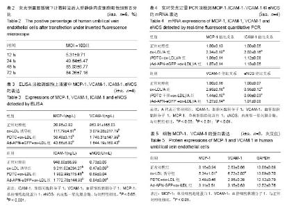
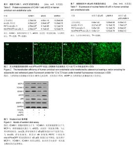
2.1 重组腺病毒体外转染效率 倒置荧光显微镜下观察细胞的绿色荧光蛋白表达,病毒转染12 h后,MOI= 100 pfu/cell组的细胞开始表达eGFP;eGFP的表达强度 随着时间延长、转染复数的增加逐渐增强;人脐静脉内皮细胞在转染后48 h达到高峰。至转染后72 h,eGFP在人脐静脉内皮细胞仍处于高表达,见表2,图2。 2.2 内皮细胞体外损伤模型的建立 2.2.1 rAd-APN-eGFP降低人脐静脉内皮细胞上清液中的促炎因子表达,增加抗炎因子表达 ox-LDL 30 mg/L刺激48 h后,MCP-1、VCAM-1和ICAM-1表达急剧增高。单因素方差分析结果显示,与ox-LDL诱导组相比,分别用PDTC干预和rAd-APN-eGFP转染诱导处理后,培养液中的MCP-1、VCAM-1和ICAM-1水平均下降,低于ox-LDL诱导组。而PDTC干预和rAd-APN-eGFP处理组两组间差异没有显著性。说明PDTC干预和rAd-APN-eGFP对由ox-LDL诱导的人脐静脉内皮细胞分泌产生促炎因子都有抑制,且作用相似。ox-LDL诱导人脐静脉内皮细胞48 h后,ox-LDL诱导组的人脐静脉内皮细胞上清液中的内皮型一氧化氮合酶活性低于空白对照组,差异具有显著性;而PDTC干预+ ox-LDL诱导组细胞上清液中的内皮型一氧化氮合酶活性变化,差异无显著性意义(P=0.426),rAd-APN-eGFP转染+ ox-LDL诱导 48 h后细胞上清液中内皮型一氧化氮合酶活性和对照组相比升高。证明了rAd-APN-eGFP转导能够增加细胞分泌内皮型一氧化氮合酶,促进内皮细胞产生一氧化氮,减轻内皮细胞损伤,加快内皮细胞修复,见表3。 2.2.2 rAd-APN-eGFP对核因子κB p65核蛋白和通路下游促炎因子表达的作用 和ox-LDL诱导组相比,rAd-APN-eGFP转染增加了抗炎因子内皮型一氧化氮合酶的基因表达,降低了促炎因子MCP-1、VCAM-1、ICAM-1的基因表达。rAd-APN-eGFP转染细胞和PDTC具有相近的作用,能够抑制ox-LDL诱导的炎症反应,减轻内皮细胞的损伤(见表4,5)。与正常对照组相比,ox-LDL诱导组核因子κB p65核蛋白水平升高,差异有显著性表达;与ox-LDL诱导组相比,核因子κB抑制剂PDTC干预组和rAd-APN-eGFP转染组,核因子κB p65核蛋白水平降低,促炎因子MCP-1、VCAM-1、ICAM-1的表达降低,抗炎因子内皮型一氧化氮合酶的表达增加,差异有显著性表达;PDTC干预组和rAd-APN-eGFP转染组,2组之间差异没有显著性表达。提示rAd-APN-eGFP转染具有和PDTC相似的抑制核因子κB信号通路激活的作用,同时降低促炎因子表达,增加抗炎因子的表达。见表4-7,图3。"

"

| [1] Paneni F, Costantino S, Volpe M, et al. Epigenetic signatures and vascular risk in type 2 diabetes: a clinical perspective. Atherosclerosis. 2013;230(2):191-197.[2] Ekmekci H,Ekmekci OB.The role of adiponectin in atherosclerosis and thrombosis. ClinApplThrombHemost. 2006; 12(2):163-168.[3] Suzuki M, Minami A, Nakanishi A, et al.Atherosclerosis and tumor suppressor molecules (review). Int J Mol Med. 2014 Oct;34(4):934-940.[4] Bao MH, Zhang YW, Zhou HH. Paeonol suppresses oxidized low-density lipoprotein induced endothelial cell apoptosis via activation of LOX-1/p38MAPK/NF-κB pathway. J Ethnopharmacol.2013;146(2):543-551. [5] Kim S, Kim CK, Lee KS, et al. Aqueous extract of unripe Rubuscoreanus fruit attenuates atherosclerosis by improving blood lipid profile and inhibiting NF-κB activation via phase II gene expression.J Ethnopharmacol. 2013;146(2):515-524.[6] Ku CS, Pham TX, Park Y, et al. Edible blue-green algae reduce the production of pro-inflammatory cytokines by inhibiting NF-κB pathway in macrophages and splenocytes. BiochimBiophysActa.2013;1830(4):2981-2988. [7] Ozkanlar S, Akcay F. Antioxidant vitamins in atherosclerosis—animal experiments and clinical studies. AdvClinExp Med. 2011;10(2):591-608.[8] Bailey-Downs LC,Tucsek Z,Toth P,et al.Aging exacerbates obesity-inducedoxidative stress and inflammation in perivascular adipose tissue in mice: a paracrine mechanism contributing to vascular redox dysregulation and inflammation. J Gerontol A BiolSci Med Sci. 2013;68(7): 780-792. [9] Guo H, Chen Y, Liao L, et al. Resveratrol protects HUVECs from oxidized-LDL induced oxidative damage by autophagy upregulation via the AMPK/SIRT1 pathway.Cardiovasc Drugs Ther.2013;27(3):189-198.[10] Zhang HP, Zheng FL, Zhao JH, et al. Genistein inhibits ox-LDL-induced VCAM-1, ICAM-1 and MCP-1 expression of HUVECs through heme oxygenase-1. Arch Med Res. 2013; 44(1):13-20.[11] Qin B, Xiao B, Liang D, et al. MicroRNA let-7c inhibits Bcl-xl expression and regulates ox-LDL-induced endothelial apoptosis. BMB Rep. 2012; 45(8):464-469. [12] Zhang L, Zhou G, Song W, et al.Pterostilbene protects vascular endothelial cells against oxidized low-density lipoprotein-induced apoptosis in vitro and in vivo. Apoptosis. 2012;17(1):25-36.[13] 曹政,杨勇,华先平,等.脂联素对内皮祖细胞增殖和迁移能力的影响[J].中华老年心脑血管病杂志, 20l2,14(9):977-979.[14] 韩同英,马路一.肥胖大鼠血清脂联素水平与血管内皮NF-κB, VCAM-1,ICAM-1关系的研究[J].临床儿科杂志,2011, 29(7): 674-677.[15] Feng Y,Cai ZR,Tang Y,et al.TLR4/NF-κB signaling pathway-mediated andoxLDL-induced up-regulation of LOX-1, MCP-1, and VCAM-1 expressions inhuman umbilical vein endothelial cells. Genet Mol Res.2014;13(1):680-695.[16] Mackesy DZ, Goalstone ML.Insulin augments tumor necrosis factor-alpha stimulated expression of vascular cell adhesion molecule-1 in vascular endothelial cells. J Inflamm (Lond) . 2011; 8:34.[17] Mitra S, Khaidakov M, Lu J, et al. Prior exposure to oxidized low-density lipoprotein limits apoptosis in subsequent generations of endothelial cells by altering promoter methylation.Am J Physiol Heart Circ Physiol. 2011;301(2): H506-513. [18] Kuo MY, Ou HC, Lee WJ, et al.Ellagic acid inhibits oxidized low-density lipoprotein induced metalloproteinase (MMP) expression by modulatingthe protein kinase C-α/extracellular signal-regulated kinase/peroxisome proliferator-activated receptor γ/nuclear factor-κB (PKC-α/ERK/PPAR-γ/NF-κB) signalingpathway in endothelial cells.J Agric Food Chem.2011; 59(9):5100-5108.[19] Melgarejo E, Medina MA, Sanchez-Jimenez F, et al.Monocyte chemoattractant protein-1: a key mediator in inflammatory processes.Int J Biochem Cell Biol. 2009; 41:998-1001.[20] Wang X,Pu H, Ma C,et al.Adiponectin abates atherosclerosis by reducing oxidative stress. Med SciMonit. 2014;20: 1792-1800.[21] Blum A. HMG-CoA reductase inhibitors (statins), inflammation, and endothelial progenitor cells-New mechanistic insights of atherosclerosis. Biofactors. 2014; 40:295-302.[22] Lee S, Zhang H, Chen J, et al.Adiponectin abates diabetes-induced endothelial dysfunction by suppressing oxidative stress, adhesion molecules, and inflammation in type 2 diabetic mice. Am J Physiol Heart Circ Physiol.2012; 303:H106-115.[23] Kempe S,Kestler H,Lasar A,et al.NF-kappa B controls the global pro-inflammatory response in endothelial cells: evidence for the regulation of a pro-atherogenic program. Nucleic Acids Research.2005;33:5308-5319.[24] Heo KS, Cushman HJ, Akaike M, et al. ERK5 activation in macrophages promotes efferocytosis and inhibits atherosclerosis. Circulation. 2014; 130:180-191.[25] Ishibashi S, Brown MS, Goldstein JL, et al. Hypercholesterolemia in low density lipoprotein receptor knockout mice and its reversal by adenovirus-mediated gene delivery. J Clin Invest.1993; 92:883-893.[26] Khan R, Spagnoli V, Tardif JC, et al.Novel anti-inflammatory therapies for the treatment of atherosclerosis. Atherosclerosis. 2015; 240:497-509.[27] 张立功,王立俊,汪翼,等.OxLDL/LOX-1系统及NF-κB通路在糖尿病血管内皮功能障碍中的作用机制[J].中华内分泌代谢杂志, 2012, 28(7):589-592.[28] 刘婷,赵丽玲,廖二元,等.脂联素对主动脉平滑肌细胞体外钙化的影响[J].中国现代医学杂志,2010,20(9):2910-2913.[29] 王雪梅,蒲红伟,姜涛,等.脂联素通过减轻氧化应激治疗动脉粥样硬化的研究[J].生物医学工程研究, 2014,33(2):103-107.[30] Wang X, Chen Q, Pu H, et al. Adiponectin improves NF-κB-mediated inflammation and abates atherosclerosis progression in apolipoprotein E-deficient mice. Lipids in Health and Disease. 2016; 15(1):33.[31] Pateras I, Giaginis C, Tsigris C, et al. NF-κB signaling at the crossroads of inflammation and atherogenesis: searching for new therapeutic links. Expert OpinTher Targets. 2014; 18(9): 1089-1101.[32] Shi J, Sun X, Lin Y, et al. Endothelial cell injury and dysfunction induced by silver nanoparticles through oxidative stress via IKK/NF-κB pathways. Biomaterials. 2014; 35(24): 6657-6666. [33] Sui Y, Park SH, Xu J, et al. IKKβ links vascular inflammation to obesity and atherosclerosis. J Exp Med. 2014; 211(5):869- 886.[34] Ren S, Fan X, Peng L, et al.Expression of NF-κB, CD68 and CD105 in carotid atherosclerotic plaque. J Thorac Dis. 2013; 5(6):771-776. |
| [1] | Yao Xiaoling, Peng Jiancheng, Xu Yuerong, Yang Zhidong, Zhang Shuncong. Variable-angle zero-notch anterior interbody fusion system in the treatment of cervical spondylotic myelopathy: 30-month follow-up [J]. Chinese Journal of Tissue Engineering Research, 2022, 26(9): 1377-1382. |
| [2] | Zhang Lichuang, Xu Hao, Ma Yinghui, Xiong Mengting, Han Haihui, Bao Jiamin, Zhai Weitao, Liang Qianqian. Mechanism and prospects of regulating lymphatic reflux function in the treatment of rheumatoid arthritis [J]. Chinese Journal of Tissue Engineering Research, 2022, 26(9): 1459-1466. |
| [3] | Zhu Bingbing, Deng Jianghua, Chen Jingjing, Mu Xiaoling. Interleukin-8 receptor enhances the migration and adhesion of umbilical cord mesenchymal stem cells to injured endothelium [J]. Chinese Journal of Tissue Engineering Research, 2022, 26(7): 1045-1050. |
| [4] | Zhang Yujie, Yang Jiandong, Cai Jun, Zhu Shoulei, Tian Yuan. Mechanism by which allicin inhibits proliferation and promotes apoptosis of rat vascular endothelial cells [J]. Chinese Journal of Tissue Engineering Research, 2022, 26(7): 1080-1084. |
| [5] | Zhang Jinglin, Leng Min, Zhu Boheng, Wang Hong. Mechanism and application of stem cell-derived exosomes in promoting diabetic wound healing [J]. Chinese Journal of Tissue Engineering Research, 2022, 26(7): 1113-1118. |
| [6] | An Weizheng, He Xiao, Ren Shuai, Liu Jianyu. Potential of muscle-derived stem cells in peripheral nerve regeneration [J]. Chinese Journal of Tissue Engineering Research, 2022, 26(7): 1130-1136. |
| [7] | He Yunying, Li Lingjie, Zhang Shuqi, Li Yuzhou, Yang Sheng, Ji Ping. Method of constructing cell spheroids based on agarose and polyacrylic molds [J]. Chinese Journal of Tissue Engineering Research, 2022, 26(4): 553-559. |
| [8] | He Guanyu, Xu Baoshan, Du Lilong, Zhang Tongxing, Huo Zhenxin, Shen Li. Biomimetic orientated microchannel annulus fibrosus scaffold constructed by silk fibroin [J]. Chinese Journal of Tissue Engineering Research, 2022, 26(4): 560-566. |
| [9] | Chen Xiaoxu, Luo Yaxin, Bi Haoran, Yang Kun. Preparation and application of acellular scaffold in tissue engineering and regenerative medicine [J]. Chinese Journal of Tissue Engineering Research, 2022, 26(4): 591-596. |
| [10] | Kang Kunlong, Wang Xintao. Research hotspot of biological scaffold materials promoting osteogenic differentiation of bone marrow mesenchymal stem cells [J]. Chinese Journal of Tissue Engineering Research, 2022, 26(4): 597-603. |
| [11] | Shen Jiahua, Fu Yong. Application of graphene-based nanomaterials in stem cells [J]. Chinese Journal of Tissue Engineering Research, 2022, 26(4): 604-609. |
| [12] | Zhang Tong, Cai Jinchi, Yuan Zhifa, Zhao Haiyan, Han Xingwen, Wang Wenji. Hyaluronic acid-based composite hydrogel in cartilage injury caused by osteoarthritis: application and mechanism [J]. Chinese Journal of Tissue Engineering Research, 2022, 26(4): 617-625. |
| [13] | Li Hui, Chen Lianglong. Application and characteristics of bone graft materials in the treatment of spinal tuberculosis [J]. Chinese Journal of Tissue Engineering Research, 2022, 26(4): 626-630. |
| [14] | Gao Cangjian, Yang Zhen, Liu Shuyun, Li Hao, Fu Liwei, Zhao Tianyuan, Chen Wei, Liao Zhiyao, Li Pinxue, Sui Xiang, Guo Quanyi. Electrospinning for rotator cuff repair [J]. Chinese Journal of Tissue Engineering Research, 2022, 26(4): 637-642. |
| [15] | Guan Jian, Jia Yanfei, Zhang Baoxin , Zhao Guozhong. Application of 4D bioprinting in tissue engineering [J]. Chinese Journal of Tissue Engineering Research, 2022, 26(3): 446-455. |
| Viewed | ||||||
|
Full text |
|
|||||
|
Abstract |
|
|||||