Chinese Journal of Tissue Engineering Research ›› 2016, Vol. 20 ›› Issue (52): 7829-7835.doi: 10.3969/j.issn.2095-4344.2016.52.011
Previous Articles Next Articles
Biocompatibility of calcium phosphate cement/fibrin glue in filling radius defects
- 1Second People’s Hospital of Hainan Province, Wuzhishan 572299, Hainan Province, China; 2Hainan Provincial Hospital of Chinese Medicine, Haikou 570203, Hainan Province, China
-
Received:2016-09-23Online:2016-12-16Published:2016-12-16 -
About author:Huang Tian, Associate chief physician, Second People’s Hospital of Hainan Province, Wuzhishan 572299, Hainan Province, China
CLC Number:
Cite this article
Huang Tian1, Zheng Nan-sheng2, Zhang Yu-zhuan1, Wu Yong-le1, Wang Gang1.
share this article
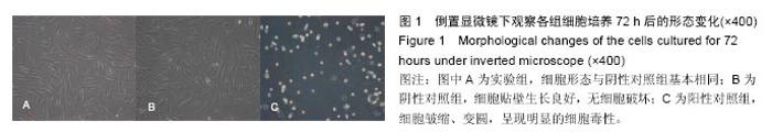
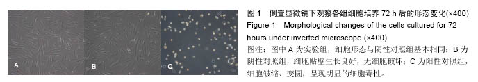
2.1 细胞毒性实验结果 倒置显微镜下观察阳性对照组细胞几乎完全破坏,呈现明显的细胞毒性;阴性对照细胞贴壁生长良好,呈长梭形,无细胞破坏;实验组细胞形态与阴性对照组基本相同(图1)。MTT检测结果显示,实验组培养24,48,72 h的细胞相对增殖率分别为95.49%、101.12%、103.12%,该磷酸钙骨水泥/纤维蛋白胶复合材料细胞毒性评级为1级或0级。实验组各时间点吸光度值与阳性对照组比较差异有显著性意义(P < 0.05),见表1。"
"

2.2 血液相容性实验结果 阳性对照组有溶血,实验组和阴性对照组无溶血发生。阴性对照组、阳性对照组、实验组的吸光度值分别为0.021 2±0.000 6,0.987 5± 0.012 9,0.345 1±0.001 8,计算磷酸钙骨水泥/纤维蛋白胶复合材料的溶血率为3.15%<5%,符合国家医疗器械生物学评价标准。 2.3 体内修复实验结果 2.3.1 一般情况分析 术后所有动物全身状况良好,饮食、活动等均正常。术后7 d内,部分术区周围有一定程度肿胀,未进行特殊处理肿胀自行消退,术后创口均未出现感染现象。无死亡,所有动物均计入实验统计。 2.3.2 各组大体标本观察结果 见图2。"


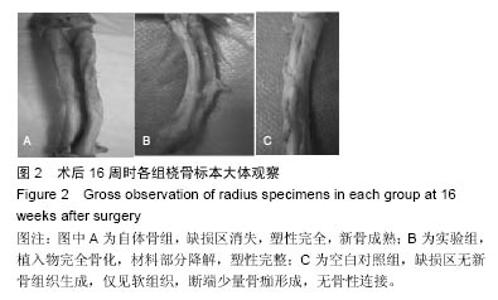
术后4周时,实验组纤维蛋白完全吸收,植入物两端有大量骨痂形成,植入物与断端界限不清;自体骨组植入的自体骨大部分吸收,也有大量骨痂形成,植入骨与断端界限不清。术后8周时实验组植入物完全骨化,被大量骨痂包埋,形成的新骨较4周明显增多;自体骨组缺损区充满新生骨,覆盖板层骨。术后16周时,实验组植入物完全骨化,材料部分降解,缺损区骨端皮质连续,塑性完整;自体骨组缺损区消失,塑性完全,新骨成熟。空白对照组至术后16周时缺损区未有新骨组织生成,仅见软组织,断端少量骨痂形成,无骨性连接。 2.3.3 影像学检查评分结果 见图3。"
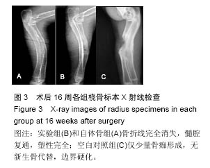
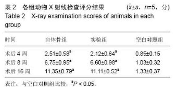
术后第4周,实验组骨缺损区已模糊,断端界限不清,新生骨痂密度增高;自体骨组骨折线模糊减少,骨痂密度增高。术后第8周,实验组骨缺损区密度较之前明显增高,骨折周围界限模糊不清;自体骨组骨折线基本消失,骨皮质形成,髓腔复通。术后16周,实验组和自体骨组骨折线完全消失,髓腔复通,塑性完全。至术后16周空白对照组仅少量骨痂形成,无新生骨代替,边界硬化。 Lane-Sandhu X射线评分析结果表明随时间增加,实验组和自体骨组X射线片评分逐渐增高,实验组、自体骨组Lane-Sandhu X射线评分高于空白对照组(P < 0.05),而实验组与自体骨组Lane-Sandhu X射线评分比较差异无显著性意义(P > 0.05),见表2。"

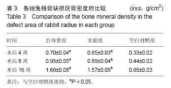
2.3.4 骨密度比较 术后4,8,16周,自体骨组、实验组缺损区骨密度大于空白对照组(P < 0.05),自体骨组和实验组缺损区骨密度比较差异无显著性意义(P > 0.05),见表3。"
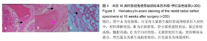
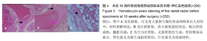
2.3.5 苏木精-伊红染色结果 见图4。 术后第4周,自体骨组骨折断端有纤维组织包裹,类骨组织开始转变为编织骨,纤维性骨痂中的软骨组织经软骨化骨演变为骨组织,形成骨性骨痂;实验组材料开始降解,在材料边缘有类骨质形成;空白对照组无新骨组织生成。 术后第8周,自体骨组骨小梁较粗大,可见骨小梁正常的排列结构正在重新恢复;实验组材料进一步降解,降解形成空隙结构并有新生骨小梁长入;空白对照组无新骨组织生成,骨折断端未愈合。 术后第16周,自体骨组新生骨进一步形成,骨小梁重建较明显,板层骨较成熟,髓腔再通;实验组可见新生骨小梁继续长入,进一步增长、增粗、增多,有大量新生编织骨成网格状长入材料中,材料降解明显,降解与骨长入同步;空白对照组同前,无新生骨变化。"

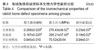
2.3.6 生物力学检测结果 术后第16周,实验组和自体骨组骨标本抗弯曲能力的最大负荷、最大应力、破坏能量值均明显高于空白对照组(P < 0.05),实验组和自体骨组骨标本抗弯曲能力的最大负荷、最大应力、破坏能量值比较差异无显著性意义(P > 0.05),见表4。 "
| [1]周磊,闫景龙,胡春杰,等.聚磷酸钙纤维/磷酸钙骨水泥复合材料修复骨缺损的实验研究[J].中国脊柱脊髓杂志, 2006,16(11):851-855.[2]Tyllianakis M,Giannikas D,Panagopoulos A, et al.Use of injectable calcium phosphate in the treatment of intra-articular distal radius fractures. Orthopedics. 2002; 25(3):311-315.[3]高俊,张曦,盛永华,等.外固定支架结合注射用磷酸钙骨水泥治疗桡骨远端不稳定骨折[J].中国骨与关节损伤杂志, 2008,23(5):407-408.[4]黄程川,马川,黄家骏,等.磷酸钙骨水泥与自体骨植骨治疗C2型桡骨远端骨折的疗效比较[J].四川医学, 2012,33(3): 505-507.[5]胡运生,范清宇,马保安,等.磷酸钙/纤维蛋白胶复合支架材料的结构及力学性能分析[J].功能材料, 2006,37(4): 607-610.[6]Tobe M,Mizutani K,Tsubuku Y.Treatment of Distal Radius Fracture With the Use of Calcium Phosphate Bone Cement as a Filler.Tech Hand Up Extrem Surg. 2004;8(2):95-101.[7]Iida K,Sudo A,Ishiguro S,et al.Clinical and radiological results of calcium phosphate cement-assisted balloon osteoplasty for Colles' fractures in osteoporotic senile female patients.J Orthop Sci.2010;15(2):204-209.[8]Bohner M,Baumgart F.Theoretical model to determine the effects of geometrical factors on the resorption of calcium phosphate bone substitutes.Biomaterials. 2004;25(17):3569-3582.[9]代宇.磷酸钙骨水泥填充治疗桡骨远端骨质疏松骨折的实验研究[D].遵义医学院,2010.[10]唐理英,刘运辉,谢诗涓,等.经皮注射磷酸钙骨水泥治疗桡骨远端骨质疏松性骨折的临床应用[J].临床医学工程, 2015,22(6):709-710.[11]Tobe M,Mizutani K,Tsubuku Y.Treatment of distal radius fracture with the use of calcium phosphate bone cement as a filler.Tech Hand Up Extrem Surg. 2004;8(2):95.[12]Leroux T,Perez-Ordonez B,von-Schroeder HP,et al. Osteolysis after the use of a silicon-stabilized tricalcium phosphate-based bone substitute in a radius fracture: a case report.J Hand Surg Am. 2007;32(4):497-500.[13]代宇,何慕舜,叶俊强,等.磷酸钙骨水泥治疗老年骨质疏松性桡骨远端骨折的疗效观察[J].中国骨与关节损伤杂志, 2010,25(3):246-247.[14]夏长所,叶发刚,洪光祥,等.磷酸钙人工骨结合骨髓基质干细胞移植修复骨缺损的实验研究[J].中华手外科杂志, 2004,20(3):174-176.[15]张才龙,夏长所,叶发刚,等.磷酸钙人工骨复合骨髓基质干细胞移植修复骨缺损的实验[J].中国临床康复, 2005, 9(46):140-142.[16]Ishiguro S,Oota Y,Sudo A,et al.Calcium phosphate cement-assisted balloon osteoplasty for a Colles' fracture on arteriovenous fistula forearm of a maintenance hemodialysis patient.J Hand Surg Am.2007;32(6):821-826.[17]邹国耀,江峰.自体游离骨膜骨髓自固化磷酸钙复合移植治疗低负重或非负重部位节段性骨缺损的实验研究[J].中国临床康复,2004,8(11):2152-2153.[18]Cross KJ,Huq NL,Stanton DP,et al.NMR studies of a novel calcium, phosphate and fluoride delivery vehicle-alpha(S1)-casein(59-79) by stabilized amorphous calcium fluoride phosphate nanocomplexes.Biomaterials.2004;25(20):5061-5069.[19]immermann R,Gabl M,Lutz M,et al.Injectable calcium phosphate bone cement Norian SRS for the treatment of intra-articular compression fractures of the distal radius in osteoporotic women.Arch Orthop Trauma Surg. 2003;123(1):22-27.[20]张志鹏,闫冰,闫景龙,等.同种异体微小颗粒骨/磷酸钙骨水泥复合物修复骨缺损的实验研究[J].中国康复医学杂志,2005,20(7):514-516.[21]Abramo A,Geijer M,Kopylov P,et al.Osteotomy of distal radius fracture malunion using a fast remodeling bone substitute consisting of calcium sulphate and calcium phosphate.J Biomed Mater Res B Appl Biomater. 2010;92B(1):281-286.[22]王剑龙,米雷,候光辉,等.新型组织工程化骨磷酸钙骨水泥/聚乳酸-聚羟基乙酸复合骨髓间充质干细胞修复兔桡骨缺损[J].中国组织工程研究与临床康复, 2008,12(41): 8001-8005.[23]廖红兴,刘展亮,邹学农.磷酸钙骨水泥复合骨形态发生蛋白6和血管内皮生长因子修复骨缺损[J].中国组织工程研究,2014,18(8):1155-1160.[24]李光宇,董洲,刘鑫,等.明胶-壳聚糖-羟基磷灰石-米诺环素仿生纳米复合材料修复桡骨缺损[J].中国组织工程研究,2015,19(30):4757-4763.[25]陈盈君,王南翔,周磊,等.丝素纤维/磷酸钙骨水泥复合材料修复兔桡骨骨缺损的实验研究[J].中华创伤骨科杂志, 2014,16(1):62-66.[26]郝伟,姜明,王新,等.重组骨形态发生蛋白2、碱性成纤维细胞生长子共转染骨髓间充质干细胞复合纳米羟基磷灰 石/重组类人胶原基/聚乳酸复合支架材料支架修复桡骨缺损研究[J].中华实验外科杂志,2013,30(5):1016-1019.[27]马佳滨,周磊,闫景龙,等.磷酸钙骨水泥/聚磷酸钙纤维复合不同配比的颗粒骨修复兔桡骨缺损[J].中华创伤杂志, 2011,27(8):737-741.[28]梁建平,刘兴炎,葛宝丰,等.磷酸钙骨水泥复合微小颗粒骨修复兔桡骨缺损[J].中国矫形外科杂志, 2010,18(6): 486-488.[29]刘洪,臧小方,赵自平,等.磷酸钙骨水泥与外源性神经生长因子复合移植修复免桡骨缺损[J].中国组织工程研究与临床康复,2008,12(41):8037-8041.[30]Hidaka N,Yamano Y,Kadoya Y,et al.Calcium phosphate bone cement for treatment of distal radius fractures: a preliminary report.J Orthop Sci. 2002;7(2): 182-187.[31]Seeherman HJ,Li XJ,Smith E,et al.rhBMP-2/calcium phosphate matrix induces bone formation while limiting transient bone resorption in a nonhuman primate core defect model.J Bone Joint Surg Am. 2012;94/A(19): 1765-1776. |
| [1] | Zhang Tongtong, Wang Zhonghua, Wen Jie, Song Yuxin, Liu Lin. Application of three-dimensional printing model in surgical resection and reconstruction of cervical tumor [J]. Chinese Journal of Tissue Engineering Research, 2021, 25(9): 1335-1339. |
| [2] | Zeng Yanhua, Hao Yanlei. In vitro culture and purification of Schwann cells: a systematic review [J]. Chinese Journal of Tissue Engineering Research, 2021, 25(7): 1135-1141. |
| [3] | Xu Dongzi, Zhang Ting, Ouyang Zhaolian. The global competitive situation of cardiac tissue engineering based on patent analysis [J]. Chinese Journal of Tissue Engineering Research, 2021, 25(5): 807-812. |
| [4] | Wu Zijian, Hu Zhaoduan, Xie Youqiong, Wang Feng, Li Jia, Li Bocun, Cai Guowei, Peng Rui. Three-dimensional printing technology and bone tissue engineering research: literature metrology and visual analysis of research hotspots [J]. Chinese Journal of Tissue Engineering Research, 2021, 25(4): 564-569. |
| [5] | Chang Wenliao, Zhao Jie, Sun Xiaoliang, Wang Kun, Wu Guofeng, Zhou Jian, Li Shuxiang, Sun Han. Material selection, theoretical design and biomimetic function of artificial periosteum [J]. Chinese Journal of Tissue Engineering Research, 2021, 25(4): 600-606. |
| [6] | Liu Fei, Cui Yutao, Liu He. Advantages and problems of local antibiotic delivery system in the treatment of osteomyelitis [J]. Chinese Journal of Tissue Engineering Research, 2021, 25(4): 614-620. |
| [7] | Li Xiaozhuang, Duan Hao, Wang Weizhou, Tang Zhihong, Wang Yanghao, He Fei. Application of bone tissue engineering materials in the treatment of bone defect diseases in vivo [J]. Chinese Journal of Tissue Engineering Research, 2021, 25(4): 626-631. |
| [8] | Zhang Zhenkun, Li Zhe, Li Ya, Wang Yingying, Wang Yaping, Zhou Xinkui, Ma Shanshan, Guan Fangxia. Application of alginate based hydrogels/dressings in wound healing: sustained, dynamic and sequential release [J]. Chinese Journal of Tissue Engineering Research, 2021, 25(4): 638-643. |
| [9] | Chen Jiana, Qiu Yanling, Nie Minhai, Liu Xuqian. Tissue engineering scaffolds in repairing oral and maxillofacial soft tissue defects [J]. Chinese Journal of Tissue Engineering Research, 2021, 25(4): 644-650. |
| [10] | Xing Hao, Zhang Yonghong, Wang Dong. Advantages and disadvantages of repairing large-segment bone defect [J]. Chinese Journal of Tissue Engineering Research, 2021, 25(3): 426-430. |
| [11] | Chen Siqi, Xian Debin, Xu Rongsheng, Qin Zhongjie, Zhang Lei, Xia Delin. Effects of bone marrow mesenchymal stem cells and human umbilical vein endothelial cells combined with hydroxyapatite-tricalcium phosphate scaffolds on early angiogenesis in skull defect repair in rats [J]. Chinese Journal of Tissue Engineering Research, 2021, 25(22): 3458-3465. |
| [12] | Wang Hao, Chen Mingxue, Li Junkang, Luo Xujiang, Peng Liqing, Li Huo, Huang Bo, Tian Guangzhao, Liu Shuyun, Sui Xiang, Huang Jingxiang, Guo Quanyi, Lu Xiaobo. Decellularized porcine skin matrix for tissue-engineered meniscus scaffold [J]. Chinese Journal of Tissue Engineering Research, 2021, 25(22): 3473-3478. |
| [13] | Mo Jianling, He Shaoru, Feng Bowen, Jian Minqiao, Zhang Xiaohui, Liu Caisheng, Liang Yijing, Liu Yumei, Chen Liang, Zhou Haiyu, Liu Yanhui. Forming prevascularized cell sheets and the expression of angiogenesis-related factors [J]. Chinese Journal of Tissue Engineering Research, 2021, 25(22): 3479-3486. |
| [14] | Liu Chang, Li Datong, Liu Yuan, Kong Lingbo, Guo Rui, Yang Lixue, Hao Dingjun, He Baorong. Poor efficacy after vertebral augmentation surgery of acute symptomatic thoracolumbar osteoporotic compression fracture: relationship with bone cement, bone mineral density, and adjacent fractures [J]. Chinese Journal of Tissue Engineering Research, 2021, 25(22): 3510-3516. |
| [15] | Liu Liyong, Zhou Lei. Research and development status and development trend of hydrogel in tissue engineering based on patent information [J]. Chinese Journal of Tissue Engineering Research, 2021, 25(22): 3527-3533. |
| Viewed | ||||||
|
Full text |
|
|||||
|
Abstract |
|
|||||