Chinese Journal of Tissue Engineering Research ›› 2016, Vol. 20 ›› Issue (43): 6438-6444.doi: 10.3969/j.issn.2095-4344.2016.43.008
Previous Articles Next Articles
In vivo osseointegration of micro-arc oxidation-silane-melatonin-covered titanium alloy implant
- Key Biomedical Materials Laboratory of Colleges and Universities in Heilongjiang Province, Jiamusi University, Jiamusi 154007, Heilongjiang Province, China
-
Received:2016-07-23Online:2016-10-21Published:2016-10-21 -
Contact:Li Mu-qin, Professor, Key Biomedical Materials Laboratory of Colleges and Universities in Heilongjiang Province, Jiamusi University, Jiamusi 154007, Heilongjiang Province, China -
About author:Li De-chao, M.D., Professor, Key Biomedical Materials Laboratory of Colleges and Universities in Heilongjiang Province, Jiamusi University, Jiamusi 154007, Heilongjiang Province, China -
Supported by:the National Natural Science Foundation of China, No. 31370979; the Innovation Team Building Project in Heilongjiang Provincial Colleges, No. 2012TD010; Collaborative Innovation Center of Jiamusi University, No. 2011xtcx2016-02
CLC Number:
Cite this article
Li De-chao, Liu Hui-ping, Li Mu-qin, Liu Miao.
share this article
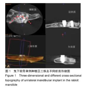
2.1 实验动物数量分析 18只兔均进入结果分析。 2.2 术后一般情况 所有实验动物均于术后2 h 左右清醒,进食、行动良好,没有其他不适现象。定期清创,1 d/次,创口区域愈合良好,未见明显感染及种植体脱落,双侧颌下区无异常情况。取材后所有植体保存完好,均未松动及脱落,种植体骨结合良好区域顶部有骨质覆盖,周围无软组织化脓、感染及坏死现象。 2.3 种植体锥形束CT检查结果 图1是兔下颌骨单侧种植区三维及不同断面形貌图,用于宏观观察种植体置入后与周围骨组织的结合情况,用锥形束CT在不破坏样本下进行的扫描,取得三维形貌,观察种植体的冠状面、横断面和矢状面。矢状面上能够清晰观察到种植体周围的骨结合情况。种植体置入的方向、位置适当,兔下颌骨均未出现低密度影像,无置入部位的排斥及感染现象,2个种植体之间的距离合适,无骨吸收及坏死现象。"


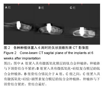
图2是置入6周后3组植体矢状面锥形束CT影像,A组靠近种植体周围两侧均有低密度阴影区,种植体两侧的密度低于正常区域最为明显,说明种植体两侧骨结合不紧密,新骨未在两侧完全生长;B组种植体周围密度明显比A组升高,但植体一侧仍存在低密度影,说明植体一侧有骨结合;C组种植体的周围未见到低密度影,与下颌骨结合紧密,且植体周围密度高于A、B组,相对比较,C组的骨结合最好。"
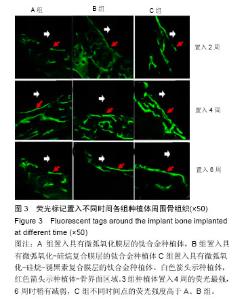
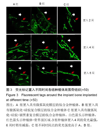
2.4 激光共聚焦镜下观察结果 骨组织中的Ca2+能与荧光标记物特异性结合,在特定波长激发光下的荧光显微镜中观察到的特殊颜色可以显示骨组织,实验中种植体-骨界面新生骨着色饱和、亮度高、呈绿色;而兔下颌骨(即宿主骨)着色较浅,为淡绿色,软组织在显微镜下没有显示颜色,见图3。 置入2周时,A组种植体周围的荧光条带成点状、断续排列且强度较弱;B组弱的荧光形成细条线状排列在种植体-骨界面周围;C组植体-骨界面的荧光强度较A、B组强,但不连续排列,只有少部分成亮绿色。置入4周时,相比较而言,3组种植体周围的荧光标记明显比2周强,呈现亮绿色荧光。其中A组的荧光带较细、狭窄、断断续续;B组较A组来说荧光带明显增宽;而C组的荧光强度最强且较多,连续状,靠近种植体-骨界面处的强荧光带最宽。置入6周时,荧光带明显减少,强度减弱;但仍可以观察到C组的条带较A、B组宽;A组为片状排列,B组为细条带状。"
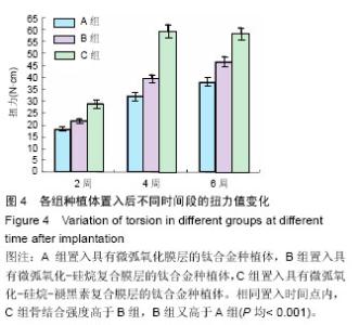
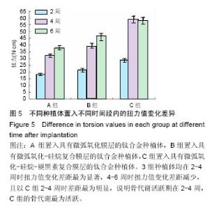
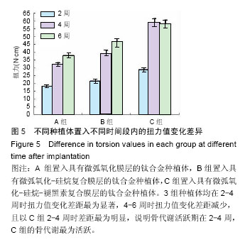
2.5 生物力学检测结果 图4为置入不同时间3组种植体的扭力值变化图。3组种植体的骨结合力存在着明显差异,随着置入时间的延长力值逐渐增加;相同置入时间点内,C组骨结合强度高于B组,B组又高于A组(P均< 0.001),说明C组的骨结合力优于A、B组;C组置入2,4,6周时的骨结合强度分别为(29.58±0.56),(59.53± 1.52),(58.44±1.93) N•cm,说明C组种植体置入4周时的骨结合力达到峰值,表明骨结合时间少于A、B组 (P < 0.05)。 图5为3组种植体在各时间段内的骨结合力变化差异图,3组种植体均在2-4周时扭力值变化差距最为显著,4-6周时扭力值变化差距减少,且以C组2-4周时差距最为明显,说明骨代谢活跃期在2-4周,C组的骨代谢最为活跃。"

"
| [1] Deng Z,Wang L,Zhang D,et al.Lanthanum-containing hydroxyapatite coating on ultrafine-grained titanium by micro-arc oxidation: A promising strategy to enhance overall performance of titanium.Med Sci Monit. 2014; 20(3):163-166.
[2] Oshida Y,Tuna EB,Aktören O,et al.Dental Implant Systems.Int J Mol Sci.2010;11(1):1580-1678.
[3] 王丹宁,赵宝红.钛种植体表面微弧氧化技术研究进展[J].中国实用口腔科杂志,2010,3(9):570-573.
[4] 樊志斌,阎峰云,邵敬涛,等.基于微弧氧化技术的复合涂层的研究现状[J].特种铸造及有色合金,2014,34(1):66-70.
[5] Zagury R,Harari ND,Conz MB,et al. Histomorphometric analyses of bone interface with titanium-aluminum- vanadium and hydroxyapatite- coated implants by biomimetic process.Implant Dent. 2007;16(3):290-296.
[6] 封伟,赵宝红.兔下颌骨双侧种植体植入动物模型的建立[J].口腔医学研究,2013,29(5):393-396.
[7] 武秀萍,李冰.改进超硬组织切片技术用于钛种植体骨界面研究[J].中国实用口腔科杂志,2013,6(11):661-663.
[8] 谭见容,杨小红.应用甲基丙烯酸甲酯包埋不脱钙骨组织方法的探讨[J].中国矫形外科杂志, 2009,17(7): 534-536.
[9] 陈昭宏,陈舜.钛种植体植入兔管状骨愈合界面的变化[J].福建医科大学学报,2008,42(4):324-326.
[10] 齐进,黄萍.EXAKT切磨系统在带金属种植体软硬组织切片中的应用[J].中国组织工程研究, 2012,16(35): 6555-6559.
[11] 戚娟娟,刘天涛.带种植体软硬组织磨片手工制作方法的体会[J].现代口腔医学杂志, 2012,26(3):186-189.
[12] 朱安棣,容明灯.硬组织锯割机制作带种植体不脱钙骨磨片的方法研究[J].广东牙病防治,2009,17(6):258-259.
[13] Kotlarczyk MP,Lassila HC,O'Neil CK,et al.Melatonin osteoporosis prevention study (MOPS): a randomized, double-blind, placebo-controlled study examining the effects of melatonin on bone health and quality of life in perimenopausal women.J Pineal Res. 2012;52(4): 414-426.
[14] Cutando A,Gómez-Moreno G,Arana C,et al.Melatonin stimulates osteointegration of dental implants.J Pineal Res.2008;45(2):174-179.
[15] Cutando A,Gómez-Moreno G,Arana C,et al.Melatonin: potential functions in the oral cavity.J Periodontol. 2007;78(6):1094-1102.
[16] Koyama H,Nakade O,Takada Y,et al.Melatonin at pharmacologic doses increases bone mass by suppressing resorption through down-regulation of the RANKL-mediated osteoclast formation and activation.J Bone Miner Res.2002;17:1219-1229.
[17] Behairy RE,Hamed M,Ahmed I,et al.Validity of Growth hormone and Melatonin mixture locally applied around immediate implants: Aclinical study.Nat Sci. 2013;11(8): 54-58.
[18] Koyama H,Nakade O, Takada Y,et al.Melatonin at pharmacologic doses increases bone mass by suppressing resorption through down-regulation of the RANKL-mediated osteoclast formation and activation.J Bone Miner Res.2002;17(7):1219-1229.
[19] Calvo-Guirado JL,Ramírez-Fernández MP, Gómez-Moreno G,et al.Melatonin stimulates the growth of new bone around implants in the tibia of rabbits.J Pineal Res.2010;49(4):356-363.
[20] Nakade O,Koyama H,Ariji H,et al.Melatonin stimulates proliferation and type I collagen synthesis in human bone cells in vitro.J Pineal Res.1999;27:106-110.
[21] Calvo-Guirado JL,Gómez-Moreno G,Barone A,et al. Melatonin plus porcine bone on discrete calcium deposit implant surface stimulates osteointegration in dental implants.J Pineal Res.2009;47(2):164-172.
[22] 古丽巴努•依马木,徐国强,迪丽努尔•阿吉,等.硅烷偶联剂对纯钛表面改性及细胞相容性的影响[J].中国组织工程研究,2014,18(12):1864-1869.
[23] 李建福,李世荣.活体顺序性荧光标记技术在实验性骨折愈合研究中的应用[J].中国实用美容整形外科杂志, 2005, 16(3):179-181.
[24] 余娜,肖丽.钛种植体-骨间隙愈合界面成骨方式的激光共聚焦显微镜观察[J].口腔颌面修复学杂志2008,9(4): 274-276.
[25] Sun QY,Yu ZT,Zhu RH.Dynamic fracture toughness of Ti–2.5Cu alloy strengthened with nano-scale particles at room and low temperatures.Mater Sci Eng. 2008;3: 131-134.
[26] Zhu L,Ye X,Tang G,et al.Corrosion test, cell behavior test, and in vivo study of gradient TiO2 layers produced by compound electrochemical oxidation.J Biomed Mater Res A.2006;78(3):515-522.
[27] 王磊,闫凤英.微弧氧化时间对纯钛表面膜层微观结构的影响[J].中国组织工程研究,2012,16(51):9546-9550.
[28] 张薷文,周延民.种植体骨结合相关检测手段研究现状[J].实用口腔医学杂志2013,29(5):730-733.
[29] 李晓萌,史久慧.酸蚀加碱热处理钛合金种植体早期诱导成骨作用的观察[J].口腔医学研究2010,26(5): 661-663.
[30] 郭泽鸿,周磊.纯钛种植体激光-微弧氧化表面处理对早期骨结合的影响[J].口腔颌面外科杂志2014,24(3):208- 213.
[31] 井文森,韩建业.多孔β型钛合金及其微弧氧化涂层的体内植入研究[J].稀有金属材料与工程2012,41(9):1657- 1660.
[32] Xiu P,Jia Z,Lv J,et al.Tailored Surface Treatment of 3D Printed Porous Ti6Al4V by Microarc Oxidation for Enhanced Osseointegration via Optimized Bone In-Growth Patterns and Interlocked Bone/Implant Interface.ACS Appl Mater Interfaces. 2016;8(28): 17964-17975.
[33] Li G,Cao H,Zhang W,et al.Enhanced Osseointegration of Hierarchical Micro/Nanotopographic Titanium Fabricated by Microarc Oxidation and Electrochemical Treatment.ACS Appl Mater Interfaces. 2016;8(6): 3840-3852.
[34] Wang Z,Wu G,Feng Z,et al.Microarc-oxidized titanium surfaces functionalized with microRNA-21-loaded chitosan/hyaluronic acid nanoparticles promote the osteogenic differentiation of human bone marrow mesenchymal stem cells.Int J Nanomedicine. 2015; 10:6675-6687.
[35] Cheng Y,Zhao X,Liu X,et al.Antibacterial activity and biological performance of a novel antibacterial coating containing a halogenated furanone compound loaded poly(L-lactic acid) nanoparticles on microarc-oxidized titanium.Int J Nanomedicine.2015;10:727-737.
[36] Fang K,Song W,Wang L,et al.Immobilization of chitosan film containing semaphorin 3A onto a microarc oxidized titanium implant surface via silane reaction to improve MG63 osteogenic differentiation.Int J Nanomedicine.2014;9:4649-4657.
[37] Cheng Y,Wu J,Gao B,et al.Fabrication and in vitro release behavior of a novel antibacterial coating containing halogenated furanone-loaded poly(L-lactic acid) nanoparticles on microarc-oxidized titanium.Int J Nanomedicine.2012;7:5641-5652.
[38] Nozaki K,Wang W,Horiuchi N,et al.Enhanced osteoconductivity of titanium implant by polarization-induced surface charges.J Biomed Mater Res A.2014;102(9):3077-3086.
[39] Marques Ida S,Alfaro MF,Saito MT,et al.Biomimetic coatings enhance tribocorrosion behavior and cell responses of commercially pure titanium surfaces. Biointerphases.2016;11(3):031008. |
| [1] | Zhang Tongtong, Wang Zhonghua, Wen Jie, Song Yuxin, Liu Lin. Application of three-dimensional printing model in surgical resection and reconstruction of cervical tumor [J]. Chinese Journal of Tissue Engineering Research, 2021, 25(9): 1335-1339. |
| [2] | Zeng Yanhua, Hao Yanlei. In vitro culture and purification of Schwann cells: a systematic review [J]. Chinese Journal of Tissue Engineering Research, 2021, 25(7): 1135-1141. |
| [3] | Xu Dongzi, Zhang Ting, Ouyang Zhaolian. The global competitive situation of cardiac tissue engineering based on patent analysis [J]. Chinese Journal of Tissue Engineering Research, 2021, 25(5): 807-812. |
| [4] | Wu Zijian, Hu Zhaoduan, Xie Youqiong, Wang Feng, Li Jia, Li Bocun, Cai Guowei, Peng Rui. Three-dimensional printing technology and bone tissue engineering research: literature metrology and visual analysis of research hotspots [J]. Chinese Journal of Tissue Engineering Research, 2021, 25(4): 564-569. |
| [5] | Chang Wenliao, Zhao Jie, Sun Xiaoliang, Wang Kun, Wu Guofeng, Zhou Jian, Li Shuxiang, Sun Han. Material selection, theoretical design and biomimetic function of artificial periosteum [J]. Chinese Journal of Tissue Engineering Research, 2021, 25(4): 600-606. |
| [6] | Liu Fei, Cui Yutao, Liu He. Advantages and problems of local antibiotic delivery system in the treatment of osteomyelitis [J]. Chinese Journal of Tissue Engineering Research, 2021, 25(4): 614-620. |
| [7] | Li Xiaozhuang, Duan Hao, Wang Weizhou, Tang Zhihong, Wang Yanghao, He Fei. Application of bone tissue engineering materials in the treatment of bone defect diseases in vivo [J]. Chinese Journal of Tissue Engineering Research, 2021, 25(4): 626-631. |
| [8] | Zhang Zhenkun, Li Zhe, Li Ya, Wang Yingying, Wang Yaping, Zhou Xinkui, Ma Shanshan, Guan Fangxia. Application of alginate based hydrogels/dressings in wound healing: sustained, dynamic and sequential release [J]. Chinese Journal of Tissue Engineering Research, 2021, 25(4): 638-643. |
| [9] | Chen Jiana, Qiu Yanling, Nie Minhai, Liu Xuqian. Tissue engineering scaffolds in repairing oral and maxillofacial soft tissue defects [J]. Chinese Journal of Tissue Engineering Research, 2021, 25(4): 644-650. |
| [10] | Xing Hao, Zhang Yonghong, Wang Dong. Advantages and disadvantages of repairing large-segment bone defect [J]. Chinese Journal of Tissue Engineering Research, 2021, 25(3): 426-430. |
| [11] | Chen Siqi, Xian Debin, Xu Rongsheng, Qin Zhongjie, Zhang Lei, Xia Delin. Effects of bone marrow mesenchymal stem cells and human umbilical vein endothelial cells combined with hydroxyapatite-tricalcium phosphate scaffolds on early angiogenesis in skull defect repair in rats [J]. Chinese Journal of Tissue Engineering Research, 2021, 25(22): 3458-3465. |
| [12] | Wang Hao, Chen Mingxue, Li Junkang, Luo Xujiang, Peng Liqing, Li Huo, Huang Bo, Tian Guangzhao, Liu Shuyun, Sui Xiang, Huang Jingxiang, Guo Quanyi, Lu Xiaobo. Decellularized porcine skin matrix for tissue-engineered meniscus scaffold [J]. Chinese Journal of Tissue Engineering Research, 2021, 25(22): 3473-3478. |
| [13] | Mo Jianling, He Shaoru, Feng Bowen, Jian Minqiao, Zhang Xiaohui, Liu Caisheng, Liang Yijing, Liu Yumei, Chen Liang, Zhou Haiyu, Liu Yanhui. Forming prevascularized cell sheets and the expression of angiogenesis-related factors [J]. Chinese Journal of Tissue Engineering Research, 2021, 25(22): 3479-3486. |
| [14] | Liu Chang, Li Datong, Liu Yuan, Kong Lingbo, Guo Rui, Yang Lixue, Hao Dingjun, He Baorong. Poor efficacy after vertebral augmentation surgery of acute symptomatic thoracolumbar osteoporotic compression fracture: relationship with bone cement, bone mineral density, and adjacent fractures [J]. Chinese Journal of Tissue Engineering Research, 2021, 25(22): 3510-3516. |
| [15] | Liu Liyong, Zhou Lei. Research and development status and development trend of hydrogel in tissue engineering based on patent information [J]. Chinese Journal of Tissue Engineering Research, 2021, 25(22): 3527-3533. |
| Viewed | ||||||
|
Full text |
|
|||||
|
Abstract |
|
|||||