Chinese Journal of Tissue Engineering Research ›› 2016, Vol. 20 ›› Issue (38): 5684-5690.doi: 10.3969/j.issn.2095-4344.2016.38.009
Previous Articles Next Articles
Biocompatibility of magnetic ferrosoferric oxide nanoparticles in preosteoblasts
- Guanghua School of Stomatology, Hospital of Stomatology, Sun Yat-sen University, Guangdong Provincial Key Laboratory of Stomatology, Guangzhou 510055, Guangdong Province, China
-
Received:2016-07-08Online:2016-09-16Published:2016-09-16 -
Contact:Zeng Rong-sheng, Master, Doctoral supervisor, Chief physician, Guanghua School of Stomatology, Hospital of Stomatology, Sun Yat-sen University, Guangdong Provincial Key Laboratory of Stomatology, Guangzhou 510055, Guangdong Province, China -
About author:Guan Chen-yu, Studying for master’s degree, Guanghua School of Stomatology, Hospital of Stomatology, Sun Yat-sen University, Guangdong Provincial Key Laboratory of Stomatology, Guangzhou 510055, Guangdong Province, China -
Supported by:the National Natural Science Foundation of China, No. 81070818
CLC Number:
Cite this article
Guan Chen-yu, Hou Shi-da, Zhou Yang,Zeng Rong-sheng.
share this article
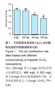
2.1 细胞增殖率 图1可知,随着材料质量浓度升高,细胞增殖率下降,其中200 mg/L磁性Fe3O4纳米颗粒组吸光度值为1.91±0.16,与对照组(0 mg/L)相比差异无显著性意义(P=0.600),400 mg/L和800 mg/L磁性Fe3O4纳米颗粒组吸光度值明显减少,分别为1.57±0.05,1.31±0.14,细胞增殖率显著降低(P=0.001,P=0.000)。提示400 mg/L和800 mg/L磁性Fe3O4纳米颗粒可抑制细胞增殖。"
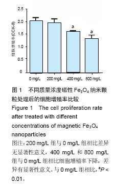
2.2 碱性磷酸酶活性/总蛋白比值测试 由图2可知,200 mg/L磁性四氧化三铁纳米颗粒作用于小鼠前成骨细胞后,碱性磷酸酶活性/总蛋白比值与对照组相比差异无显著性意义(P=0.065)。400 mg/L,800 mg/L组与对照组相比比值升高,差异有显著性意义(P=0.02,P=0.03)。"

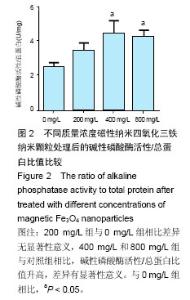
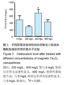
2.3 骨钙素水平测试 磁性四氧化三铁纳米颗粒处理小鼠前成骨细胞24 h后,可见上清中骨钙素质量浓度在 200 mg/L组降低(P=0.08),但差异无显著性意义,见图3。400 mg/L组升高(P=0.015),而800 mg/L与对照组相比差异无显著性意义(P=0.316)。故此,可见400 mg/L磁性纳米四氧化三铁纳米颗粒在24 h时具有一定的促进骨钙素表达的作用。"
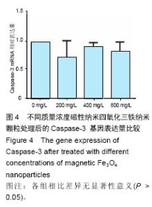
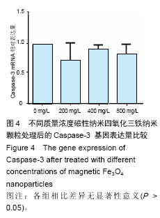
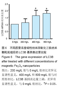
2.4 细胞凋亡及自噬基因的表达量 采用2-??Ct法将实时荧光定量 PCR结果进行分析,检测细胞凋亡及自噬基因的Caspase-3、LC3A及LC3B的mRNA相对表达量。结果提示,经不同质量浓度磁性四氧化三铁纳米颗粒处理后,小鼠前成骨细胞Caspase-3及LC3A基因表达量与对照组差异无显著性意义(P > 0.05)(图4,5),而 400 mg/L及800 mg/L组与对照组相比,LC3B基因表达升高,差异有显著性意义(P=0.025,P=0.028)(图6)。提示高质量浓度磁性四氧化三铁纳米颗粒对成骨细胞的相关功能损伤,可能借由细胞自噬方式产生。"

"


"
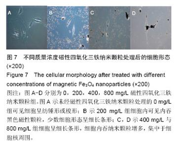
2.5 细胞形态观察 倒置显微镜下可见,随着磁性四氧化三铁纳米颗粒浓度增加,小鼠前成骨细胞形态发生改变,由纺锤形变为长梭形、不规则形,细胞内可见内吞的纳米颗粒,部分细胞呈黑色,细胞数量减少(图7)。"

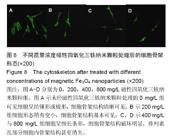
2.6 细胞骨架形态观察 随着磁性四氧化三铁纳米颗粒浓度增加,小鼠前成骨细胞骨架发生改变,纺锤形、规则的细胞骨架结构,变为长梭形、不规则,细胞骨架结构排列紊乱,细胞缩小(图8)。"
| [1]孙涛,王光辉,陆安慧,等.磁性氧化铁纳米颗粒的研究进展[J].化工进展, 2010,30(7):1241-1250. [2]郭大伟,朱玲英,顾宁. 磁性纳米颗粒作为基因递送载体的研究进展[J]. 中国材料进展, 2013,32(10):605-610. [3]陈庆梅,宗小林.磁性纳米材料及其在癌症诊疗中的应用[J].微纳电子技术, 2009,13(6):340-345. [4]李慧,王大新,顾健.超顺磁性纳米颗粒治疗肿瘤的应用进展[J].中国组织工程研究与临床康复, 2009,13(51): 10133-10136. [5]黄江鸿,段莉,陈洁琳,等.磁性纳米多孔复合支架材料的细胞相容性研究[J].生物骨科材料与临床研究, 2014,12(5):16-19. [6]许振,孟洁,张宇,等.磁性纳米纤维复合材料原位诱导体内成骨的研究(英文)[J].东南大学学报(医学版), 2011,30(1):1-6. [7]Yun HM, Ahn SJ, Park KR, et al. Magnetic nanocomposite scaffolds combined with static magnetic field in the stimulation of osteoblastic differentiation and bone formation. Biomaterials. 2016;85:88-98. [8]丁玲,刘鹏,李世迁.纳米材料毒性和安全性研究进展[J].材料导报, 2010,24(5):29-32. [9]Li X, Wei J, Aifantis KE, et al. Current investigations into magnetic nanoparticles for biomedical applications. J Biomed Mater Res A. 2016. [10]Valdiglesias V, Kilic G, Costa C, et al. Effects of iron oxide nanoparticles: cytotoxicity, genotoxicity, developmental toxicity, and neurotoxicity. Environ Mol Mutagen. 2015;56(2):125-148. [11]Prijic S, Scancar J, Romih R, et al. Increased cellular uptake of biocompatible superparamagnetic iron oxide nanoparticles into malignant cells by an external magnetic field. J Membr Biol. 2010;236(1):167-179. [12]Yeh C, Hsiao J, Wang J, et al. Immunological impact of magnetic nanoparticles (Ferucarbotran) on murine peritoneal macrophages. J Nan Res. 2010;12(1SI):151-160. [13]罗宁,李晓杰,闫鸿浩,等.爆轰合成碳包覆钴、镍磁性纳米颗粒的探索[J].高压物理学报, 2009, 23(6):415-420. [14]林晓芬,陈爱政,王士斌.磁性氧化铁纳米颗粒的生物相容性研究进展[J].科学通报, 2011,56(26):2223-2228. [15]Chatterjee S, Sarkar S, Bhattacharya S. Toxic metals and autophagy. Chem Res Toxicol. 2014;27(11):1887-1900. [16]Hajba L, Guttman A. The use of magnetic nanoparticles in cancer theranostics: Toward handheld diagnostic devices. Biotechnol Adv. 2016;34(4):354-361. [17]Ai F, Ferreira CA, Chen F, et al. Engineering of radiolabeled iron oxide nanoparticles for dual-modality imaging. Wiley Interdiscip Rev Nanomed Nanobiotechnol. 2016;8(4):619-630. [18]Zhang C, Yan Y, Zou Q, et al. Superparamagnetic iron oxide nanoparticles for MR imaging of pancreatic cancer: Potential for early diagnosis through targeted strategies. Asia Pac J Clin Oncol. 2016;12(1):13-21. [19]周洋,曾融生,官晨雨,等.不同强度恒定磁场对小鼠成骨细胞增殖及功能的影响[J].中华口腔医学研究杂志(电子版), 2015,9(3):185-192. [20]赵煜,李冰雁,巢永烈,等.磁性附着体模拟静磁场对成骨细胞增殖活性和周期分布及凋亡率的影响[J]. 华西口腔医学杂志, 2007,25(5):437-440. [21]Meng J, Zhang Y, Qi X, et al. Paramagnetic nanofibrous composite films enhance the osteogenic responses of pre-osteoblast cells. Nanoscale. 2010;2(12):2565-2569. [22]Harush-Frenkel O, Debotton N, Benita S, et al. Targeting of nanoparticles to the clathrin-mediated endocytic pathway. Biochem Biophys Res Commun. 2007;353(1):26-32. [23]Raynal I, Prigent P, Peyramaure S, et al. Macrophage endocytosis of superparamagnetic iron oxide nanoparticles: mechanisms and comparison of ferumoxides and ferumoxtran-10. Invest Radiol. 2004;39(1):56-63. [24]贾光,郑玉新.充分认识纳米材料安全性研究的重要意义[J].中华预防医学杂志, 2007,41(2):83-84. [25]Chou L, Firth JD, Uitto VJ, et al. Substratum surface topography alters cell shape and regulates fibronectin mRNA level, mRNA stability, secretion and assembly in human fibroblasts. J Cell Sci. 1995;108( Pt 4): 1563-1573. [26]Zhang L, Wang X, Miao Y, et al. Magnetic ferroferric oxide nanoparticles induce vascular endothelial cell dysfunction and inflammation by disturbing autophagy. J Hazard Mater. 2015;304:186-195. [27]Tian L, Chen BA, Cheng J, et al. Effects of magnetic nanoparticles of Fe3O4 combinated with gambogic acid on apoptosis of SMMC-7721 cells. Onco Targets Ther. 2015;8:2285-2290. [28]王文静,孙昱皓,戴敏超,等.氧化应激对脑缺血再灌注损伤后自噬调控机制[J].中国微侵袭神经外科杂志, 2013, 18(6):275-279. [29]邓正亭,李湧健,程彬彬.自噬与凋亡的相互关系在肿瘤方面的研究进展[J].现代肿瘤医学, 2015,23(10):1467-1471. [30]李冉,李磊,刘江,等. Beclin 1与LC3蛋白在腮腺多形性腺瘤及腮腺癌在多形性腺瘤中的表达和意义[J]. 第三军医大学学报, 2012,34(13):1293-1296. [31]Yang Z, Klionsky DJ. An overview of the molecular mechanism of autophagy. Curr Top Microbiol Immunol. 2009;335:1-32. [32]袁聿军.细胞骨架的基本成分与功能[J].生物学教学, 2006, 31(4):5-8. [33]Klein-Nulend J, Bacabac R G, Bakker AD. Mechanical loading and how it affects bone cells: the role of the osteocyte cytoskeleton in maintaining our skeleton. Eur Cell Mater. 2012;24:278-291. [34]Charras GT, Horton MA. Single cell mechanotransduction and its modulation analyzed by atomic force microscope indentation. Biophys J. 2002;82(6):2970-2981. [35]黄帅帅,王宇多,王萍.细胞骨架和cAMP/PKA信号通路的互动效应调控细胞行为的研究进展[J].中国细胞生物学学报, 2014,36(7):1018-1026. [36]夏伟,周建伟.细胞骨架与细胞凋亡及细胞内信息通路的关系[J].细胞生物学杂志, 2001,23(4):205-209. [37]董萍,王英杰.细胞骨架在跨膜信号传递中的作用[J].生物学杂志, 1995,12(2):2-4. [38]Noel C, Simard JC, Girard D. Gold nanoparticles induce apoptosis, endoplasmic reticulum stress events and cleavage of cytoskeletal proteins in human neutrophils. Toxicol In Vitro. 2016;31:12-22. [39]汤莹,范晓燕,羊富光,等.四氧化三铁磁性纳米颗粒对人角质形成细胞超微结构的影响[J].第二军医大学学报, 2012, 33(7):703-706. [40]王晓娜,唐萌,张婷,等.磁性三氧化二铁纳米粒子对小鼠腹腔巨噬细胞的氧化损伤作用[J].中国组织工程研究与临床康复, 2007,11(13):2575-2577, 2585. [41]陆敏玉,唐萌,夏婷,等. Fe_2O_3纳米颗粒对人肝癌细胞游离钙的影响[J].环境与职业医学, 2009,26(2):133-135. |
| [1] | Zhang Tongtong, Wang Zhonghua, Wen Jie, Song Yuxin, Liu Lin. Application of three-dimensional printing model in surgical resection and reconstruction of cervical tumor [J]. Chinese Journal of Tissue Engineering Research, 2021, 25(9): 1335-1339. |
| [2] | Zeng Yanhua, Hao Yanlei. In vitro culture and purification of Schwann cells: a systematic review [J]. Chinese Journal of Tissue Engineering Research, 2021, 25(7): 1135-1141. |
| [3] | Xu Dongzi, Zhang Ting, Ouyang Zhaolian. The global competitive situation of cardiac tissue engineering based on patent analysis [J]. Chinese Journal of Tissue Engineering Research, 2021, 25(5): 807-812. |
| [4] | Wu Zijian, Hu Zhaoduan, Xie Youqiong, Wang Feng, Li Jia, Li Bocun, Cai Guowei, Peng Rui. Three-dimensional printing technology and bone tissue engineering research: literature metrology and visual analysis of research hotspots [J]. Chinese Journal of Tissue Engineering Research, 2021, 25(4): 564-569. |
| [5] | Chang Wenliao, Zhao Jie, Sun Xiaoliang, Wang Kun, Wu Guofeng, Zhou Jian, Li Shuxiang, Sun Han. Material selection, theoretical design and biomimetic function of artificial periosteum [J]. Chinese Journal of Tissue Engineering Research, 2021, 25(4): 600-606. |
| [6] | Liu Fei, Cui Yutao, Liu He. Advantages and problems of local antibiotic delivery system in the treatment of osteomyelitis [J]. Chinese Journal of Tissue Engineering Research, 2021, 25(4): 614-620. |
| [7] | Li Xiaozhuang, Duan Hao, Wang Weizhou, Tang Zhihong, Wang Yanghao, He Fei. Application of bone tissue engineering materials in the treatment of bone defect diseases in vivo [J]. Chinese Journal of Tissue Engineering Research, 2021, 25(4): 626-631. |
| [8] | Zhang Zhenkun, Li Zhe, Li Ya, Wang Yingying, Wang Yaping, Zhou Xinkui, Ma Shanshan, Guan Fangxia. Application of alginate based hydrogels/dressings in wound healing: sustained, dynamic and sequential release [J]. Chinese Journal of Tissue Engineering Research, 2021, 25(4): 638-643. |
| [9] | Chen Jiana, Qiu Yanling, Nie Minhai, Liu Xuqian. Tissue engineering scaffolds in repairing oral and maxillofacial soft tissue defects [J]. Chinese Journal of Tissue Engineering Research, 2021, 25(4): 644-650. |
| [10] | Xing Hao, Zhang Yonghong, Wang Dong. Advantages and disadvantages of repairing large-segment bone defect [J]. Chinese Journal of Tissue Engineering Research, 2021, 25(3): 426-430. |
| [11] | Chen Siqi, Xian Debin, Xu Rongsheng, Qin Zhongjie, Zhang Lei, Xia Delin. Effects of bone marrow mesenchymal stem cells and human umbilical vein endothelial cells combined with hydroxyapatite-tricalcium phosphate scaffolds on early angiogenesis in skull defect repair in rats [J]. Chinese Journal of Tissue Engineering Research, 2021, 25(22): 3458-3465. |
| [12] | Wang Hao, Chen Mingxue, Li Junkang, Luo Xujiang, Peng Liqing, Li Huo, Huang Bo, Tian Guangzhao, Liu Shuyun, Sui Xiang, Huang Jingxiang, Guo Quanyi, Lu Xiaobo. Decellularized porcine skin matrix for tissue-engineered meniscus scaffold [J]. Chinese Journal of Tissue Engineering Research, 2021, 25(22): 3473-3478. |
| [13] | Mo Jianling, He Shaoru, Feng Bowen, Jian Minqiao, Zhang Xiaohui, Liu Caisheng, Liang Yijing, Liu Yumei, Chen Liang, Zhou Haiyu, Liu Yanhui. Forming prevascularized cell sheets and the expression of angiogenesis-related factors [J]. Chinese Journal of Tissue Engineering Research, 2021, 25(22): 3479-3486. |
| [14] | Liu Chang, Li Datong, Liu Yuan, Kong Lingbo, Guo Rui, Yang Lixue, Hao Dingjun, He Baorong. Poor efficacy after vertebral augmentation surgery of acute symptomatic thoracolumbar osteoporotic compression fracture: relationship with bone cement, bone mineral density, and adjacent fractures [J]. Chinese Journal of Tissue Engineering Research, 2021, 25(22): 3510-3516. |
| [15] | Liu Liyong, Zhou Lei. Research and development status and development trend of hydrogel in tissue engineering based on patent information [J]. Chinese Journal of Tissue Engineering Research, 2021, 25(22): 3527-3533. |
| Viewed | ||||||
|
Full text |
|
|||||
|
Abstract |
|
|||||