Chinese Journal of Tissue Engineering Research ›› 2016, Vol. 20 ›› Issue (28): 4196-4202.doi: 10.3969/j.issn.2095-4344.2016.28.014
Previous Articles Next Articles
Influences of CD133+ cells on human umbilical cord blood mononuclear cell transplantation for treating heart failure
Ma Li, Xie Yi-xu, Chang Yu, Yao Lei
- Department of Cardiothoracic Surgery, Zhengzhou Central Hospital Affiliated to Zhengzhou University, Zhengzhou 450000, Henan Province, China
-
Revised:2016-05-03Online:2016-07-01Published:2016-07-01 -
About author:Ma Li, Associate chief physician, Department of Cardiothoracic Surgery, Zhengzhou Central Hospital Affiliated to Zhengzhou University, Zhengzhou 450000, Henan Province, China
CLC Number:
Cite this article
Ma Li, Xie Yi-xu, Chang Yu, Yao Lei . Influences of CD133+ cells on human umbilical cord blood mononuclear cell transplantation for treating heart failure[J]. Chinese Journal of Tissue Engineering Research, 2016, 20(28): 4196-4202.
share this article
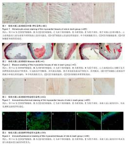
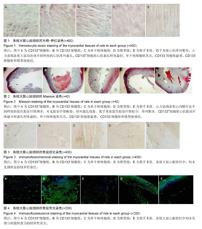
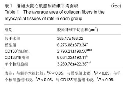
2.1 实验动物数量分析 40只SD大鼠均进入结果分析,中途无脱落。 2.2 各组大鼠心脏组织苏木精-伊红染色结果 模型组大鼠出现心肌排列紊乱,假手术组心肌排列整齐,CD133+细胞组、CD133-细胞组和单个核细胞组的心肌紊乱程度介于模型组和对照组之间。CD133+细胞组、CD133-细胞组和单个核细胞组比较,CD133+细胞组心肌紊乱程度最轻,单个核细胞组其次,CD133-细胞组最重,CD133-细胞组和模型组接近,见图1。 2.3 各组大鼠心脏组织的Masson染色结果 Masson染色后红细胞、纤维素和肌纤维呈红色,弹力纤维呈棕色,胶原纤维呈蓝绿色。镜下观察发现,模型组出现胶原纤维增多,排列紊乱,胶原纤维断裂等现象,假手术组胶原纤维很少,且排列整齐,CD133+细胞组、CD133-细胞组和单个核细胞组的胶原纤维增多和紊乱程度介于模型组和对照组之间。CD133+细胞组、CD133-细胞组和单个核细胞组比较,CD133+细胞组心肌胶原纤维减少,紊乱程度最轻,单个核细胞组其次,CD133-细胞组最重,CD133-细胞组和模型组接近。 模型组、CD133+细胞组、CD133-细胞组和单个核细胞组的胶原纤维面积均明显高于假手术组(P < 0.05),与模型组比较,CD133+细胞组和单个核细胞组胶原纤维面积减小(P < 0.05),模型组和CD133-细胞组相近,差异无显著性意义(P > 0.05)。CD133+细胞组胶原纤维面积少于单个核细胞组(P < 0.05),见表1,图2。 2.4 各组大鼠心脏组织的免疫组化染色结果 免疫组化染色后,深棕色为特异性染色,蓝色为细胞核,显微镜下观察发现,各组大鼠心肌组织中均未发现特异性染色细胞,考虑脐血CD133细胞没有在心肌组织中定植,见图3。 2.5 各组大鼠心脏组织的免疫荧光染色结果 免疫荧光染色后,特异性抗体经FITC标记后,在荧光显微镜下可见到翠绿色荧光,深蓝色为细胞核,显微镜下观察各组大鼠心肌组织中均未发现与细胞核重合的特异性荧光细胞,与免疫组化结果一致,脐血CD133细胞没有在心肌组织中定植,见图4。"
"

| [1] Go AS, Mozaffarian D, Roger VL, et al. Heart disease and stroke statistics--2013 update: a report from the American Heart Association. Circulation. 2013;127(1): e6-e245. [2] Rohde LE, Bertoldi EG, Goldraich L, et al. Cost-effectiveness of heart failure therapies. Nat Rev Cardiol. 2013;10(6):338-354. [3] Heidenreich PA, Trogdon JG, Khavjou OA, et al. Forecasting the future of cardiovascular disease in the United States: a policy statement from the American Heart Association. Circulation. 2011;123(8):933-944. [4] Braunschweig F, Cowie MR, Auricchio A. What are the costs of heart failure. Europace. 2011;13 Suppl 2:ii13-17. [5] Clifford DM, Fisher SA, Brunskill SJ, et al. Stem cell treatment for acute myocardial infarction. Cochrane Database Syst Rev. 2012;2:CD006536. [6] Wollert KC, Drexler H. Cell therapy for the treatment of coronary heart disease: a critical appraisal. Nat Rev Cardiol. 2010;7(4):204-215. [7] Rehman J. Bone marrow tinctures for cardiovascular disease: lost in translation. Circulation. 2013;127(19): 1935-1937. [8] Strauer BE, Steinhoff G. 10 years of intracoronary and intramyocardial bone marrow stem cell therapy of the heart: from the methodological origin to clinical practice. J Am Coll Cardiol. 2011;58(11):1095-1104. [9] Traverse JH, Henry TD, Pepine CJ, et al. Effect of the use and timing of bone marrow mononuclear cell delivery on left ventricular function after acute myocardial infarction: the TIME randomized trial. JAMA. 2012;308(22):2380-2389. [10] Perin EC, Willerson JT, Pepine CJ, et al. Effect of transendocardial delivery of autologous bone marrow mononuclear cells on functional capacity, left ventricular function, and perfusion in chronic heart failure: the FOCUS-CCTRN trial. JAMA. 2012;307(16): 1717-1726. [11] Traverse JH, Henry TD, Ellis SG, et al. Effect of intracoronary delivery of autologous bone marrow mononuclear cells 2 to 3 weeks following acute myocardial infarction on left ventricular function: the LateTIME randomized trial. JAMA. 2011;306(19): 2110-2119. [12] 郝牧,漆佩静,李刚,等.人脐带间充质干细胞对脐血CD34+细胞在NOD/SCID小鼠体内造血重建的影响[J].中国医学科学院学报,2010,32(1):71-75. [13] 车京津,张承宗,陈元禄,等.缬沙坦、苯那普利及合用时对心肌梗死后心室重构影响的比较[J].中国心血管杂志, 2002,7(6):381-384. [14] 张军霞,李瑞,赵鹏,等.CD133免疫磁珠分选肪血内皮祖细胞的培养及鉴定[J].中国肿瘤生物治疗杂志,2008, 15(2):159-162. [15] 中华医学会心血管病学分会,中华心血管病杂志编辑委员会.中国心力衰竭诊断和治疗指南2014 [J].中华心血管病杂志,2014,42(2):98-122. [16] Ferrario CM, Schiffrin EL. Role of mineralocorticoid receptor antagonists in cardiovascular disease. Circ Res. 2015;116(1):206-213. [17] Kim HY, Bae EH, Ma SK, et al. Effects of spironolactone in combination with angiotensin-converting enzyme inhibitors or Angiotensin receptor blockers in patients with proteinuria. Kidney Blood Press Res. 2014;39(6): 573-580. [18] Zannad F, McMurray JJ, Krum H, et al. Eplerenone in patients with systolic heart failure and mild symptoms. N Engl J Med. 2011;364(1):11-21. [19] Bisping E, Wakula P, Poteser M, et al. Targeting cardiac hypertrophy: toward a causal heart failure therapy. J Cardiovasc Pharmacol. 2014;64(4):293-305. [20] Tendera M, Talajic M, Robertson M, et al. Safety of ivabradine in patients with coronary artery disease and left ventricular systolic dysfunction (from the BEAUTIFUL Holter Substudy). Am J Cardiol. 2011; 107(6):805-811. [21] Swedberg K, Komajda M, Böhm M, et al. Effects on outcomes of heart rate reduction by ivabradine in patients with congestive heart failure: is there an influence of beta-blocker dose?: findings from the SHIFT (Systolic Heart failure treatment with the I(f) inhibitor ivabradine Trial) study. J Am Coll Cardiol. 2012;59(22):1938-1945. [22] 崔炜.2012欧洲心脏病学会心力衰竭指南更新要点[J].中国心血管杂志,2012,17(5): 324-326. [23] Shaffer JA, Thompson JL, Cheng B, et al. Association of quality of life with anticoagulant control in patients with heart failure: the Warfarin and Aspirin in Reduced Cardiac Ejection Fraction (WARCEF) trial. Int J Cardiol. 2014;177(2):715-717. [24] McMurray J, Packer M, Desai A, et al. A putative placebo analysis of the effects of LCZ696 on clinical outcomes in heart failure. Eur Heart J. 2015;36(7): 434-439. [25] 张艳,杨辉,冯志强.益气活血中药对慢性心力衰竭临床疗效及预后分析研究[J].辽宁中医杂志,2014,41(2):193-195. [26] 张治祥,马宏秀,张康.芪黄颗粒治疗慢性心力衰竭60例[J].陕西中医,2014,35(10):1377-1379. [27] 赵瑞刚,蔡俊彦,王军力.N端脑钠肽在心力衰竭诊治中的应用进展[J].河北医药,2013,35(2):268-270. [28] 魏艳静.血浆NT-proBNP 水平对慢性心力衰竭患者的临床诊断意义[J].中国医药指南,2013,11(1):559-560. [29] 黄佩花.难治性心力衰竭临床治疗进展研究[J].中国美容医学,2012,21(8):387-388. [30] 魏瑾.干细胞移植有望治疗扩张型心肌病[J].临床心血管病杂志,2013,29(2):81-82. [31] Zimmet H, Porapakkham P, Porapakkham P, et al. Short- and long-term outcomes of intracoronary and endogenously mobilized bone marrow stem cells in the treatment of ST-segment elevation myocardial infarction: a meta-analysis of randomized control trials. Eur J Heart Fail. 2012;14(1):91-105. [32] Moccetti T, Sürder D, Klersy C, et al. Sustained improvement in left ventricular function after bone marrow derived cell therapy in patients with acute ST elevation myocardial infarction. A 5-year follow-up from the Stem Cell Transplantation in Ischaemic Myocardium Study. Swiss Med Wkly. 2012;142: w13632. [33] 任晓庆,浦介麟,张澍,等.自体骨髓间叶干细胞诱导分化移植重建窦房结起搏功能的实验研究[J].中国循环杂志, 2004,19(6):462-465. [34] Huang XP, Sun Z, Miyagi Y, et al. Differentiation of allogeneic mesenchymal stem cells induces immunogenicity and limits their long-term benefits for myocardial repair. Circulation. 2010;122(23): 2419-2429. [35] Hirsch A, Nijveldt R, van der Vleuten PA, et al. Intracoronary infusion of mononuclear cells from bone marrow or peripheral blood compared with standard therapy in patients after acute myocardial infarction treated by primary percutaneous coronary intervention: results of the randomized controlled HEBE trial. Eur Heart J. 2011;32(14):1736-1747. [36] 邱小华,戴育成,胡葵葵.脐带间充质干细胞的研究进展[J].中华临床医师杂志:电子版,2012, 6(6):1494-1497. [37] Ye L, Zhang S, Greder L, et al. Effective cardiac myocyte differentiation of human induced pluripotent stem cells requires VEGF. PLoS One. 2013;8(1): e53764. [38] Perin EC, Willerson JT. CD34+ autologous human stem cells in treating refractory angina. Circ Res. 2011;109(4):351-352. [39] Losordo DW, Henry TD, Davidson C, et al. Intramyocardial, autologous CD34+ cell therapy for refractory angina. Circ Res. 2011;109(4):428-436. [40] Irollo E, Pirozzi G. CD133: to be or not to be, is this the real question. Am J Transl Res. 2013;5(6):563-581. [41] Donovan LK, Pilkington GJ. CD133: holy of grail of neuro-oncology or promiscuous red-herring. Cell Prolif. 2012;45(6):527-537. [42] Ma N, Ladilov Y, Moebius JM, et al. Intramyocardial delivery of human CD133+ cells in a SCID mouse cryoinjury model: Bone marrow vs. cord blood-derived cells. Cardiovasc Res. 2006;71(1):158-169. |
| [1] | Zhang Tongtong, Wang Zhonghua, Wen Jie, Song Yuxin, Liu Lin. Application of three-dimensional printing model in surgical resection and reconstruction of cervical tumor [J]. Chinese Journal of Tissue Engineering Research, 2021, 25(9): 1335-1339. |
| [2] | Zeng Yanhua, Hao Yanlei. In vitro culture and purification of Schwann cells: a systematic review [J]. Chinese Journal of Tissue Engineering Research, 2021, 25(7): 1135-1141. |
| [3] | Xu Dongzi, Zhang Ting, Ouyang Zhaolian. The global competitive situation of cardiac tissue engineering based on patent analysis [J]. Chinese Journal of Tissue Engineering Research, 2021, 25(5): 807-812. |
| [4] | Wu Zijian, Hu Zhaoduan, Xie Youqiong, Wang Feng, Li Jia, Li Bocun, Cai Guowei, Peng Rui. Three-dimensional printing technology and bone tissue engineering research: literature metrology and visual analysis of research hotspots [J]. Chinese Journal of Tissue Engineering Research, 2021, 25(4): 564-569. |
| [5] | Chang Wenliao, Zhao Jie, Sun Xiaoliang, Wang Kun, Wu Guofeng, Zhou Jian, Li Shuxiang, Sun Han. Material selection, theoretical design and biomimetic function of artificial periosteum [J]. Chinese Journal of Tissue Engineering Research, 2021, 25(4): 600-606. |
| [6] | Liu Fei, Cui Yutao, Liu He. Advantages and problems of local antibiotic delivery system in the treatment of osteomyelitis [J]. Chinese Journal of Tissue Engineering Research, 2021, 25(4): 614-620. |
| [7] | Li Xiaozhuang, Duan Hao, Wang Weizhou, Tang Zhihong, Wang Yanghao, He Fei. Application of bone tissue engineering materials in the treatment of bone defect diseases in vivo [J]. Chinese Journal of Tissue Engineering Research, 2021, 25(4): 626-631. |
| [8] | Zhang Zhenkun, Li Zhe, Li Ya, Wang Yingying, Wang Yaping, Zhou Xinkui, Ma Shanshan, Guan Fangxia. Application of alginate based hydrogels/dressings in wound healing: sustained, dynamic and sequential release [J]. Chinese Journal of Tissue Engineering Research, 2021, 25(4): 638-643. |
| [9] | Chen Jiana, Qiu Yanling, Nie Minhai, Liu Xuqian. Tissue engineering scaffolds in repairing oral and maxillofacial soft tissue defects [J]. Chinese Journal of Tissue Engineering Research, 2021, 25(4): 644-650. |
| [10] | Xing Hao, Zhang Yonghong, Wang Dong. Advantages and disadvantages of repairing large-segment bone defect [J]. Chinese Journal of Tissue Engineering Research, 2021, 25(3): 426-430. |
| [11] | Chen Siqi, Xian Debin, Xu Rongsheng, Qin Zhongjie, Zhang Lei, Xia Delin. Effects of bone marrow mesenchymal stem cells and human umbilical vein endothelial cells combined with hydroxyapatite-tricalcium phosphate scaffolds on early angiogenesis in skull defect repair in rats [J]. Chinese Journal of Tissue Engineering Research, 2021, 25(22): 3458-3465. |
| [12] | Wang Hao, Chen Mingxue, Li Junkang, Luo Xujiang, Peng Liqing, Li Huo, Huang Bo, Tian Guangzhao, Liu Shuyun, Sui Xiang, Huang Jingxiang, Guo Quanyi, Lu Xiaobo. Decellularized porcine skin matrix for tissue-engineered meniscus scaffold [J]. Chinese Journal of Tissue Engineering Research, 2021, 25(22): 3473-3478. |
| [13] | Mo Jianling, He Shaoru, Feng Bowen, Jian Minqiao, Zhang Xiaohui, Liu Caisheng, Liang Yijing, Liu Yumei, Chen Liang, Zhou Haiyu, Liu Yanhui. Forming prevascularized cell sheets and the expression of angiogenesis-related factors [J]. Chinese Journal of Tissue Engineering Research, 2021, 25(22): 3479-3486. |
| [14] | Liu Chang, Li Datong, Liu Yuan, Kong Lingbo, Guo Rui, Yang Lixue, Hao Dingjun, He Baorong. Poor efficacy after vertebral augmentation surgery of acute symptomatic thoracolumbar osteoporotic compression fracture: relationship with bone cement, bone mineral density, and adjacent fractures [J]. Chinese Journal of Tissue Engineering Research, 2021, 25(22): 3510-3516. |
| [15] | Liu Liyong, Zhou Lei. Research and development status and development trend of hydrogel in tissue engineering based on patent information [J]. Chinese Journal of Tissue Engineering Research, 2021, 25(22): 3527-3533. |
| Viewed | ||||||
|
Full text |
|
|||||
|
Abstract |
|
|||||