Chinese Journal of Tissue Engineering Research ›› 2016, Vol. 20 ›› Issue (19): 2831-2837.doi: 10.3969/j.issn.2095-4344.2016.19.014
Previous Articles Next Articles
Bone marrow mononuclear cell transplantation for cerebral hemorrhage
Yang Yu-ye1, Wang Jing-feng1, Zhang Hong-yi1, Wang Yue-wu1, Yang Shu-quan2
- 1Department of Neurosurgery, Tangshan Worker’s Hospital, Tangshan 063000, Hebei Province, China
2Department of Neurosurgery, Affiliated Hospital of Hebei University, Baoding 071000, Hebei Province, China
-
Received:2016-03-18Online:2016-05-06Published:2016-05-06 -
About author:Yang Yu-ye, Attending physician, Department of Neurosurgery, Tangshan Worker’s Hospital, Tangshan 063000, Hebei Province, China -
Supported by:the Medical Science Research Project of Hebei Province, No. 20130617
CLC Number:
Cite this article
Yang Yu-ye, Wang Jing-feng, Zhang Hong-yi, Wang Yue-wu, Yang Shu-quan. Bone marrow mononuclear cell transplantation for cerebral hemorrhage[J]. Chinese Journal of Tissue Engineering Research, 2016, 20(19): 2831-2837.
share this article

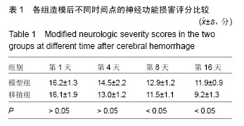
2.1 实验动物数量分析 36只SD大鼠均进入结果分析。 2.2 各组大鼠mNSS评分结果比较 正常对照组大鼠mNSS评分为0;模型组大鼠mNSS评分结果比较稳定,一直处于较高水平;移植组大鼠脑出血第8,16天的mNSS评分显著低于模型组(P < 0.05),表明骨髓单个核细胞可以促进脑出血大鼠神经功能的恢复,见表1。"
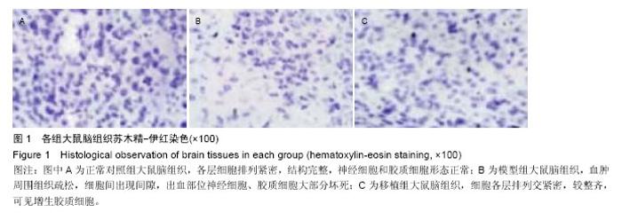
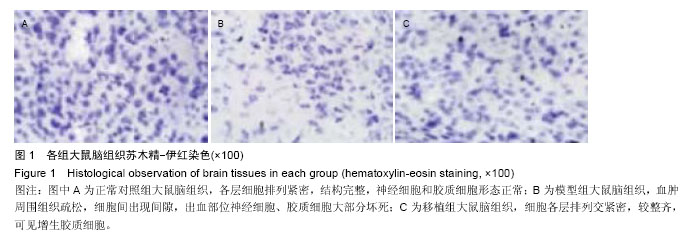
2.3 各组大鼠脑组织苏木精-伊红染色结果 光镜下发现,正常对照组大鼠脑组织各层细胞排列紧密,结构完整,神经细胞和胶质细胞形态正常;模型组大鼠脑组织血肿周围组织疏松,细胞间出现间隙,出血部位神经细胞、胶质细胞大部分坏死;移植组大鼠脑组织细胞各层排列交紧密,较整齐,可见增生胶质细胞,见图1。"

2.4 各组大鼠血肿脑组织内神经元细胞凋亡率的比较 流式细胞仪检测结果显示,模型组大鼠在移植2 d和4 d时血肿周围组织中的细胞凋亡率较高;移植组大鼠在2 d和4 d时血肿周围组织中的细胞凋亡率显著低于模型组(P < 0.05),见表2。"

| [1] Qureshi AI,Tuhrim S,Broderick JP,et al. Spontaneous in tracerebral hemorrhage. N Engl J Med.2001; 344 (19):1450-1460.[2] 刘太平.自拟方治疗高血压性脑出血120 例疗效观察[J].中医学报,2001,16(6):30-31.[3] Schellinger PD,Fiebach JB,Hoffmann K,et al.Stroke MRI in intracerebral hemorrhage: is there a perihemorrhagic penumbra. Stroke. 2003;34(7): 1674-1679.[4] Jiang B,Wang WZ,Chen H,et al.Incidence and trends of stroke and its subtypes in China:Results from three large eities.Stroke.2006;37(1):63-68.[5] Elliott J,Smith M.The acute management of intracerebral hemorrhage: a clinical review.Anesth Analg.2010;110(5):1419-14271.[6] Eriksson PS, Perfilieva E, Björk-Eriksson T, et al. Neurogenesis in the adult human hippocampus.Nat Med.1998;4(11):1313-1317.[7] Curtis MA,Kam M,Nannmark U,et al.Human neuroblasts migrate to the olfactory bulb via a lateral ventricular extension. Science. 2007;315(5816): 1243-1249.[8] Wang SP,Wang ZH,Peng DY,et al.Therapeutic effect of mesenchymal stem cells in rats with intracerebral hemorrhage:reduced apoptosis and enhanced neuroprotection. Mol Med Report.2012;6(4):848- 854.[9] Pittenger MF,Mackay AM,Beck SC,et al.Multilineage potential of adult human mesenchymal stem cells. Science.1999;284(5411):143-147.[10] Woodburg D,Schwarz EJ,Prockp DJ,et al.Adult rat human bone marrow stromal cells differentiate into neurons.J Neurosci Res.2000;61(4):364-370.[11] 何远宏,李魁,王建平,等.骨髓单个核细胞移植对脑出血大鼠的影响[J].郑州大学学报(医学版),2014,49(2):264-268.[12] Gerdoni E,Gallo B,Casazza S,et al.Mesenchymal stem cells effectively modulate pathogenic immune response in experimental autoimmune encephalomyelitis. Ann Neurol.2007;61:219-227.[13] 张雪莹,卢宏,王建平,等.骨髓单个核细胞移植对脑出血大鼠神经元样细胞分化和GDNF的影响[J].中华神经医学杂志,2014,13(1):22-25.[14] 王建平,余列,蒋超,等.正常与脑梗死后大鼠来源的骨髓单个核细胞治疗缺血性脑卒中的效果对比[J].中华行为医学与脑科学杂志,2013,22(2):103.[15] 陈君.骨髓单个核细胞移植治疗大鼠脑出血后脑损伤的实验研究[D].郑州大学,2014.[16] Peeling J,Yan HJ,Corbett D,et al.Effect of FK- 506 on inflammation and behavioral outcome following intracerebral hemorrhage in rat.Exp Neurol. 2001; 167(2):341-347.[17] Zhang H,Huang A,Xu Y,et al.Differentiation and neurological benefit of the mesenchymal stem cells transplanted into the rat brain following intracerebral hemorrhage.Neurol Res.2006;28(1):10.[18] Lee SH,Kim BJ,Ryu WS,et al.White matter lesions and poor outcome after intracerebral hemorrhage: a nationwide co-hort study. Neurology. 2010; 74(19): 1502-15101.[19] Steiner T,Vincent C,Morris S,et al.Neurosurgical outcomes after intracerebral hemorrhage: results of the factor seven for acute hemorrhagic stroke trial (FAST).J Stroke Cerebrovasc Dis.2011;20(4): 287-294.[20] Sakamoto S,Shibukawa M,Tani I,et al.Open-cell Stent Deployment across the Wide Neck of a Large Middle Cerebral Aneurysm Using the Stent Anchor Technique. J Cerebrovasc Endovasc Neurosurg. 2016;18(1):38-41.[21] Jiang H,Qin Y,Liu T,et al.Nao-Xue-Shu Oral Liquid Protects and Improves Secondary Brain Insults of Hypertensive Cerebral Hemorrhage.Evid Based Complement Alternat Med.2016;2016:9121843.[22] Zijlstra IA,Gathier CS,Boers AM,et al.Association of Automatically Quantified Total Blood Volume after Aneurysmal Subarachnoid Hemorrhage with Delayed Cerebral Ischemia.AJNR Am J Neuroradiol. 2016. [Epub ahead of print][23] Mutoh T,Mutoh T,Sasaki K,et al.Isoflurane postconditioning with cardiac support promotes recovery from early brain injury in mice after severe subarachnoid hemorrhage. Life Sci.2016.pii: S0024-3205(16)30241-30247.[24] Tsivgoulis G,Zand R,Katsanos AH,et al.Risk of Symptomatic Intracerebral Hemorrhage After Intravenous Thrombolysis in Patients With Acute Ischemic Stroke and High Cerebral Microbleed Burden: A Meta-analysis.JAMA Neurol.2016.doi: 10.1001/jamaneurol.2016.0292. [Epub ahead of print][25] Ciccone A,Pozzi M,Motto C,et al.Epidemiological, clinical, and therapeutic aspects of primary intracerebral hemorrhage.Neurol Sci.2008;29(Suppl 2):S256-S2571.[26] 呼铁民,孙瓅贤,王维兴,等.青年与中老年急性脑出血的危险因素及预后比较[J].中国全科医学,2010,13(14): 1537-1540.[27] Thrift AG,McNeil JJ,Donnan GA.The risk of intracerebral hemorrhage with smoking. The Melbourne Risk Factor Study Group.Cerebrovasc Dis.2004;99(10):34-39.[28] Monforte R,Estruch R,Graus F,et al. High ethanol consumption as risk factor for intracerebral hemorrhage in young and middle-aged people. Stroke.1990;21(11):1529-1532.[29] Ruiz-Sandoval JL,Cantu C,Barinagarrementeria F.Intracerebral Hemorrhage in Young people. Stroke. 1999;30:537-541.[30] Willmot M,Leonardi BeeJ,Bath PMW.High blood pressure in acute stroke and subsequent outcome: a systematic review.Hypertension.2004;43(1):18-24.[31] 刘运海,奉俊敏,杨期东,等.高血压性脑出血住院患者的近期预后影响因素回顾性分析[J].卒中与神经疾病,2004, 11(6):364-367.[32] Sturgeon JD,Folsom AR,Longstreth WT Jr,et al.Risk factors for intracerebral hemorrhage in a pooled prospective study.Stroke.2007;8(10):2718-2725.[33] Iribarren C,Jacobs DR,Sadler M,et al.Low total serum cholesterol and intracerebral hemorrhagic stroke: is the association conlined to the elderly men ? the Kaiser permanent medical care program. Stroke. 1996; 27:1993-1998.[34] SSegal AZ, Chiu RI, Eggleston-Sexton PM, et al.Low cholesterolas a risk factor for primary intracerebral hemorrhage: A case-controi study.Neuroepidemiology. 2003;18(4):185-193.[35] 罗晶,王永红,钟小妮,等.158 例青年脑出血病因、危险因素及预后分析[J].现代预防医学,2008,8(35):1587-1589.[36] Andersen KK,Olsen TS,Dehlendorff C,et al. Hemorrhagic and ischemic strokes compared: stroke severity, mortality, and risk factors. Stroke. 2009,40: 2068-2072.[37] Wang Z,Cui C,Li Q,et al.Intracerebral transplantation of foetal neural stem cells improeves brain dysfunction induced by intracerebral haemorrhage stroke in mice. J Cell Mol Med.2011;15(12):2624-2633.[38] Wang SP,Wang ZH,Peng DY,et al.Therapeutic effect of mesenchymal stem cells in rats with intracerebral hemorrhage:reduced apoptosis and enhanced neuroprotection.Mol Med Report.2012;6(4):848-854.[39] 李秀云,张国华.脐血干细胞移植治疗脑出血的疗效分析[J].中华全科医学,2011,9(11):1724-1725.[40] Liao W,Xie J,Zhong J,et al.Therapeutic effect of human umbilical cord multipotent mesenchymal stromal cells in a rat model of stroke. Transplantation. 2009; 87(3): 350-359.[41] 张化彪,许予明,张苏明.丁新生骨髓间质干细胞移植治疗大鼠脑出血的实验研究[J].中华神经科杂志,2003,36(6): 432.[42] Lee HJ,Lim IJ,Lee MC,et al.Human neural stem cells genetically modified to over-express brain- derived neurotrophic factor promote functional recovery and neuro-protection in a mouse stroke model.Neurosci Res.2010;88(15):3282-3294.[43] Mimtra T,Dezawa M,Kamo H,et al.Peripheral nerve regeneration by transplantation of bone marrow stromal cell-derived Schwann cells in adult rats.J Neurosurg. 2004;10l:806-812.[44] Mahmood A,Lu D,Chopp M.Intravenous administration of marrow stromal cells(MSCs) increases the expression of growth factors in rat brain after traumatic brain injury. J Neurotrauma.2004; 21:33-39.[45] Ma XL,Liu KD,Li FC,et al.Human mesenchymal stem cells increases expression of α‐tubulin and angiopoietin1and2 in focal cerebral ischemia and reperfusion.Curr Neurovasc Res.2013;10(2):103-111. |
| [1] | Pu Rui, Chen Ziyang, Yuan Lingyan. Characteristics and effects of exosomes from different cell sources in cardioprotection [J]. Chinese Journal of Tissue Engineering Research, 2021, 25(在线): 1-. |
| [2] | Lin Qingfan, Xie Yixin, Chen Wanqing, Ye Zhenzhong, Chen Youfang. Human placenta-derived mesenchymal stem cell conditioned medium can upregulate BeWo cell viability and zonula occludens expression under hypoxia [J]. Chinese Journal of Tissue Engineering Research, 2021, 25(在线): 4970-4975. |
| [3] | Zhang Tongtong, Wang Zhonghua, Wen Jie, Song Yuxin, Liu Lin. Application of three-dimensional printing model in surgical resection and reconstruction of cervical tumor [J]. Chinese Journal of Tissue Engineering Research, 2021, 25(9): 1335-1339. |
| [4] | Geng Qiudong, Ge Haiya, Wang Heming, Li Nan. Role and mechanism of Guilu Erxianjiao in treatment of osteoarthritis based on network pharmacology [J]. Chinese Journal of Tissue Engineering Research, 2021, 25(8): 1229-1236. |
| [5] | Zhang Xiumei, Zhai Yunkai, Zhao Jie, Zhao Meng. Research hotspots of organoid models in recent 10 years: a search in domestic and foreign databases [J]. Chinese Journal of Tissue Engineering Research, 2021, 25(8): 1249-1255. |
| [6] | Hou Jingying, Yu Menglei, Guo Tianzhu, Long Huibao, Wu Hao. Hypoxia preconditioning promotes bone marrow mesenchymal stem cells survival and vascularization through the activation of HIF-1α/MALAT1/VEGFA pathway [J]. Chinese Journal of Tissue Engineering Research, 2021, 25(7): 985-990. |
| [7] | Shi Yangyang, Qin Yingfei, Wu Fuling, He Xiao, Zhang Xuejing. Pretreatment of placental mesenchymal stem cells to prevent bronchiolitis in mice [J]. Chinese Journal of Tissue Engineering Research, 2021, 25(7): 991-995. |
| [8] | Liang Xueqi, Guo Lijiao, Chen Hejie, Wu Jie, Sun Yaqi, Xing Zhikun, Zou Hailiang, Chen Xueling, Wu Xiangwei. Alveolar echinococcosis protoscolices inhibits the differentiation of bone marrow mesenchymal stem cells into fibroblasts [J]. Chinese Journal of Tissue Engineering Research, 2021, 25(7): 996-1001. |
| [9] | Fan Quanbao, Luo Huina, Wang Bingyun, Chen Shengfeng, Cui Lianxu, Jiang Wenkang, Zhao Mingming, Wang Jingjing, Luo Dongzhang, Chen Zhisheng, Bai Yinshan, Liu Canying, Zhang Hui. Biological characteristics of canine adipose-derived mesenchymal stem cells cultured in hypoxia [J]. Chinese Journal of Tissue Engineering Research, 2021, 25(7): 1002-1007. |
| [10] | Geng Yao, Yin Zhiliang, Li Xingping, Xiao Dongqin, Hou Weiguang. Role of hsa-miRNA-223-3p in regulating osteogenic differentiation of human bone marrow mesenchymal stem cells [J]. Chinese Journal of Tissue Engineering Research, 2021, 25(7): 1008-1013. |
| [11] | Lun Zhigang, Jin Jing, Wang Tianyan, Li Aimin. Effect of peroxiredoxin 6 on proliferation and differentiation of bone marrow mesenchymal stem cells into neural lineage in vitro [J]. Chinese Journal of Tissue Engineering Research, 2021, 25(7): 1014-1018. |
| [12] | Zhu Xuefen, Huang Cheng, Ding Jian, Dai Yongping, Liu Yuanbing, Le Lixiang, Wang Liangliang, Yang Jiandong. Mechanism of bone marrow mesenchymal stem cells differentiation into functional neurons induced by glial cell line derived neurotrophic factor [J]. Chinese Journal of Tissue Engineering Research, 2021, 25(7): 1019-1025. |
| [13] | Duan Liyun, Cao Xiaocang. Human placenta mesenchymal stem cells-derived extracellular vesicles regulate collagen deposition in intestinal mucosa of mice with colitis [J]. Chinese Journal of Tissue Engineering Research, 2021, 25(7): 1026-1031. |
| [14] | Pei Lili, Sun Guicai, Wang Di. Salvianolic acid B inhibits oxidative damage of bone marrow mesenchymal stem cells and promotes differentiation into cardiomyocytes [J]. Chinese Journal of Tissue Engineering Research, 2021, 25(7): 1032-1036. |
| [15] | Guan Qian, Luan Zuo, Ye Dou, Yang Yinxiang, Wang Zhaoyan, Wang Qian, Yao Ruiqin. Morphological changes in human oligodendrocyte progenitor cells during passage [J]. Chinese Journal of Tissue Engineering Research, 2021, 25(7): 1045-1049. |
| Viewed | ||||||
|
Full text |
|
|||||
|
Abstract |
|
|||||