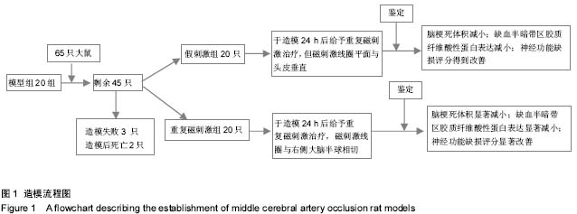
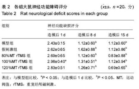
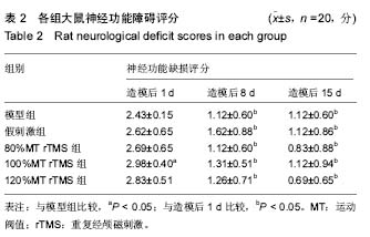
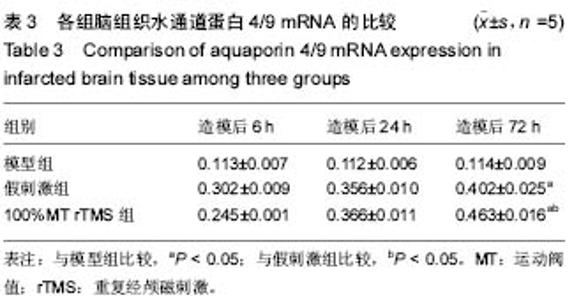
| [1] Leonardo CC, Hall AA, Collier LA, et al. Human umbilical cord blood cell therapy blocks the morphological change and recruitment of CD11b-expressing, isolectin-binding proinflammatory cells after middle cerebral artery occlusion. J Neurosci Res. 2010;88(6):1213-1222.
[2] 周国庆,金怡,张鹏.沉默RhoA基因对骨髓间充质干细胞静脉移植治疗脑梗死大鼠的作用[J].中国组织工程研究与临床康复, 2010,14(45):8416-8420.
[3] Phuc PV, Nhung TH, Loan DT, et al. Differentiating of banked human umbilical cord blood-derived mesenchymal stem cells into insulin-secreting cells. In Vitro Cell Dev Biol Anim. 2011; 47(1):54-63.
[4] 陈雪扉,莫万斌.注射用七叶皂苷钠联合脐带间充质干细胞移植改善脑梗死大鼠神经功能[J].中国组织工程研究与临床康复, 2011,27(15):5080-5084.
[5] Zhao T, Li Y, Tang L, et al. Protective effects of human umbilical cord blood stem cell intravitreal transplantation against optic nerve injury in rats. Graefes Arch Clin Exp Ophthalmol. 2011;249(7):1021-1028.
[6] Mutlu O, Ulak G, Celikyurt IK, et al. Effects of olanzapine, sertindole and clozapine on learning and memory in the Morris water maze test in naive and MK-801-treated mice. Pharmacol Biochem Behav. 2011;98(3):398-404.
[7] Chen Y, Swanson RA. Astrocytes and brain injury. J Cereb Blood Flow Metab. 2003;23(2):137-149.
[8] 张小乔,梅元武,刘传玉,等.经颅磁刺激对脑梗死大鼠神经功能恢复与皮层BDNF表达的影响[J].卒中与神经疾病,2006,13(5): 259-261.
[9] Ogiue-Ikeda M, Kawato S, Ueno S. The effect of repetitive transcranial magnetic stimulation on long-term potentiation in rat hippocampus depends on stimulus intensity. Brain Res. 2003;993(1-2):222-226.
[10] Seo JP, Jang SH.Disappearance of unaffected motor cortex activation by repetitive transcranial magnetic stimulation in a patient with cerebral infarct. Neural Regen Res. 2014;9 (7): 761-762
[11] Thal SC, Mebmer K, Schmid-Elsaesser R, et al. Neurological impairment in rats after ubarachnoid hemorrhage-a comparison of functional tests. J Neurol Sci. 2008;268(1-2): 150-159.
[12] Yozbatiran N, Alonso-Alonso M, See J, et al. Safety and behavioral effects of high-frequency repetitive transcranial magnetic stimulation in stroke. Stroke. 2009;40(1):309-312.
[13] 毕妍贞,胡可慧,姜伟,等.运动训练对脑梗死大鼠学习记忆能力和半暗带突触结构的影响[J].中华物理医学与康复杂志, 2008, 30(2):76-79.
[14] Kim HS, Jang SH, Lee ZI,et al.Therapeutic benefit of repetitive transcranial magnetic stimulation for severe mirror movements A case report.Neural Regen Res. 2013;8 (6): 569-574
[15] 严莉,经颅重频磁刺激对Wistar大鼠大脑中动脉梗塞再灌流损伤模型影响初步探讨[D].北京:中国医学科学院,2005.
[16] Aydin-Abidin S, Trippe J, Funke K, et al. High- and low-frequency repetitive transcranial magnetic stimulation differentially activates c-Fos and zif268 protein expression in the rat brain. Exp Brain Res. 2008;188(2):249-261.
[17] 王晓明,龙存国,吴碧华,等.经颅磁刺激安全性的实验研究[J].临床神经电生理学杂志,2002,11(4):227-229.
[18] 严婷婷,何明利,顾正天,等.高频及低频重复经颅磁刺激对脑卒中后抑郁的疗效对比研究[J].青岛医药卫生,2010,42(2):81-85.
[19] 孙毅,汤晓芙,郭玉璞.经颅磁刺激安全性的实验研究[J].中华神经科杂志,1996,29(4):217-221.
[20] Nowak DA, Grefkes C, Ameli M, et al. Interhemispheric competition after stroke:brain stimulation to enhance recovery of function of the affected hand. Neurorehabil Neural Repair. 2009;23(7):641-656.
[21] 廖维靖,刘淑红,范明,等.线栓阻断大鼠大脑中动脉制作缺血性脑损伤模型的改良[J].中华物理医学与康复杂志,2002,24(6):321- 324.
[22] Luft AR, Kaelin-Lang A, Hauser TK, et al. Transcranial magnetic stimulation in therat. Exp Brain Res. 2001;140(1): 112-121.
[23] 吴小丽,吴宏胜,谢德丰,等.重复经颅磁刺激在脑梗塞运动功能康复中的作用[J].海南医学院学报,2010,16(6):799-80.
[24] Mahmood A, Lu D, Qu C, et al. Long-term recovery after bone marrow stromal cell treatment of traumatic brain injury in rats. J Neurosurg. 2006;104(2):272-277.
[25] Liu YR, Cardamone L, Hogan RE, et al. Progressive metabolic and structural cerebral perturbations after traumatic brain injury: an in vivo imaging study in the rat. J Nucl Med. 2010;51(11):1788-1795.
[26] 张洪连,吴晓牧.骨髓间充质干细胞移植治疗脑梗死及存在问题[J].中华脑血管病杂志,2010,4(4):300-307.
[27] 严莉,丰宏林,崔丽英.经颅重频磁刺激对大鼠脑缺血损伤早期运动皮质兴奋性神经功能的影响[J].中国临床康复,2005,9(25): 243-245.
[28] Feng HL, Yan L, Guan YZ, et al. Effects of transcranial magnetic stimulation on motorcortical excitability and neuro function after cerebral ischemia reperfusion injury inrats. Chin Med Sci J. 2005;20(4):226-230.
[29] 马玉娟,黄杰,方征字,等.不同强度20 Hz重复经颅磁刺激对脑梗死大鼠神经行为学及胶质纤维酸性蛋白的影响[J].中华物理医学与康复杂志,2012,34(2):85-87.
[30] Yozbatiran N, Alonso-Alonso M, See J, et al. Safety and behavioral effects of high-frequency repetitive transcranial magnetic stimulation in stroke. Stroke. 2009;40(1):309-312.
[31] 小乔,梅元武,刘传玉,等.经颅磁刺激对脑梗死大鼠神经功能恢复与皮层BDNF表达的影响[J].卒中与神经疾病,2006,13:259-261.
[32] 赵合庆,孙永安,戴永萍,等.经颅磁刺激对脑缺血大鼠皮层脑源性神经营养因子表达及梗死体积的影响[J].中华神经科杂志, 2005, 38(5):330-331.
[33] Paulo SB, Alonso-Alonso M, Carlos GM, et al. Hand function improvement with lowfrequency repetitive transcranialmagnetic stimulation of the unaffected hemisphere in a severe ase of stroke. Am J PhysicalMed Rehabilit. 2006;85:927.
[34] 刘传玉,梅元武,张小乔.经颅磁刺激对局灶性脑缺血大鼠梗死周边区GAP-43和Syp表达的影响[J].卒中与神经疾病, 2006,13(1): 15-18.
[35] 黄晓琳,韩肖华,郭铁成,等.电针联合经颅磁刺激对急性脑缺血大鼠VEGF及其受体Flk-表达的影响[J].中华物理医学与康复杂志, 2004,26(10):581-584.
[36] 冯新红.经颅重复磁刺激的临床应用研究进展[J].国际神经病学神经外科学杂志,2010,37(6):535-538.
[37] 吴冰洁,记宏,岳崴,等.高频重复经颅磁刺激对脑梗死患者运动功能的影响[J].中国全科医学,2014,23(17):2751-2760.
|