Chinese Journal of Tissue Engineering Research ›› 2015, Vol. 19 ›› Issue (17): 2625-2630.doi: 10.3969/j.issn.2095-4344.2015.17.001
Significance of Ranawat’s triangle method in determining the rotation center of normal hip joint
Liu Ya-bin1, 2, Ma Xin-long1, 3
- 1Graduate School of Tianjin Medical University, Tianjin 300070, China; 2 2nd Department of Limb Orthopedic and Reconstructive Surgery, Tianjin Hospital, Tianjin 300210, China; 3General Hospital of Tianjin Medical University, Tianjin 300052, China
-
Online:2015-04-23Published:2015-04-23 -
Contact:Ma Xin-long, Chief physician, Doctoral supervisor, Graduate School of Tianjin Medical University, Tianjin 300070, China; General Hospital of Tianjin Medical University, Tianjin 300052, China -
About author:Liu Ya-bin, Studying for master’s degree, Graduate School of Tianjin Medical University, Tianjin 300070, China; 2nd Department of Limb Orthopedic and Reconstructive Surgery, Tianjin Hospital, Tianjin 300210, China
CLC Number:
Cite this article
Liu Ya-bin, Ma Xin-long. Significance of Ranawat’s triangle method in determining the rotation center of normal hip joint[J]. Chinese Journal of Tissue Engineering Research, 2015, 19(17): 2625-2630.
share this article
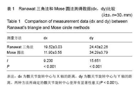
2.1 参与者数量分析 按意向性处理,纳入30例发育正常的创伤性股骨颈骨折单侧人工全髋关节置换后患者做为观察对像,采用Ranawat三角法及Mose圆法分别对同一观察对象的X射线片进行测量。全部进入结果分析,无脱落。 2.2 测量结果 测得的数据经统计分析发现:髋关节旋转中心与X轴的距离(dx)、及与Y轴的距离(dy),Ranawat三角法和Mose圆法之间差异有显著性意义(P < 0.001, 表1);使用Ranawat三角法确定的髋关节旋转中心更偏向内上方[dx1-dx2=(7.62±4.52) mm,dy2-dy1=(9.86± 3.45) mm]。"

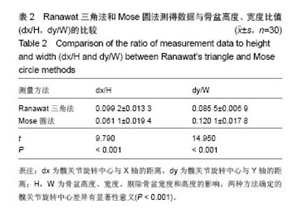
将测得的数据与骨盆的高度和宽度做比(dx1/H,dy1/W;dx2/H,dy2/W)并分析两组数据(dx1/H,dx2/H;dy1/W,dy2/W)发现,二者之间差异仍有显著性意义(P < 0.01,表2)。"

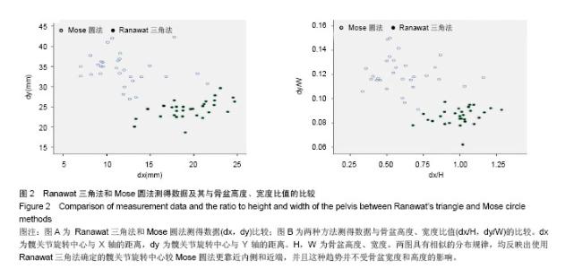
使用散点图对数据进行分析发现,所得的数据(dx,dy;dx/H,dy/W)具有相似的分布规律,并且这种规律并不受骨盆高度和宽度的影响(图2)。"
| [1]Abolqhsaemian M, Samiezadeh S, Jafari D, et al. Displacement of the hip center of rotation after arthroplasty of Crowe III and IV dysplasia: a radiological and biomechanical study. J Arthroplasty. 2013;28(6): 1031-1035.
[2]Grunert R, Kretzschmar C, Rotsch C, et al. 2014. Development of an optical measurement system for hip implant surgery to evaluate the leg length and the hip rotation center. Middle East Conference on Biomedical Engineering (MECBME), Hilton Hotel, Doha, Qatar.17-20.
[3]Ha YC, Yoo JJ, Lee YK, et al. Acetabular component positioning using anatomic landmarks of the acetabulum. Clin Orthop Relat Res.2012;470: 3515-3523.
[4]Charissoux JL, Asloum Y, Marcheix PS. Surgical management of recurrent dislocation after total hip arthroplasty. Orthop Traumatol Surg Res. 2014;100(1 Suppl):S25-34.
[5]Goel A, Lau EC, Ong KL. Dislocation Rates Following Primary Total Hip Arthroplasty Have Plateaued in the Medicare Population. J Arthroplasty. 2014.
[6]Robbins GM, Masri BA, Garbuz DS, et al. Treatment of hip instability. Orthop Clin North Am. 2001;32:593.
[7]Khalifa AR, Marzouk AR, Alam M, et al. Short-term results of primary total hip arthroplasty for Protrusio Acetabuli. Int J. 2014;2(4): 538-542.
[8]Bicanic G, Barbaric K, Bohacek I, et al. Current concept in dysplastic hip arthroplasty: Techniques for acetabular and femoral reconstruction. World J Orthop. 2014;5(4):412-424.
[9]Wu X, Lou LM, Li SH, et al. Soft tissue balancing in total hip arthroplasty for patients with adult dysplasia of the hip. Orthop Surg. 2009;1(3):212-215.
[10]Holzapfel BM, Greimel F, Prodinger PM,et al. Total hip replacement in developmental dysplasia using an oval-shaped cementless press-fit cup. Int Orthop. 2012;36(7): 1355-1361.
[11]Delp SL, Komattu AV, Wixson RL. Superior displacement of the hip in total joint replacement: effects of prosthetic neck length, neck-stem angle, and anteversion angle on the moment-generating capacity of the muscles. J Orthop Res. 1994;12:860-870.
[12]Ranawat CS, Dorr LD, Inglis AE. Total hip arthroplasty in protrusio acetabuli of rheumatoid arthritis. J Bone Joint Surg (Am). 1980;62(7):1059-1065.
[13]Bell AL, Pedersen DR, Brand RA. A comparison of the accuracy of several hip center location prediction methods. J Biomech,1990;23(6):617-621.
[14]Bouffard V, Beqon M, Farhadnia P, et al. Hip joint center localisation: A biomechanical application to hip arthroplasty population.World J Orthop. 2012;3(8):131-136.
[15]Krishnan SP, Carrington RW, Mohiyaddin S, et al. Common misconceptions of normal hip joint relations on pelvic radiographs. J Arthroplasty. 2006;21(3);409-412.
[16]Wu X, Li SH, Lou LM, et al. The techniques of soft tissue release and true socket reconstruction in total hip arthroplasty for patients with severe developmental dysplasia of the hip. Int Orthop. 2012;36(9):1795-1801.
[17]Boudriot U, Hilgert J, Hinrichs F. Determination of the rotational center of the hip. Arch Orthop Trauma Surg. 2006; 126(6):417-420.
[18]Bicanic G, Delimar D, Delimar M, et al. Influence of the acetabular cup position on hip load during arthroplasty in hip dysplasia. Int Orthop. 2009;33(2): 397-402.
[19]Fousek J, Indrakova P. Total hip arthroplasty in post-dysplastic hip arthritis. Can type and position of the acetabular component influence longevity of the prosthesis? Acta Chir Orthop Traumatol Cech. 2007;74(1): 47-54.
[20]Kim DH, Cho SH, Jeong ST, et al. Restoration of the center of rotation in revision total hip arthroplasty. Arthroplasty. 2010; 25(7):1041-1046.
[21]Edurado GR, Ricardo FF, David D, et al. Reconstruction of the rotation center of the hip after oblong cups in revision total hip arthroplasty. J Orthop Traumatol. 2013;14(1):39-49.
[22]Bouffard V, Begon M, Champagne A, et al. Hip joint center location: a biomechanical application to hip arthroplasty population. World J Orthop. 2012;3(8): 131-136.
[23]Lazarinis S, Milbrink J, Mattsson P, et al. Bone loss around a stable, partly threaded hydroxyapatite-coated cup: prospective cohort study using RSA and DXA. Hip Int. 2014; 24(2):155-166.
[24]王坤正.初次全髋关节置换术选择骨水泥或生物型固定方式的比较[J].中华关节外科杂志(电子版),2012,6(4):492-495.
[25]刘明,王岩,陈继营,等.1436髋Ribbed假体单中心应用分析[J].中国矫形外科杂志,2008,16(14):1051-1053.
[26]史庆轩,李佩佳,孙磊,等.662髋Ribbed假体中远期临床疗效观察[J].中国矫形外科志,2012,20(15):1370-1373.
[27]Dalury DF, Kelley TC, Adams MJ. Modern proximally tapered uncemented sems can be safely used in Dorr Type C femoral bone. J Arthroplasty. 2012;27(6):1014-1018.
[28]Dandachli W, Nakhla A, Iranpour F, et al. Can the acetabular position be derived from a pelvic frame of reference? Clin Orthop Relat Res. 2009;467(4):886-893.
[29]Abolqhsaemian M, Samiezadeh S, Jafari D, et al. Displacement of the hip center of rotation after arthroplasty of Crowe III and IV dysplasia: a radiological and biomechanical study. J Arthroplasty. 2013;28(6):1031-1035.
[30]Dib Z, Dardenne G, Poirier N, et al. Detection of the hip center in computer-assisted surgery: An in vitro assessment study. IRBM. 2013;34(4):319-321.
[31]García-Rey E, Fernández-Fernández R, Durán D, et al. Reconstruction of the rotation center of the hip after oblong cups in revision total hip arthroplasty. J Orthop Traumatol. 2013;14(1): 39-49.
[32]Lenaerts G, Groote DF, Demeulenaere B, et al. Subject-specific hip geometry affects predicted hip joint contace forces during gait. J Biomech. 2008;41: 1243-1252.
[33]Heller MO, Schröder JH, Matziolis G, et al. Musculoskeletal load analysis. A biomechanical explanation for clinical results-and more? (in German). Orthopade. 2007;36(3):188, 190-194.
[34]Nawabi DH, Meftal M, Nam D, et al. Durable fixation achieved with medialized, high hip center cementless THAs for Crowe II and III dysplasia. Clin Orthop Relat Res. 2014;472(2): 630-636.
[35]Kaneuji A , Suqimori T, Ichiseki T, et al. Minimum ten-year results of a porous acetabular component for Crowe I to III hip dysplasia using an elevated hip center. J Arthroplasty. 2009; 24(2): 187-194.
[36]Baqhdadi YM, Larson AN, Sierra RJ. Restoration of the hip center during THA performed for protrusio acetabuli is associated with better implant survival. Clin Orthop Relat Res. 2013;471(10):3251-3259.
[37]Kaneuji A, Sugimori T, Ichiseki T, et al. Minimum ten-year results of a porous acetabular component for Crowe I to III hip dysplasia using an elevated hip center. J Arthroplasty. 2009; 24:187- 194 .
[38]Bicanic G, Delimar D, Delimar M, et al. Influence of the acetabular cup position on hip load during arthroplasty in hip dysplasia. Int Orthop. 2009;33:397-402.
[39]Hartofilakidis G, Karachalios T. Total hip arthroplasty for congenital hip disease. J Bone Joint Surg Am. 2004;86-A: 242-250.
[40]Sknk EL, Zaltz I, Heare T, et al. Acetabular cartilage and labral damage observed during surgical hip dislocation for stable slipped capital femoral epiphysis. J Pediatr Orthop. 2010; 30(1): 26-30.
[41]Shi HF, Xiong J, Chen YX, et al. Radiographic analysis of the restoration of hip joint center following open reduction and internal fixation of acetabular fractures: a retrospective cohort study. BMC Musculoskelet Disord. 2014;15:277.
[42]The B, Kootstra JW, Hosman AH, et al. Comparison of the techniques for correction of magnification of pelvic X-ray for hip surgery planning. J Digit Imaging. 2007; 20(4): 329-335. |
| [1] | Zhang Chao, Lü Xin. Heterotopic ossification after acetabular fracture fixation: risk factors, prevention and treatment progress [J]. Chinese Journal of Tissue Engineering Research, 2021, 25(9): 1434-1439. |
| [2] | Chen Junming, Yue Chen, He Peilin, Zhang Juntao, Sun Moyuan, Liu Youwen. Hip arthroplasty versus proximal femoral nail antirotation for intertrochanteric fractures in older adults: a meta-analysis [J]. Chinese Journal of Tissue Engineering Research, 2021, 25(9): 1452-1457. |
| [3] | Chen Lu, Zhang Jianguang, Deng Changgong, Yan Caiping, Zhang Wei, Zhang Yuan. Finite element analysis of locking screw assisted acetabular cup fixation [J]. Chinese Journal of Tissue Engineering Research, 2021, 25(3): 356-361. |
| [4] | Fan Yinuo, Guan Zhiying, Li Weifeng, Chen Lixin, Wei Qiushi, He Wei, Chen Zhenqiu. Research status and development trend of bibliometrics and visualization analysis in the assessment of femoroacetabular impingement [J]. Chinese Journal of Tissue Engineering Research, 2021, 25(3): 414-419. |
| [5] | Cheng Chongjie, Yan Yan, Zhang Qidong, Guo Wanshou. Diagnostic value and accuracy of D-dimer in periprosthetic joint infection: a systematic review and meta-analysis [J]. Chinese Journal of Tissue Engineering Research, 2021, 25(24): 3921-3928. |
| [6] | Fu Panfeng, Shang Wei, Kang Zhe, Deng Yu, Zhu Shaobo. Efficacy of anterolateral minimally invasive approach versus traditional posterolateral approach in total hip arthroplasty: a meta-analysis [J]. Chinese Journal of Tissue Engineering Research, 2021, 25(21): 3409-3415. |
| [7] | Yang Kun, Fei Chen, Wang Pengfei, Zhang Binfei, Yang Na, Tian Ding, Zhuang Yan, Zhang Kun . Meta-analysis of the efficacy of robot-assisted and traditional manual implantation of cannulated screws in the treatment of femoral neck fracture [J]. Chinese Journal of Tissue Engineering Research, 2021, 25(18): 2938-2944. |
| [8] | Liu Yongyu, Xu Jingli, Lin Tianye, Wu Feng, Shen Chulong, Xiong Binglang, Zou Qizhao, Lai Qizhong, Zhang Qingwen . Medium-and long-term follow-up of Salter pelvic osteotomy and implant fixation for children with developmental hip dislocation [J]. Chinese Journal of Tissue Engineering Research, 2021, 25(15): 2380-2384. |
| [9] | Yang Youwei, Feng Wei . Quadrilateral plate fractures: research progress of implant treatment [J]. Chinese Journal of Tissue Engineering Research, 2021, 25(15): 2416-2422. |
| [10] | Zhang Haifeng, Zhao Can, Liu Meixiao, Song Cuirong, Pang Yin. Analysis of various degrees of freedom of the hip movement based on motion capture technology [J]. Chinese Journal of Tissue Engineering Research, 2021, 25(12): 1815-1819. |
| [11] | Zhang Nianjun, Liu Xiaofang, Zhou Guanming, Su Yao, Hong Shi. Three-dimensional-printing assisted total hip arthroplasty for individualized treatment of adult developmental dysplasia of the hip [J]. Chinese Journal of Tissue Engineering Research, 2021, 25(12): 1820-1825. |
| [12] | Cui Keze, Guo Xiang, Han Guibin, Chen Yuanliang, Liu Yiheng, Zhong Haibo. Learning curve and early clinical results of total hip arthroplasty with MAKO robot assisted posterolateral approach [J]. Chinese Journal of Tissue Engineering Research, 2020, 24(9): 1313-1317. |
| [13] | Deng Biyong, Deng Bixiang. Differences in relative parameters of acetabulum in hip arthroplasty between CT horizontal plane and CT true pelvic plane [J]. Chinese Journal of Tissue Engineering Research, 2020, 24(9): 1405-1409. |
| [14] | Ai Di. Absorbable suture anchors for treating femoroacetabular impingement syndrome combined with acetabular labral tears [J]. Chinese Journal of Tissue Engineering Research, 2020, 24(4): 499-504. |
| [15] | Kang Peng, Zhang Fangxin, Maihemuti·Yakufu, Wei Weitao, Yilihamu·Toheti, Aierken·Amudong. True acetabulum morphology of Crowe type IV developmental dysplasia of the hip based on three-dimensional surgical simulation [J]. Chinese Journal of Tissue Engineering Research, 2020, 24(36): 5749-5754. |
| Viewed | ||||||
|
Full text |
|
|||||
|
Abstract |
|
|||||