Chinese Journal of Tissue Engineering Research ›› 2015, Vol. 19 ›› Issue (3): 399-404.doi: 10.3969/j.issn.2095-4344.2015.03.013
Previous Articles Next Articles
Influence of different concentrations of enamel matrix proteins on bioactivity of human periodontal ligament cells
Qu Zhe1, Zhang Jing-ying2, Guo Ying1, Ma Wei-dong1, Ma Lan1
- 1Department of Dental Implantation, Dalian Stomatological Hospital, Dalian 116021, Liaoning Province, China; 2Medicine Department, Dalian University, Dalian 116622, Liaoning Province, China
-
Online:2015-01-15Published:2015-01-15 -
Contact:Qu Zhe, Department of Dental Implantation, Dalian Stomatological Hospital, Dalian 116021, Liaoning Province, China -
About author:Qu Zhe, M.D., Associate professor, Department of Dental Implantation, Dalian Stomatological Hospital, Dalian 116021, Liaoning Province, China -
Supported by:the Start Fund for Returnees of Ministry of Human Resources and Social Security of China, No. 2011508; the Scientific Research Project in High Education of Liaoning Provincial Education Department, No. 2008027; the Science and Technology Project for People’s Livelihood in Dalian, No. 2013E15SF169
CLC Number:
Cite this article
Qu Zhe, Zhang Jing-ying, Guo Ying, Ma Wei-dong, Ma Lan. Influence of different concentrations of enamel matrix proteins on bioactivity of human periodontal ligament cells[J]. Chinese Journal of Tissue Engineering Research, 2015, 19(3): 399-404.
share this article
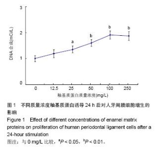
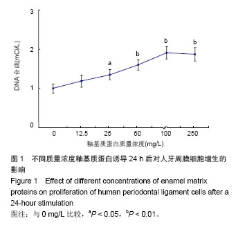
2.1 釉基质蛋白对人牙周膜细胞增殖的影响 通过3H-胸腺嘧啶核苷掺入法检测细胞DNA合成,评估不同质量浓度釉基质蛋白对于牙周膜细胞增生的影响,见图1。各实验组细胞DNA合成明显高于阴性对照组(P < 0.05,P < 0.01),而且随着釉基质蛋白质量浓度从12.25 mg/L增加到 100 mg/L,细胞增殖明显提高;釉基质蛋白质量浓度从 100 mg/L到250 mg/L,细胞增殖趋势有所降低。"
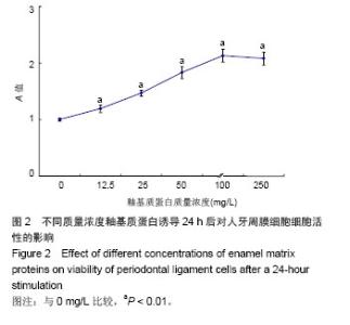
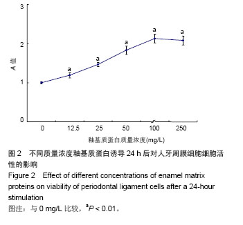
2.2 釉基质蛋白对人牙周膜细胞活性的影响 通过MTT检测细胞活性及统计学处理,不同质量浓度釉基质蛋白对牙周膜细胞活性的影响,见图2。各实验组细胞的细胞活性明显高于阴性对照组(P < 0.01),而且随着釉基质蛋白质量浓度从12.25 mg/L增加到100 mg/L,细胞活性明显提高;釉基质蛋白质量浓度从100 mg/L到250 mg/L,细胞活性增长趋势有所降低。"
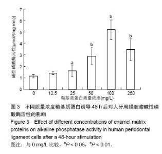
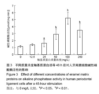
2.3 细胞碱性磷酸酶活性检测 通过检测细胞碱性磷酸酶活性及统计学处理,评估不同质量浓度釉基质蛋白对牙周膜细胞碱性磷酸酶活性的影响,见图3。除12.25 mg/L实验组外,其余各实验组细胞活性明显高于阴性对照组(P < 0.05, P < 0.01),而且随着釉基质蛋白质量浓度从12.25 mg/L增加到100 mg/L,细胞碱性磷酸酶活性明显提高;釉基质蛋白质量浓度从100 mg/L到250 mg/L,细胞碱性磷酸酶活性趋势有所降低。"
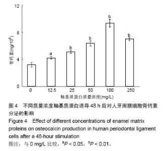
2.4 细胞骨钙素分泌 通过检测细胞骨钙素分泌及统计学处理,评估不同质量浓度釉基质蛋白对人牙周膜细胞骨钙素分泌的影响,见图4。各实验组细胞细胞活性明显高于阴性对照组(P < 0.05,P < 0.01),而且随着釉基质蛋白质量浓度从12.25 mg/L增加到100 mg/L,细胞骨钙素分泌明显提高;釉基质蛋白质量浓度从100 mg/L到250 mg/L,细胞骨钙素分泌趋势有所降低。"

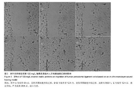
2.5 体外细胞创伤模型 在初始观察6 h时,25,50, 100 mg/L釉基质蛋白实验组创缘周围的细胞开始向中心生长(图5A),而其他组没有细胞迁移生长;在观察8 h后, 12.5 mg/L釉基质蛋白实验组和对照组创缘周围的细胞开始向中心生长,250 mg/L釉基质蛋白实验组直到培养10 h后才有创缘周围的细胞开始向中心生长;在培养8-14 h中,细胞逐渐想创缘中心生长(图5B-D);待培养12 h时, 100 mg/L釉基质蛋白实验组创缘两侧细胞开始融合(图5E),而25,50 mg/L釉基质蛋白实验组14 h后创缘两侧细胞开始融合,12.5 mg/L釉基质蛋白实验组16 h后创缘两侧细胞开始融合,对照组在培养18 h后创缘两侧细胞开始融合;培养20 h后,25,50,100 mg/L釉基质蛋白实验组创缘两侧细胞融合完全,创缘完全关闭(图5F);12.5 mg/L釉基质蛋白实验组创缘两侧细胞融合完全,直到24 h对照组和250 mg/L 釉基质蛋白实验组仍未出现细胞融合完全。"

| [1]蒋少云,束蓉.釉基质蛋白促进牙周组织再生机制的研究进展[J].牙体牙髓牙周病学杂志,2008,17(9):535-538.
[2]邹慧儒,秦宗长,张兰成.釉基质蛋白各组成成分及其作用[J].国际口腔医学杂志,2013,40(3):355-258.
[3]Qu Z,Laky M,Ulm C,et al. Effect of Emdogain on proliferation and migration of different periodontal tissue-associated cells[J].Oral Surg Oral Med Oral Pathol Oral Radiol Endod. 2010;109(6):924-931.
[4]DiPietro LA.Oral Stem Cells: The Fountain of Youth for Epithelialization and Wound Therapy? Adv Wound Care(New Rochelle).2014;3(7):465-467.
[5]Rodionova NV.The dynamics of proliferation and differentiation of osteogenic cells under supportive unloading. Tsitol Genet.2011;45(2):22-27.
[6]Rausch-fan X,Qu Z,Wieland M,et al.Differentiation and cytokine synthesis of human alveolar osteoblasts compared to osteoblast-like cells (MG63) in response to titanium surfaces. Dent Mater.2008;24(1):102-110.
[7]Bertl K,An N,Bruckmann C,et al.Effects of enamel matrix derivative on proliferation/viability, migration, and expression of angiogenic factor and adhesion molecules in endothelial cells in vitro.J Periodontol.2009;80(10):1622-1630.
[8]Filippi A,Pohl Y,von Arx T.Treatment of replacement resorption by intentional replantation, resection of the ankylosed sites, and Emdogain--results of a 6-year survey.Dent Traumatol.2006;22(6):307-311.
[9]侯小丽,吴织芬,万玲,等.釉基质蛋白对骨髓基质细胞分泌TGF-β1的影响[J].牙体牙髓牙周病学杂志,2004,14(4):194-196.
[10]Miron RJ ,Wei L,Yang S,et al. Effect of an Enamel Matrix Derivative on Periodontal Wound Healing/Regeneration in an Osteoporotic Model.J Periodontol.2014;26(3):1-12.
[11]Ninomiya M,Kamata N,Fujimoto R,et al.Application of enamel matrix derivative in autotransplantation of an impacted maxillary premolar: a case report.J Periodontol. 2002;73(3): 346-351.
[12]Döri F,Arweiler NB,Szántó E,et al.Ten-year results following treatment of intrabony defects with an enamel matrix protein derivative combined with either a natural bone mineral or a β-tricalcium phosphate.J Periodontol.2013;84(6):749-757.
[13]项陈洋,张凌琳,李伟.釉基质蛋白在口腔医学领域中的应用进展[J].国际口腔医学杂志,2012,39(6):766-769.
[14]Obregon-Whittle MV,Stunes AK,Almqvist S,et al.Enamel matrix derivative stimulates expression and secretion of resistin in mesenchymal cells.Eur J Oral Sci.2011;119 Suppl 1:366-372.
[15]Grandin HM,Gemperli AC,Dard M.Enamel matrix derivative: a review of cellular effects in vitro and a model of molecular arrangement and functioning.Tissue Eng Part B Rev.2012; 18(3):181-202.
[16]Matsumoto N,Minakami M,Hatakeyama J,et al.Histologic Evaluation of the Effects of Emdogain Gel on Injured Root Apex in Rats.J Endod.2014;40(12):1989-1994.
[17]Ferreira MM,Filomena BM,Lina C,et al.The effect of Emdogain gel on periodontal regeneration in autogenous transplanted dog's teeth.Indian J Dent Res. 2014;25(5): 589-593.
[18]Izumi Y,Aoki A,Yamada Y,et al.Current and future periodontal tissue engineering. Periodontol 2000.2011;56(1):166-187.
[19]宋忠臣,束蓉,宋爱梅,等.釉基质蛋白对人骨髓基质细胞生长和黏附的影响[J].上海口腔医学,2006,15(6):601-604.
[20]宋忠臣,束蓉,谢玉峰.釉基质蛋白对人骨髓基质细胞增殖和矿化的影响[J].上海口腔医学,2008,24(6):831-834.
[21]won YD,Choi HJ,Lee H,et al.Cellular viability and genetic expression of human gingival fibroblasts to zirconia with enamel matrix derivative (Emdogain®).J Adv Prosthodont. 2014;6(5):406-414.
[22]de Oliveira MT,Bentregani LG,Pasternak B,et al.Histometric study of resorption on replanted teeth with enamel matrix-derived protein.J Contemp Dent Pract. 2013;14(3): 468-472.
[23]Jabbari E.Osteogenic peptides in bone regeneration(R).Curr Pharm Des. 2013;19(19):3391-402.
[24]Amin HD,Olsen I,Knowles JC,et al.Effects of enamel matrix proteins on multi-lineage differentiation of periodontal ligament cells in vitro.Acta Biomater. 2013;9(1):4796-4805.
[25]Wada Y,Mizuno M,Nodasaka Y,et al.The effect of enamel matrix derivative on spreading, proliferation, and differentiation of osteoblasts cultured on zirconia.Int J Oral Maxillofac Implants.2012;27(4):849-858.
[26]张凤秋,孟焕新,韩劫,等.釉基质蛋白对人牙周膜细胞生物学影响的体外研究[J].北京大学学报:医学版,2012,44(1):6-10.
[27]刘兰宁,刘宏伟,金岩,等.釉基质蛋白对牙周膜成纤维细胞合成蛋白的影响[J].中国临床康复,2005,9(30):104-106.
[28]李玲,王茜.3种根尖倒充填材料对体外人牙周韧带细胞创伤模型创面愈合的影响[J].上海口腔医学,2011,20(1):41-45.
[29]Yan XZ,Rathe F,Gilissen C,et al.The effect of enamel matrix derivative (Emdogain®) on gene expression profiles of human primary alveolar bone cells.J Tissue Eng Regen Med.2014; 8(6):463-472.
[30]Miron RJ,Bosshardt DD,Zhang Y,et al.Gene array of primary human osteoblasts exposed to enamel matrix derivative in combination with a natural bone mineral.Clin Oral Investig. 2013;17(2):405-410. |
| [1] | Yang Junhui, Luo Jinli, Yuan Xiaoping. Effects of human growth hormone on proliferation and osteogenic differentiation of human periodontal ligament stem cells [J]. Chinese Journal of Tissue Engineering Research, 2021, 25(25): 3956-3961. |
| [2] | Yang Caihui, Liu Qicheng, Dong Ming, Wang Lina, Zuo Meina, Lu Ying, Niu Weidong. Serine/threonine protein kinases can promote bone destruction in mouse models of chronic periapical periodontitis [J]. Chinese Journal of Tissue Engineering Research, 2021, 25(23): 3654-3659. |
| [3] | Liu Yan, Gao Xiang, Zhao Xiaoxia, Chen Qingyu, Gao Junwu. Effects of conditioned medium of human periodontal ligament stem cells on proliferation and osteogenic differentiation of inflammatory tissue-derived human periodontal ligament stem cells [J]. Chinese Journal of Tissue Engineering Research, 2021, 25(13): 2005-2010. |
| [4] | Long Qian, Guan Xiaoyan, Wang Qian, Hu Huan, Liu Jianguo. Transcriptome sequencing technology and its application in oral diseases, dental implants and regeneration [J]. Chinese Journal of Tissue Engineering Research, 2021, 25(11): 1791-1798. |
| [5] | Liu Yan, Chen Qingyu, Gao Xiang, Gao Junwu. Effect of collagen scaffold on proliferation and osteogenic differentiation of human periodontal ligament stem cells treated by Eucommia ulmoides Oliver leaf extract [J]. Chinese Journal of Tissue Engineering Research, 2020, 24(16): 2537-2543. |
| [6] | Wang Xiaobin, Jiang Hongxin, Qu Changhong, Wu Dongmei, Zhang Rongsheng, Xu Bin. Time-dose effects of Taohong Siwu Decoction on number and functional activity of peripheral blood endothelial progenitor cells [J]. Chinese Journal of Tissue Engineering Research, 2019, 23(9): 1354-1358. |
| [7] | Lin Yangdong, Yin Kai. Mechanism underlying adiponectin intervention on the apoptosis of human periodontal ligament cells mediated by high glucose [J]. Chinese Journal of Tissue Engineering Research, 2019, 23(7): 1097-1102. |
| [8] | Liu Dayong, Wang Yingchengyao, Sun Ruixin, Liu Fan, Jia Zhi. Effects of insulin-like growth factor-binding protein 5 on osteogenic differentiation of periodontal ligament stem cells stimulated by inflammatory cytokines [J]. Chinese Journal of Tissue Engineering Research, 2019, 23(33): 5256-5262. |
| [9] |
Zhang Yun, Chang Qun’an, Li Shengmei, Zhang Yuan.
Tumor necrosis factor-alpha inhibits osteogenic differentiation of periodontal ligament stem cells through activating nuclear factor-kappa B signaling pathway
[J]. Chinese Journal of Tissue Engineering Research, 2019, 23(25): 3993-3997.
|
| [10] | Wu Yan, Yang Yaoyao, Wang Yao. Effects of red light-emitting diodes with different light energies on proliferation of human periodontal ligament stem cells and apical papillary stem cells [J]. Chinese Journal of Tissue Engineering Research, 2019, 23(21): 3289-3293. |
| [11] | Xia Rongjun, Ou Yingfu, Xing Weishan, Zheng Linlin, Yu Shengjin, Lin Lijuan. Long non-coding RNA H19 promotes proliferation and invasion of gastric cancer stem cells [J]. Chinese Journal of Tissue Engineering Research, 2019, 23(13): 2022-2027. |
| [12] | Feng Qihao, Shen Mengjie, Yang Kun, Liu Qi. Heterogeneity of periodontal ligament stem cells: current status and prospects [J]. Chinese Journal of Tissue Engineering Research, 2019, 23(13): 2113-2120. |
| [13] | Zhou Tian, Wu Kai, Li Min, Lu Haili, Tang Haifang, Kang Na. Effects of interleukin-17 and human periodontal ligament fibroblasts on the production of osteoclast-like cells in vitro [J]. Chinese Journal of Tissue Engineering Research, 2019, 23(11): 1680-1686. |
| [14] | Tian Xinli, Jiang Bo, Yan Hong. Effect of adipose-derived mesenchymal stem cell exosomes on the proliferation and migration of keratinocytes and the underlying mechanisms [J]. Chinese Journal of Tissue Engineering Research, 2019, 23(1): 68-73. |
| [15] | Zhao Wei, Tang Hui, Shao Chuan, Qiao Fei. Insulin-like growth factor binding protein-2 promotes the proliferation, migration and invasion of glioma stem cells [J]. Chinese Journal of Tissue Engineering Research, 2018, 22(29): 4631-4636. |
| Viewed | ||||||
|
Full text |
|
|||||
|
Abstract |
|
|||||