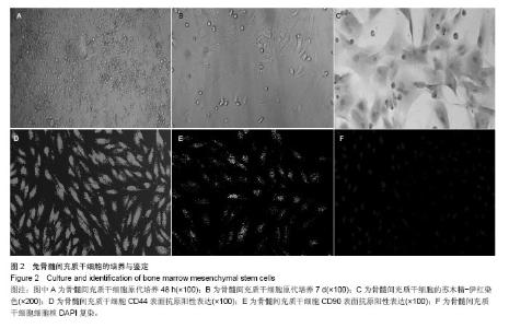
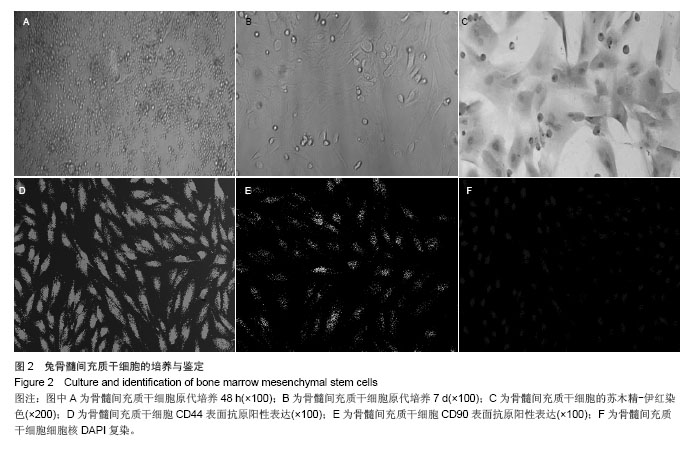
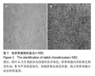
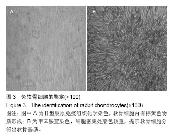
| [1]McNickle AG,Provencher MT,Cole BJ.Overview of existing cartilage repair technology.Sports Med Arthrose.2008;16(4): 196-201.
[2]漆白文,喻爱喜,祝少博,等.CS/PVA凝胶负载Ad-hTGF-1转染的BMSCs移植修复兔关节软骨缺损[J].中华显微外科,2011,34(3): 206.
[3]Garcia-Castro J,Trigueros C,Madrenas J,et al.Mesenchymal stem cells and their use as cell replacement therapy and disease modelling tool.J Cell Mol Med.2008;12(6B): 2552-2565.
[4]Kumar S,Chanda D,Ponnazhagan S.Therapeutic potential of genetically modified mesenchymal stem cells.Gene Ther. 2008;15(10):711-715.
[5]Andrews EM,Tsai SY,Johnson SC,et al.Human adult bone marrow-derived somatic cell therapy results in functional recovery and axonal plasticity following stroke in the rat.Exp Neurol.2008;211(2):588-592.
[6]Dulchavsky D,Gao X,Liu YB,et al.Bone marrow-derived stromalcells(BMSCs) interact with fibro-blats in accelerating wound healing.J Invest Surg.2008;21(5):270-279.
[7]Chen FH,Rousche KT,Tuan RS.Technology Insight:adult stem cells in cartilage regeneration and tissue engineering.Nat Clin Pract Rheumatol.2006;2(7):373-382.
[8]Iskovich S,Kaminitz A,Yafe MP,et al.Participation of adult bone marrow-derived stem cells in pancreatic regeneration: neogenesis versus endogenesis.Curr Stem Cell Res Ther. 2007; 2(4):272-279.
[9]Charbord P.Bone marrow mesenchymal stem cells:historical overview and concepts. Hum Gene Ther.2010;21(9): 1045-1056.
[10]Mauck RL,Byers BA,Yuan X,et al.Regulation of cartilaginous ECM gene transcription by chondrocytes and MSCs in 3D cuture in reponse to dynamic loading.Biomech Model Mechanobiol.2007;6(2):113-125.
[11]Liu K,Zhou GD,Liu W,et al.The dependence of in vivo stable ectopic chondrogenesis by human mesenchymal stem cells on ehondrngenie differentiation in vitro. Biomaterial. 2008; 29(14):2183-2192.
[12]Mo XT,Guo SC,Xie HQ,et al.Variations in the ratios of cocultured mesenchymal stem ceils and chondrocytes regulate the expression of car-tilaginous and OSSEOUS phenotype in alginate constructs.Bone.2009;45(1):42-51.
[13]Tsuchiya IL,Chen G,Ushida T,et al.The effect of coculture of chondrocytes witll mesenchymal stem eeHs on their cartilaginous phen-otype in vitro.Materials Sci Eng.2004; C24:391-396.
[14]陈刚,崔维顶,范卫民.软骨细胞和骨髓间质干细胞混合培养构建组织工程软骨的实验研究[J].中华骨科杂志,2010,30(7): 684-688.
[15]孙明林,吕丹,朱雷,等.共培养系统下软骨细胞对骨髓间质干细胞向软骨细胞分化的诱导作用[J].中华骨科杂志,2011,31(9): 976-982.
[16]O’Driscoll SW,Fitzsimmons JS.The role of periosteum in cartilage repair. Clin Orthop Relat Res.2001;(391 Suppl): 190-207.
[17]Pittenger MF.Mesenchymal stem cells from adult bone marrow.Methods Mol Biol. 2008;449:28-45.
[18]Ohgushi H,Calplan AI.Stem cell techonology and biocramics: from cell to gene engineering.J Biomed Mater Res.1999; 48(6):913.
[19]李德强,戴克戎,汤亭亭,等.适宜组织工程化骨构建的流体剪切力和物质转运速度的研究[J].中华骨科杂志,2011,31(5):545-547 |