Chinese Journal of Tissue Engineering Research ›› 2015, Vol. 19 ›› Issue (1): 78-84.doi: 10.3969/j.issn.2095-4344.2015.01.014
Previous Articles Next Articles
Granulosa cells with stem cell properties in the rat ovary
Liu Hui-ping, Lin Zheng-hua, Zhang Guo-min, Yu Rong, Liu Wen-e, Li Ling, Gu Xu-yu
- Prescription and Syndrome Research and Translational Medicine Laboratory of Hunan University of Traditional Chinese Medicine, Changsha 410208, Hunan Province, China
-
Revised:2014-12-07Online:2015-01-01Published:2015-01-01 -
Contact:Zhang Guo-min, M.D., Associate professor, Master’s supervisor, Prescription and Syndrome Research and Translational Medicine Laboratory of Hunan University of Traditional Chinese Medicine, Changsha 410208, Hunan Province, China -
About author:Liu Hui-ping, M.D., Associate professor, Prescription and Syndrome Research and Translational Medicine Laboratory of Hunan University of Traditional Chinese Medicine, Changsha 410208, Hunan Province, China -
Supported by:the National Natural Science Foundation of China, No. 81303123; China Postdoctoral Science Foundation, No. 2013T60770, 2012M511382; Specialized Research Fund for the Doctoral Program of Higher Education, No. 20114323120007; the Hunan Provincial Key Laboratory Foundation, No. 2014XNFZ06
CLC Number:
Cite this article
Liu Hui-ping, Lin Zheng-hua, Zhang Guo-min, Yu Rong, Liu Wen-e, Li Ling, Gu Xu-yu. Granulosa cells with stem cell properties in the rat ovary[J]. Chinese Journal of Tissue Engineering Research, 2015, 19(1): 78-84.
share this article
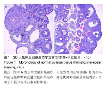
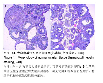
2.1 卵巢组织形态学观察结果 注射孕马血清促性腺激素大鼠的卵巢卵泡明显发育,体积增大,优势卵泡增多,有利于卵泡穿刺法提取成熟卵泡或次级卵泡中的颗粒细胞(图1A,B)。"
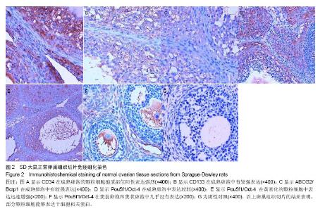
2.2 卵巢切片免疫组化CD34,CD133,ABCG2/Bcrp1,Pou5f1/Oct-4的表达 由石蜡切片免疫组化结果可见,CD34,CD133,ABCG2/Bcrp1,Pou5f1/Oct-4在颗粒细胞中均有所表达。其中,CD34阳性表达强烈(图2A),CD133和ABCG2/Bcrp1在成熟卵泡中均有较强表达(图2B,2C),而Pou5f1/Oct-4在成熟卵泡中表达较弱(图2D),窦前卵泡和窦状卵泡则几乎没有表达(图2F)。卵子排出后,黄素化的颗粒细胞Pou5f1/Oct-4表达迅速增强(图2E),退化至白体后,Pou5f1/Oct-4表达消失。 MIAS医学图像系统分析各图片灰度值见表2,灰度值越高表示阳性越强。结果表明,Pou5f1/Oct-4作为一种典型的干细胞标志物,在颗粒细胞中具有周期性表达的倾向。 值得注意的是,相比于阴性表达部位,无论是哪一种阳性表达的颗粒细胞都具有细胞排列疏松,细胞核扁平或呈三角形,细胞团块内部之间排列紊乱的特点。"

"

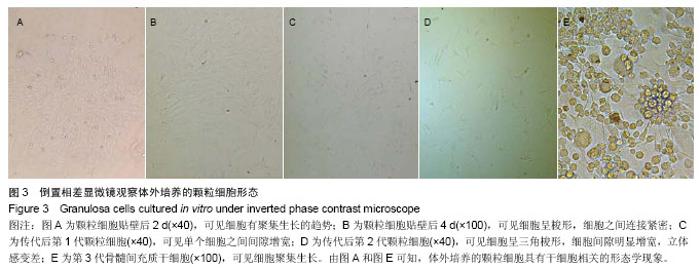
2.3 细胞增殖状态的观察 倒置相差显微镜下连续观察颗粒细胞的增殖情况,发现初提取的颗粒细胞在培养后半个小时左右即发生贴壁,其形态呈扁椭圆形,24 h后基本贴壁完全,48 h后贴壁牢固,细胞形态则由扁椭圆形转向三角形、梭形或多角棱形变化。此时可开始第1次换液,以除去尚未贴壁的其他细胞。 镜下观察发现,初提取细胞中,尚有少量悬浮红细胞,其形态圆,细胞中央透光度高,但换液后可及时除去,否则将影响颗粒细胞的生长状态。镜下观察可见,体外培养后数日颗粒细胞即可出现聚集生长的现象(图3A),但随着培养时间的延长,细胞的进一步增殖,聚集现象逐渐消失(图3B)。而聚集生长的现象为干细胞或肿瘤细胞所特有的一种现象,提示部分颗粒细胞具有类似于干细胞(图3E)或肿瘤细胞的生长特性。 随着传代次数的增加,单个颗粒细胞的体积逐渐变大,立体感变差,细胞间隙增宽,提示细胞之间接触抑制效应增强(图3C,3D)。"


2.4 细胞爬片免疫组化FSHR,CD44和C-Kit的表达 FSHR为特异性的表达于颗粒细胞胞浆的蛋白,免疫组化阳性结果为胞浆黄染,经苏木精复染以后,其细胞核呈蓝紫色(图4A)。根据阳性表达在细胞中的比例,测得原代提取的颗粒细胞纯度在95%以上。而CD44(图4B,阳性比例约60%)和C-Kit(图4C,阳性比例20%-40%)在部分细胞中有较强阳性表达,提示体外培养的颗粒细胞具有干细胞特性。"

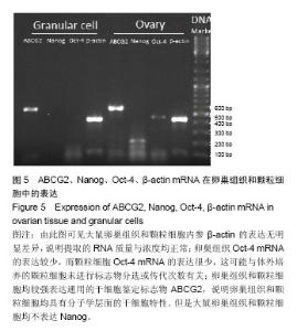
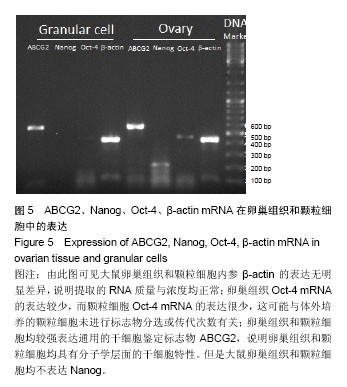
2.5 卵巢组织与体外培养的颗粒细胞ABCG2/Bcrp1,Pou5f1/Oct-4,Nanog基因的表达 卵巢组织与体外培养的颗粒细胞均表达β-actin,ABCG2/Bcrp1,Pou5f1/Oct-4基因,但是均不表达Nanog基因。其中,β-actin和ABCG2/Bcrp1表达均明显高于Pou5f1/Oct-4,而卵巢组织的表达量要明显高于颗粒细胞。但是体外培养的颗粒细胞中Pou5f1/Oct-4仅有极微弱的表达,见图5。 从卵巢组织Pou5f1/Oct-4的免疫组化结果来看,这种低表达的情况正好与Pou5f1/Oct-4的周期性表达特性相一致,因为卵泡穿刺法获取的细胞基本源于成熟卵泡中颗粒细胞。"
| [1] 龙海红.卵巢相关干细胞研究进展[J].现代生物医学进展,2012, 12(31): 6188-6191. [2] Bukovsky A, Svetlikova M, Caudle MR.Oogenesis in cultures derived from adult human ovaries.Reprod Biol Endocrinol. 2005;3:17. [3] Honda A, Hirose M, Hara K,et al.Isolation, characterization, and in vitro and in vivo differentiation of putative thecal stem cells.Proc Natl Acad Sci U S A. 2007;104(30):12389-12394. [4] Reya T, Morrison SJ, Clarke MF,et al.Stem cells, cancer, and cancer stem cells.Nature. 2001;414(6859):105-111. [5] Johnson J, Canning J, Kaneko T, et al.Germline stem cells and follicular renewal in the postnatal mammalian ovary. Nature. 2004;428(6979):145-150. [6] Kossowska-Tomaszczuk K, De Geyter C, De Geyter M, et al. The multipotency of luteinizing granulosa cells collected from mature ovarian follicles.Stem Cells. 2009;27(1):210- 219. [7] 陈毅斐,许程洁,王鑫泉,等.小鼠卵巢颗粒细胞适宜培养体系及激素干预的影响[J].中国组织工程研究,2012,16(2):282-286. [8] Dzafic E, Stimpfel M, Virant-Klun I.Plasticity of granulosa cells: on the crossroad of stemness and transdifferentiation potential.J Assist Reprod Genet. 2013;30(10):1255-1261. [9] Pisarska MD, Barlow G, Kuo FT.Minireview: roles of the forkhead transcription factor FOXL2 in granulosa cell biology and pathology.Endocrinology. 2011;152(4):1199-1208. [10] Dzafic E, Stimpfel M, Novakovic S,et al.Expression of mesenchymal stem cells-related genes and plasticity of aspirated follicular cells obtained from infertile women.Biomed Res Int. 2014;2014:508216. [11] Guo JQ, Gao X, Lin ZJ,et al.BMSCs reduce rat granulosa cell apoptosis induced by cisplatin and perimenopause.BMC Cell Biol. 2013;14:18. [12] Wang F, Wang L, Yao X,et al.Human amniotic epithelial cells can differentiate into granulosa cells and restore folliculogenesis in a mouse model of chemotherapy-induced premature ovarian failure.Stem Cell Res Ther. 2013;4(5): 124. [13] Chen HF, Jan PS, Kuo HC,et al.Granulosa cells and retinoic acid co-treatment enrich potential germ cells from manually selected Oct4-EGFP expressing human embryonic stem cells.Reprod Biomed Online. 2014;29(3):319-332. [14] Bhartiya D, Singh J.FSH-FSHR3-stem cells in ovary surface epithelium: basis for adult ovarian biology, failure, aging, and cancer.Reproduction. 2015;149(1):R35-R48. [15] Zhou S, Schuetz JD, Bunting KD,et al.The ABC transporter Bcrp1/ABCG2 is expressed in a wide variety of stem cells and is a molecular determinant of the side-population phenotype. Nat Med. 2001;7(9):1028-1034. [16] 何志旭,林槟,黄绍良,等.体外分化胚胎干细胞为造血干细胞重建造血功能的研究[J].中国病理生理学,2008,24(8):1610-1615. [17] 高芸,商雪莹,吕男男,等. FAT10过表达促进人乳头状甲状腺癌细胞抗凋亡及集落形成能力的研究[J].实用预防医学,2013, 20(6):647-649. [18] Riva F, Omes C, Bassani R,et al.In-vitro culture system for mesenchymal progenitor cells derived from waste human ovarian follicular fluid.Reprod Biomed Online. 2014;29(4): 457-469. [19] Maleki M, Ghanbarvand F, Reza Behvarz M,et al.Comparison of mesenchymal stem cell markers in multiple human adult stem cells.Int J Stem Cells. 2014;7(2):118-126. [20] Rad SM, Bamdad T, Sadeghizadeh M,et al.Transcription factor decoy against stem cells master regulators, Nanog and Oct-4: a possible approach for differentiation therapy.Tumour Biol. 2014 Dec 3. [Epub ahead of print] [21] Mattioli M, Gloria A, Turriani M,et al.Osteo-regenerative potential of ovarian granulosa cells: an in vitro and in vivo study.Theriogenology. 2012;77(7):1425-1437. [22] Yuan J, Zhang D, Wang L,et al.No evidence for neo-oogenesis may link to ovarian senescence in adult monkey.Stem Cells. 2013;31(11):2538-2550. [23] 王研,赵晓娥,杨培先,等.小鼠卵巢颗粒细胞的体外培养[J]. 西北农林科技大学学报,2007,35(8):11-14. [24] 陈志钊,蔡凯晋. 血清干细胞因子(SCF)及其受体c-kit的研究进展[J].中外医疗,2013,32(13):186-187. [25] Parrott JA, Skinner MK.Kit-ligand/stem cell factor induces primordial follicle development and initiates folliculogenesis.Endocrinology. 1999;140(9):4262-4271. [26] 乔威,冉峰,刘长建.人外周血内皮祖细胞的分离、培养及鉴定[J].中国组织工程研究,2013,17(36):6508-6514. [27] 曹青,王飞,林继先,等. 骨髓间充质干细胞对CD117+心脏干细胞分化过程中转化生长因子βⅢ型受体表达的影响[J].中国组织工程研究与临床康复,2009,13(36):7057-7062. [28] Kossowska-Tomaszczuk K, De Geyter C.Cells with stem cell characteristics in somatic compartments of the ovary.Biomed Res Int. 2013;2013:310859. [29] Varras M, Griva T, Kalles V,et al.Markers of stem cells in human ovarian granulosa cells: is there a clinical significance in ART.J Ovarian Res. 2012;5(1):36. |
| [1] | Lin Qingfan, Xie Yixin, Chen Wanqing, Ye Zhenzhong, Chen Youfang. Human placenta-derived mesenchymal stem cell conditioned medium can upregulate BeWo cell viability and zonula occludens expression under hypoxia [J]. Chinese Journal of Tissue Engineering Research, 2021, 25(在线): 4970-4975. |
| [2] | Pu Rui, Chen Ziyang, Yuan Lingyan. Characteristics and effects of exosomes from different cell sources in cardioprotection [J]. Chinese Journal of Tissue Engineering Research, 2021, 25(在线): 1-. |
| [3] | Zhang Xiumei, Zhai Yunkai, Zhao Jie, Zhao Meng. Research hotspots of organoid models in recent 10 years: a search in domestic and foreign databases [J]. Chinese Journal of Tissue Engineering Research, 2021, 25(8): 1249-1255. |
| [4] | Hou Jingying, Yu Menglei, Guo Tianzhu, Long Huibao, Wu Hao. Hypoxia preconditioning promotes bone marrow mesenchymal stem cells survival and vascularization through the activation of HIF-1α/MALAT1/VEGFA pathway [J]. Chinese Journal of Tissue Engineering Research, 2021, 25(7): 985-990. |
| [5] | Shi Yangyang, Qin Yingfei, Wu Fuling, He Xiao, Zhang Xuejing. Pretreatment of placental mesenchymal stem cells to prevent bronchiolitis in mice [J]. Chinese Journal of Tissue Engineering Research, 2021, 25(7): 991-995. |
| [6] | Liang Xueqi, Guo Lijiao, Chen Hejie, Wu Jie, Sun Yaqi, Xing Zhikun, Zou Hailiang, Chen Xueling, Wu Xiangwei. Alveolar echinococcosis protoscolices inhibits the differentiation of bone marrow mesenchymal stem cells into fibroblasts [J]. Chinese Journal of Tissue Engineering Research, 2021, 25(7): 996-1001. |
| [7] | Fan Quanbao, Luo Huina, Wang Bingyun, Chen Shengfeng, Cui Lianxu, Jiang Wenkang, Zhao Mingming, Wang Jingjing, Luo Dongzhang, Chen Zhisheng, Bai Yinshan, Liu Canying, Zhang Hui. Biological characteristics of canine adipose-derived mesenchymal stem cells cultured in hypoxia [J]. Chinese Journal of Tissue Engineering Research, 2021, 25(7): 1002-1007. |
| [8] | Geng Yao, Yin Zhiliang, Li Xingping, Xiao Dongqin, Hou Weiguang. Role of hsa-miRNA-223-3p in regulating osteogenic differentiation of human bone marrow mesenchymal stem cells [J]. Chinese Journal of Tissue Engineering Research, 2021, 25(7): 1008-1013. |
| [9] | Lun Zhigang, Jin Jing, Wang Tianyan, Li Aimin. Effect of peroxiredoxin 6 on proliferation and differentiation of bone marrow mesenchymal stem cells into neural lineage in vitro [J]. Chinese Journal of Tissue Engineering Research, 2021, 25(7): 1014-1018. |
| [10] | Zhu Xuefen, Huang Cheng, Ding Jian, Dai Yongping, Liu Yuanbing, Le Lixiang, Wang Liangliang, Yang Jiandong. Mechanism of bone marrow mesenchymal stem cells differentiation into functional neurons induced by glial cell line derived neurotrophic factor [J]. Chinese Journal of Tissue Engineering Research, 2021, 25(7): 1019-1025. |
| [11] | Duan Liyun, Cao Xiaocang. Human placenta mesenchymal stem cells-derived extracellular vesicles regulate collagen deposition in intestinal mucosa of mice with colitis [J]. Chinese Journal of Tissue Engineering Research, 2021, 25(7): 1026-1031. |
| [12] | Pei Lili, Sun Guicai, Wang Di. Salvianolic acid B inhibits oxidative damage of bone marrow mesenchymal stem cells and promotes differentiation into cardiomyocytes [J]. Chinese Journal of Tissue Engineering Research, 2021, 25(7): 1032-1036. |
| [13] | Guan Qian, Luan Zuo, Ye Dou, Yang Yinxiang, Wang Zhaoyan, Wang Qian, Yao Ruiqin. Morphological changes in human oligodendrocyte progenitor cells during passage [J]. Chinese Journal of Tissue Engineering Research, 2021, 25(7): 1045-1049. |
| [14] | Wang Zhengdong, Huang Na, Chen Jingxian, Zheng Zuobing, Hu Xinyu, Li Mei, Su Xiao, Su Xuesen, Yan Nan. Inhibitory effects of sodium butyrate on microglial activation and expression of inflammatory factors induced by fluorosis [J]. Chinese Journal of Tissue Engineering Research, 2021, 25(7): 1075-1080. |
| [15] | Wang Xianyao, Guan Yalin, Liu Zhongshan. Strategies for improving the therapeutic efficacy of mesenchymal stem cells in the treatment of nonhealing wounds [J]. Chinese Journal of Tissue Engineering Research, 2021, 25(7): 1081-1087. |
| Viewed | ||||||
|
Full text |
|
|||||
|
Abstract |
|
|||||