Chinese Journal of Tissue Engineering Research ›› 2015, Vol. 19 ›› Issue (1): 67-71.doi: 10.3969/j.issn.2095-4344.2015.01.012

Previous Articles     Next Articles

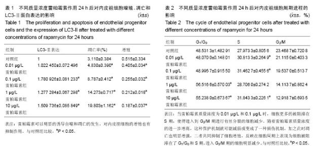
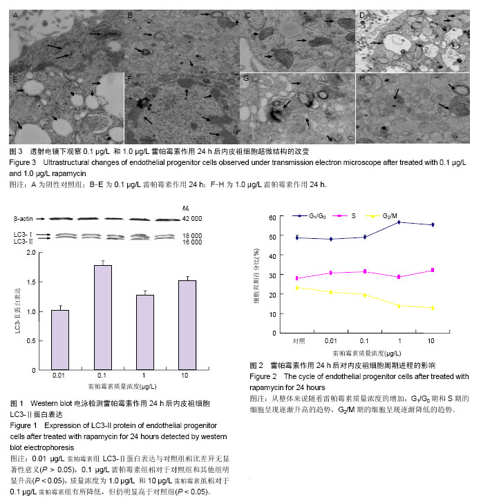
How does autophagy activation affect the apoptosis, proliferation and cycle of endothelial progenitor cells in rats?

Liu Hui1, Li Xiao-qiang2, Zhu Ren-da2, Meng Qing-you2, Lu Hui-jun1   

  1. 1Department of Vascular Surgery, Wuxi People’s Hospital Affiliated to Nanjing Medical University, Wuxi 214023, Jiangsu Province, China
    2The Second Affiliated Hospital of Soochow University, Suzhou 215004, Jiangsu Province, China
  • Revised:2014-10-20 Online:2015-01-01 Published:2015-01-01
  • Contact: Li Xiao-qiang, Professor, Doctoral supervisor, Chief physician, the Second Affiliated Hospital of Soochow University, Suzhou 215004, Jiangsu Province, China
  • About author:Liu Hui, Master, Attending physician, Department of Vascular Surgery, Wuxi People’s Hospital Affiliated to Nanjing Medical University, Wuxi 214023, Jiangsu Province, China
  • Supported by:

    the National Natural Science Foundation of China, No. 30972941

Abstract:

BACKGROUND: Previous studies have reported that rapamycin can affect the proliferation, migration and adhesion abilities of endothelial progenitor cells, but there is no report on the effect of autophagy, as well as the interaction between autophagy and apoptosis.    
OBJECTIVE: To observe the effect of rapamycin activated autophagy activation on the proliferation, apoptosis, and cycle of endothelial progenitor cells. 
METHODS: Density gradient centrifugation was used to obtain mononuclear cells from bone marrow, and the mononuclear cells were inoculated on human fibronectin-coated culture plate.Then after cultured for 7 days the adherent cells collected were the endothelial progenitor cells. Different concentrations of rapamycin (0.01, 0.1, 1 and 10 μg/L) were added and cultured for 24 hours. Western blot was used to detect the LC3-II protein expression and monitor the induction of autophagy, flow cytometry was used to observe the cell cycle progression and apoptosis changes, and methylthiazolyldiphenyl-tetrazolium bromide colorimetric assay was used to observe the proliferation ability. Meanwhile, the ultrastructural changes were observed under transmission electron microscope. 
RESULTS AND CONCLUSION: Compared with the control group, there was no significant increasing of LC3-II protein expression of endothelial progenitor cells in 0.01 μg/L rapamycin group, and the LC3-II protein expression was in the high level. The LC3-II protein expression in the 1 μg/L and 10 μg/L rapamycin groups was higher than that in the control group, but lower than that in the 0.01 μg/L rapamycin group, which indicated that autophagy was particularly active when the concentration of rapamycin was 0.01 μg/L. The apoptosis of endothelial progenitor cells was increased with the increasing of concentration of rapamycin, and the proliferation rate was decreased with the increasing of concentration of rapamycin. The results indicate that activation of autophagy by bapamycin can promote the cell apoptosis, change the cell cycle significantly, and can inhibit the proliferation of endothelial progenitor cells.


中国组织工程研究杂志出版内容重点:干细胞;骨髓干细胞;造血干细胞;脂肪干细胞;肿瘤干细胞;胚胎干细胞;脐带脐血干细胞;干细胞诱导;干细胞分化;组织工程


全文链接:

Key words: Sirolimus, Autophagy, Cell cycle, Apoptosis, Cell Proliferation

CLC Number: