Chinese Journal of Tissue Engineering Research ›› 2014, Vol. 18 ›› Issue (48): 7844-7848.doi: 10.3969/j.issn.2095-4344.2014.48.024
Previous Articles Next Articles
Application of three-dimensional models constructed using virtual simulation technique in the vertebral metastatic tumor
Liu Deng-jun1, He Xiao-bing1, Wang Ming-gui1, Li Zheng-yan1, Li Qi2, Lin Li-jun2
- 1Department of Orthopedics, Chongqing Fuling Central Hospital, Chongqing 408000, China; 2Department of Orthopedics, Zhujiang Hospital of Southern Medical University, Guangzhou 510282, Guangdong Province, China
-
Online:2014-11-26Published:2014-11-26 -
Contact:Liu Deng-jun, Department of Orthopedics, Chongqing Fuling Central Hospital, Chongqing 408000, China -
About author:Liu Deng-jun, Attending physician, Department of Orthopedics, Chongqing Fuling Central Hospital, Chongqing 408000, China
CLC Number:
Cite this article
Liu Deng-jun, He Xiao-bing, Wang Ming-gui, Li Zheng-yan, Li Qi, Lin Li-jun. Application of three-dimensional models constructed using virtual simulation technique in the vertebral metastatic tumor[J]. Chinese Journal of Tissue Engineering Research, 2014, 18(48): 7844-7848.
share this article

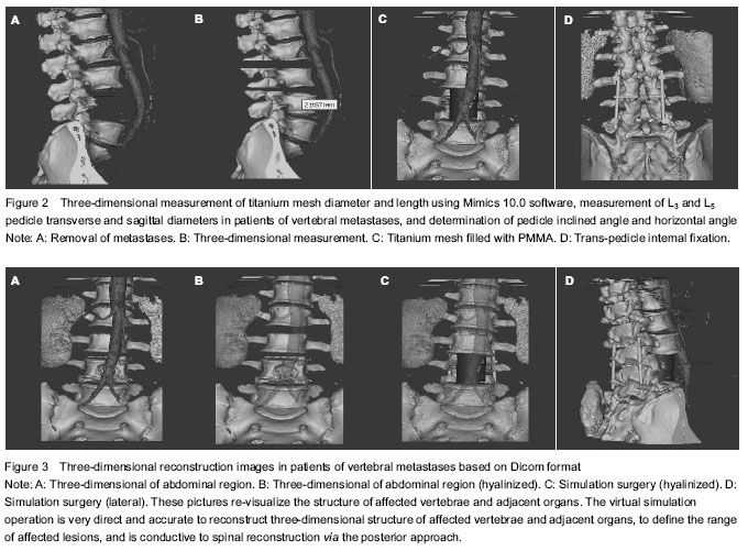
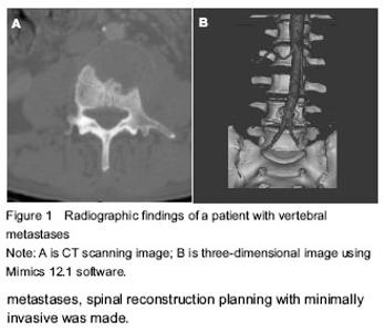
Quantitative analysis of subjects A male patient was involved in the final analysis. Three-dimensional reconstruction of the vertebrae and adjacent organs Three-dimensional reconstruction images of the vertebrae and adjacent organs were acquired from all directions and multiple angles (Figure 3A, B). After smoothing with the free-form modeling system, its structure became clearer; and all of the structures, including normal vertebrea, vertebral metastases (L4), abdominal aorta and kidneys were reconstructed in three dimensions. The three-dimensional of vertebral metastases could be amplified, rotated, and hyalinized to clarify the anatomic character of structure with omnidirectional, multiple-angle, and multilevel views. After we localized the vertebral metastases, an optimal surgical option was chosen. We obtained morphometric measurements of the apex and the axis of the pedicles of lumbal vertebrae L4 to L5, the tilt angle, chose suitable insert point of the screw, the depth and the diameter of the screw (Figure 3C, D)."
"

| [1] Borani S, Weinstein JN, Biagini R. Primary bone tumor of thespine. Spine. 1997;22(9):1036-1044. [2] Boriani S, Bandiera S, Biagini R, et al. Staging and treatment of primary tumors of the spine. Current Opinion in Orthopedics. 1999;10(2):93-100. [3] Doh JW, Halliday AL, Baldwin NG, et al. Spinal stabilization by using crossed-SCrew anterior-posterior fixation after multisegmental total Spondylectomy for thoracic chondrosarcoma: case report. J Neurosury. 2001;94(Suppl): 279-283. [4] Tomita K, Kawahara N, Kobayashi T, et al. Surgical strategy for spinal metastases. Spine. 2001;26:298-306. [5] Abe E, Kobayashi T, Murai H, et al. Total spondylectomy for primary malignant, aggressive benign, and solitary metastatic bone tumor of the thoracolumbar spine. J Spine Disorder. 2001;14:237-246. [6] Wang Q, Song Y, Zhuang H, et al. Robotic stereotactic irradiation and reirradiation for spinal metastases: safety and efficacy assessment. Chin Med J (Engl). 2014;127(2): 232-238. [7] Bohinski RJ, Rhines L. Principles and techniques of enblocvertebrectomy for bone tumors of the thoracolumbar spine: an overview. Neurosurg Focus. 2003;15(5):1-6. [8] Amiot LP, Lang K, Putzier M, et al. Comparative results between conventional and computer-assisted pedicle screw installation in the thoracic, lumbar and sacral spine. Spine. 2000;25:606-614. [9] Dickman CA, Yahiro MA, Lu HTC, et al. Surgical treatment alternatives for fixation of unstable fractures of the thoracic and lumbar spine. Spine. 1994;19(Suppl):2266-2273. [10] Ebraheim NA, Xu R, Ahmad M, et al. Projection of the thoracic pedicle and its morphometric analysis. Spine.1997; 22:233-238. [11] Esses SI, Sachs BL, Dreyzin V. Complications associated with the technique of pedicle screw fixation. Spine. 1993;18: 2231-2239. [12] Laine T, Lund T, Ylikoski M, et al. Accuracy of pedicle screw insertion with and without computer assistance. A randomised controlled clinical study in 100 consecutive patients. Eur Spine J. 2000;9:235-240. [13] Rampersaud YR, Foley KT, Shen AC, et al. Radiation exposure to the spine surgeon during fluoroscopically assisted pedicle screw insertion. Spine. 2000;25:2537- 2645. [14] Rampersaud YR, Simon DA, Foley KT. Accuracy requirements for image-guided spinal pedicle screw placement. Spine. 2001;26:352-359. [15] Robertson PA, Stewart NR. The radiologic anatomy of the lumbar and lumbosacral pedicles. Spine. 2000;25: 709-715. [16] Schlenzka D, Laine T, Lund T. Computer-assisted spine surgery. Eur Spine J. 2000;9(Suppl 1):57-64. [17] Schulze CJ, Munzinger E, Weber U. Clinical relevance of accuracy of pedicle screw placement. A computer tomographic- supported analysis. Spine. 1998;23:2215- 2220. [18] Yuan HA, Garfin SR, Dickman CA, et al. A historical cohort study of pedicle screw fixation in thoracic, lumbar, and sacral spinal fusions. Spine. 1994;(Suppl):2279-2296. [19] Vahldiek MJ, Panjabi MM. Stability potential of spinal instrumentations in tumor vertebral body replacement surgery. Spine. 1998;23:543-550. [20] Abe Y, Sato S, Kato K, et al. A novel 3D guidance system using augmented reality for percutaneous vertebroplasty: technical note. J Neurosurg Spine. 2013;19(4):492-501. [21] Cabrilo I, Bijlenga P, Schaller K. Augmented reality in the surgery of cerebral arteriovenous malformations: technique assessment and considerations. Acta Neurochir (Wien). 2014;156(9):1769-1774. [22] Shi J, Xia J, Wei Y, et al. Three-dimensional virtual reality simulation of periarticular tumors using Dextroscope reconstruction and simulated surgery: a preliminary 10-case study. Med Sci Monit. 2014;20:1043-1050. [23] Hua H, Javidi B. A 3D integral imaging optical see-through head-mounted display. Opt Express. 2014;22(11):13484- 13491. [24] Shi J, Xia J, Wei Y, et al. Three-dimensional virtual reality simulation of periarticular tumors using Dextroscope reconstruction and simulated surgery: a preliminary 10 case study. Acta Orthop Belg. 2014;80(1):132-138. [25] Hänel C, Pieperhoff P, Hentschel B, et al. Interactive 3D visualization of structural changes in the brain of a person with corticobasal syndrome. Front Neuroinform. 2014;8:42. [26] Soler L, Nicolau S, Pessaux P, et al. Real-time 3D image reconstruction guidance in liver resection surgery. Hepatobiliary Surg Nutr. 2014;3(2):73-81. [27] Eve EJ, Koo S, Alshihri AA, et al. Performance of dental students versus prosthodontics residents on a 3D immersive haptic simulator. J Dent Educ. 2014;78(4): 630-637. [28] Pessaux P, Diana M, Soler L, et al. Robotic duodenopancreatectomy assisted with augmented reality and real-time fluorescence guidance. Surg Endosc. 2014; 28(8):2493-2498. [29] Di Somma A, de Notaris M, Stagno V, et al. Extended endoscopic endonasal approaches for cerebral aneurysms: anatomical, virtual reality and morphometric study. Biomed Res Int. 2014;2014:703792. [30] Zhou Y, Bailey J, Ioannou I, et al. Constructive real time feedback for a temporal bone simulator. Med Image Comput Comput Assist Interv. 2013;16(Pt 3):315-322. [31] Duarte IC, Ferreira C, Marques J, et al. Anterior/posterior competitive deactivation/activation dichotomy in the human hippocampus as revealed by a 3D navigation task. PLoS One. 2014;9(1):e86213. [32] Uchida M. Recent advances in 3D computed tomography techniques for simulation and navigation in hepatobiliary pancreatic surgery. J Hepatobiliary Pancreat Sci. 2014; 21(4):239-245. [33] Baken L, Rousian M, Koning AH, et al. First-trimester detection of surface abnormalities: A comparison of 2- and 3-dimensional ultrasound and 3-dimensional virtual reality ultrasound. Reprod Sci. 2014;21(8):993-999. [34] Lin Q, Xu Z, Li B, Baucom R, et al. Immersive virtual reality for visualization of abdominal CT. Proc SPIE. 2013;8673. [35] Hong K, Yeom J, Jang C, et al. Full-color lens-array holographic optical element for three-dimensional optical see-through augmented reality. Opt Lett. 2014;39(1): 127-130. [36] Gong RH, Güler Ö, Kürklüoglu M, et al. Interactive initialization of 2D/3D rigid registration. Med Phys. 2013; 40(12):121911. [37] Liu YG, Zuo LX, Pei GX, et al. Establishment of Schatzker classification digital models of tibial plateau fractures and its application on virtual surgery. Zhonghua Yi Xue Za Zhi. 2013; 93(31):2478-2482. [38] Djukic T, Mandic V, Filipovic N. Virtual reality aided visualization of fluid flow simulations with application in medical education and diagnostics. Comput Biol Med. 2013; 43(12):2046-2052. [39] Rassweiler J, Rassweiler MC, Müller M, et al. Surgical navigation in urology: european perspective. Curr Opin Urol. 2014;24(1):81-97. [40] Qi S, Yan Y, Li R, et al. The impact of active versus passive use of 3D technology: a study of dental students at Wuhan University, China. J Dent Educ. 2013;77(11):1536-1542. [41] Berry F, Aider OA, Mosnier J. A visual servoing-based method for ProCam systems calibration. Sensors (Basel). 2013;13(10):13318-13323. [42] Jackson B, Lau TY, Schroeder D, et al. A lightweight tangible 3D interface for interactive visualization of thin fiber structures. IEEE Trans Vis Comput Graph. 2013;19(12): 2802-2809. [43] Hsu WH, Zhang Y, Ma KL. A multi-criteria approach to camera motion design for volume data animation. IEEE Trans Vis Comput Graph. 2013;19(12):2792-2801. [44] Zinser MJ, Mischkowski RA, Dreiseidler T, et al. Computer-assisted orthognathic surgery: waferless maxillary positioning, versatility, and accuracy of an image-guided visualisation display. Br J Oral Maxillofac Surg. 2013;51(8):827-833. [45] Dores AR, Almeida I, Barbosa F, et al. Effects of emotional valence and three-dimensionality of visual stimuli on brain activation: an fMRI study. NeuroRehabilitation. 2013;33(4):505-512. |
| [1] | Zhang Tongtong, Wang Zhonghua, Wen Jie, Song Yuxin, Liu Lin. Application of three-dimensional printing model in surgical resection and reconstruction of cervical tumor [J]. Chinese Journal of Tissue Engineering Research, 2021, 25(9): 1335-1339. |
| [2] | Zeng Yanhua, Hao Yanlei. In vitro culture and purification of Schwann cells: a systematic review [J]. Chinese Journal of Tissue Engineering Research, 2021, 25(7): 1135-1141. |
| [3] | Song Chengjie, Chang Hengrui, Shi Mingxin, Meng Xianzhong. Research progress in biomechanical stability of lateral lumbar interbody fusion [J]. Chinese Journal of Tissue Engineering Research, 2021, 25(6): 923-928. |
| [4] | Xu Dongzi, Zhang Ting, Ouyang Zhaolian. The global competitive situation of cardiac tissue engineering based on patent analysis [J]. Chinese Journal of Tissue Engineering Research, 2021, 25(5): 807-812. |
| [5] | Wu Zijian, Hu Zhaoduan, Xie Youqiong, Wang Feng, Li Jia, Li Bocun, Cai Guowei, Peng Rui. Three-dimensional printing technology and bone tissue engineering research: literature metrology and visual analysis of research hotspots [J]. Chinese Journal of Tissue Engineering Research, 2021, 25(4): 564-569. |
| [6] | Chang Wenliao, Zhao Jie, Sun Xiaoliang, Wang Kun, Wu Guofeng, Zhou Jian, Li Shuxiang, Sun Han. Material selection, theoretical design and biomimetic function of artificial periosteum [J]. Chinese Journal of Tissue Engineering Research, 2021, 25(4): 600-606. |
| [7] | Liu Fei, Cui Yutao, Liu He. Advantages and problems of local antibiotic delivery system in the treatment of osteomyelitis [J]. Chinese Journal of Tissue Engineering Research, 2021, 25(4): 614-620. |
| [8] | Li Xiaozhuang, Duan Hao, Wang Weizhou, Tang Zhihong, Wang Yanghao, He Fei. Application of bone tissue engineering materials in the treatment of bone defect diseases in vivo [J]. Chinese Journal of Tissue Engineering Research, 2021, 25(4): 626-631. |
| [9] | Zhang Zhenkun, Li Zhe, Li Ya, Wang Yingying, Wang Yaping, Zhou Xinkui, Ma Shanshan, Guan Fangxia. Application of alginate based hydrogels/dressings in wound healing: sustained, dynamic and sequential release [J]. Chinese Journal of Tissue Engineering Research, 2021, 25(4): 638-643. |
| [10] | Chen Jiana, Qiu Yanling, Nie Minhai, Liu Xuqian. Tissue engineering scaffolds in repairing oral and maxillofacial soft tissue defects [J]. Chinese Journal of Tissue Engineering Research, 2021, 25(4): 644-650. |
| [11] | Xing Hao, Zhang Yonghong, Wang Dong. Advantages and disadvantages of repairing large-segment bone defect [J]. Chinese Journal of Tissue Engineering Research, 2021, 25(3): 426-430. |
| [12] | Yang Qin, Zhou Honghai, Chen Longhao, Zhong Zhong, Xu Yigao, Huang Zhaozhi. Research status and development trend of pelvic reconstruction techniques: a bibliometric and visual analysis [J]. Chinese Journal of Tissue Engineering Research, 2021, 25(23): 3718-3724. |
| [13] | Chen Siqi, Xian Debin, Xu Rongsheng, Qin Zhongjie, Zhang Lei, Xia Delin. Effects of bone marrow mesenchymal stem cells and human umbilical vein endothelial cells combined with hydroxyapatite-tricalcium phosphate scaffolds on early angiogenesis in skull defect repair in rats [J]. Chinese Journal of Tissue Engineering Research, 2021, 25(22): 3458-3465. |
| [14] | Wang Hao, Chen Mingxue, Li Junkang, Luo Xujiang, Peng Liqing, Li Huo, Huang Bo, Tian Guangzhao, Liu Shuyun, Sui Xiang, Huang Jingxiang, Guo Quanyi, Lu Xiaobo. Decellularized porcine skin matrix for tissue-engineered meniscus scaffold [J]. Chinese Journal of Tissue Engineering Research, 2021, 25(22): 3473-3478. |
| [15] | Mo Jianling, He Shaoru, Feng Bowen, Jian Minqiao, Zhang Xiaohui, Liu Caisheng, Liang Yijing, Liu Yumei, Chen Liang, Zhou Haiyu, Liu Yanhui. Forming prevascularized cell sheets and the expression of angiogenesis-related factors [J]. Chinese Journal of Tissue Engineering Research, 2021, 25(22): 3479-3486. |
| Viewed | ||||||
|
Full text |
|
|||||
|
Abstract |
|
|||||