Chinese Journal of Tissue Engineering Research ›› 2014, Vol. 18 ›› Issue (39): 6323-6328.doi: 10.3969/j.issn.2095-4344.2014.39.016
Previous Articles Next Articles
Construction of tissue engineered adipose using human adipose stem cells with chitosan-modified silk fibroin
Kang Ting, Wang Gang, Liu Yi, Liu Gang-qiang
- PLA Center for Military Burns and Plastic Surgery, Lanzhou General Hospital, Lanzhou Command of Chinese PLA, Lanzhou 730050, Gansu Province, China
-
Online:2014-09-17Published:2014-09-17 -
Contact:Liu Yi, Chief physician, Professor, Master’s supervisor, PLA Center for Military Burns and Plastic Surgery, Lanzhou General Hospital, Lanzhou Command of Chinese PLA, Lanzhou 730050, Gansu Province, China -
About author:Kang Ting, Studying for master’s degree, PLA Center for Military Burns and Plastic Surgery, Lanzhou General Hospital, Lanzhou Command of Chinese PLA, Lanzhou 730050, Gansu Province, China -
Supported by:Key Project of Military Medical Research during the Twelfth Five-Year Plan, No. BWS11C061
CLC Number:
Cite this article
Kang Ting, Wang Gang, Liu Yi, Liu Gang-qiang . Construction of tissue engineered adipose using human adipose stem cells with chitosan-modified silk fibroin[J]. Chinese Journal of Tissue Engineering Research, 2014, 18(39): 6323-6328.
share this article
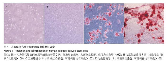
2.1 人脂肪间充质干细胞的培养及鉴定 细胞生长良好,扩增时间为四五天,倒置显微镜下可见细胞形态规则,大部分呈梭形,也可为多角形,达到80%融合时细胞可呈 “漩涡”状排列(图1A,B)。 鉴定:①成脂诱导后油红O染色:胞质内脂滴被染成红色,可清晰的反应脂化程度(图1C)。②成骨诱导后茜素红染色:可见钙化结节形成(图1D)。③表面分子鉴定: CD34、CD45为阴性;CD44、CD90为阳性[12](图2)。"

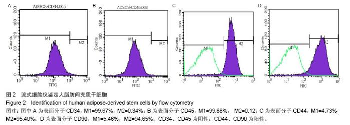
"


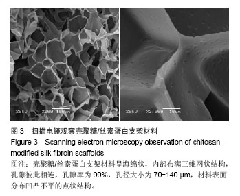
2.2 壳聚糖/丝素蛋白支架的扫描电镜观察 壳聚糖/丝素蛋白支架材料呈海绵状,内部布满三维网状结构,孔隙彼此相连,孔隙率为90%,孔径大小为70-140 μm(图3A),材料表面分布凹凸不平的点状结构(图3B)。"

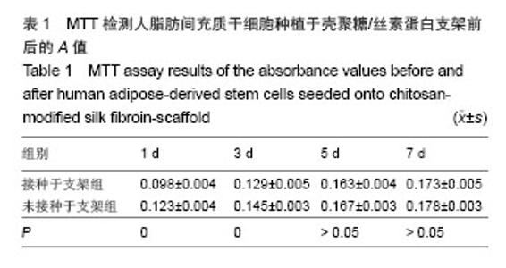
2.3 MTT法检测细胞在壳聚糖/丝素蛋白上的黏附与增殖 MTT结果显示,细胞接种后8 h,两组的A值均随时间逐渐增加,且两组之间的差距逐渐缩小(图4A)。接种后前3 d,实验组的A值显著小于对照组(P=0),第5天以后,实验组与对照组的A值差异无显著性意义(P > 0.05,图4B,表1)。"
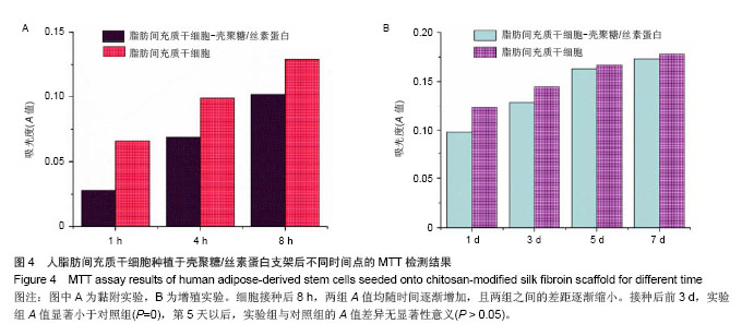
"

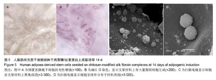
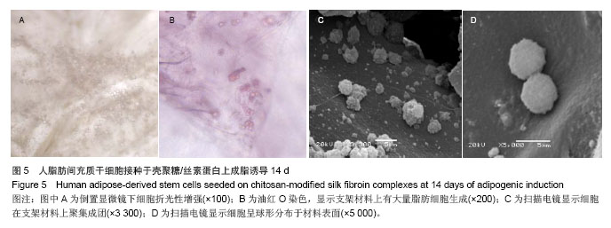
2.4 人脂肪间充质干细胞成脂诱导后在壳聚糖/丝素蛋白支架材料上的生长状况 实验组成脂诱导14 d后,光镜可见细胞折光性增强(图5A),油红O染色显示支架材料上有大量脂肪细胞生成(图5B),扫描电镜可见细胞在支架材料上聚集成团(图5C),呈球形分布于材料表面(图5D)。对照组中未见折光性强脂滴形成,油红O染色呈阴性。"
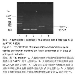
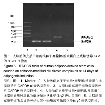
2.5 RT-PCR检测过氧化物酶增殖物活化受体γ2基因 RT-PCR结果显示,实验组细胞-支架材料复合物表达成脂特异性基因过氧化物酶增殖物活化受体γ2,而对照组过氧化物酶增殖物活化受体γ2未表达(图6),表明人脂肪间充质干细胞种植于壳聚糖/丝素蛋白后,可以体外成脂诱导分化为成熟脂肪细胞。"
| [1]Ma PX.Biomimetic materials for tissue engineering.Adv Drug Deliv Rev.2008;60(2):184-198.
[2]Metcalfe AD,Ferguson MWJ.Bioengineering skin using mechanisms of regeneration and repair. Biomaterials. 2007; 28(34):5100-5113.
[3]Luangbudnark W,Viyoch J,Laupattarakasem W,et al. Properties and biocompatibility of chitosan and silk fibroin blend films for application in skin tissue engineering.Scientific World J.2012;2012:697201.
[4]Locke M,Windsor J,Dunbar PR.Human adipose‐derived stem cells: isolation, characterization and applications in surgery.ANZ J Surg.2009;79(4):235-244.
[5]Shiffman MA,Mirrafatti S.Fat transfer techniques: the effect of harvest and transfer methods on adipocyte viability and review of the literature. Dermatol Surg.2001;27(9):819-862.
[6]Itoi Y,Takatori M,Hyakusoku H,et al.Comparison of readily available scaffolds for adipose tissue engineering using adipose-derived stem cells. J Plast Reconstr Aesthet Surg. 2010;63(5):858-864.
[7]Kim EH,Heo CY.Current applications of adipose-derived stem cells and their future perspectives.World J Stem Cells. 2014; 6:65-68.
[8]Mizuno H,Tobita M,Uysal AC.Adipose-Derived Stem Cells as a Novel Tool for Future Regenerative Medicine.Stem Cells. 2014;12:165-174.
[9]陶忠芬,蔡永国,杨仕明,等.树突状细胞扫描电镜样本制备方法[J].第三军医大学学报,2005,27(12):1299-1300.
[10]朱晓琳,徐丽丽,杨乃龙.尿酸抑制人骨髓间充质干细胞的成脂分化[J].中国组织工程研究,2012,16(36):6669-6673.
[11]唐军,刘毅,徐斌.胰岛素基因转染的人脐带间充质干细胞与丝素蛋白支架构建组织工程脂肪的研究[J].中华医学美学美容杂志, 2012,18(4):277-281.
[12]邢昕,吕蓉,李素萍,等.体外分离培养人源性脂肪间充质干细胞及其鉴定[J].中国输血杂志,2013,26(8):708-712.
[13]Dyakonov T,Yang CH,Bush D,et al.Design and characterization of a silk-fibroin-based drug delivery platform using naproxen as a model drug.J Drug Deliv. 2012;2012: 490514.
[14]Cao Y,Wang B.Biodegradation of silk biomaterials.Int J Mol Sci. 2009;10(4):1514-1224.
[15]Ju HW,Sheikh FA,Moon BM,et al.Fabrication of poly (lactic-co-glycolic acid) scaffolds containing silk fibroin scaffolds for tissue engineering applications. J Biomed Mater Res A. 2013 Sep 11. doi: 10.1002/jbm.a.34947. [Epub ahead of print]
[16]王宏昕,李敏.丝素蛋白作为组织工程生物材料的研究进展[J].中国修复重建外科杂志,2008,28(2):192-194.
[17]Li SL,Liu Y,Hui L.Construction of engineering adipose‐like tissue in vivo utilizing human insulin gene‐modified umbilical cord mesenchymal stromal cells with silk fibroin 3D scaffolds. J Tissue Eng Regen Med.2013 Mar 19. doi: 10.1002/term. 1695. [Epub ahead of print]
[18]Bhardwaj N,Kundu SC.Silk fibroin protein and chitosan polyelectrolyte complex porous scaffolds for tissue engineering applications.Carbohydr Polym. 2011;85(2): 325-333.
[19]Bhardwaj N,Nguyen QT,Chen AC,et al.Potential of 3-D tissue constructs engineered from bovine chondrocytes/silk fibroin-chitosan for in vitro cartilage tissue engineering. Biomaterials.2011;32(25):5773-5781.
[20]杨久林,谢红国,于炜婷,等.组织工程用壳聚糖研究进展[J].功能材料,2013,44(11):1521-1525.
[21]Denga JH,Zhonga DC,Luoa XZ,et al.Syntheses, crystal structures and luminescent investigations of cobalt(II) and zinc(II) complexes with 2-propyl-4,5-dicarboxylate-imidazole (H3PIDC).Inorg Chem Commun.2013;36:249-252.
[22]Wei Y,Gong K,Zheng Z,et al.Chitosan/silk fibroin-based tissue-engineered graft seeded with adipose-derived stem cells enhances nerve regeneration in a rat model.J Mater Sci Mater Med.2011;22(8):1947-1964.
[23]Kim UJ,Park J,Kim HJ,et al.Three-dimensional aqueous-derived biomaterial scaffolds from silk fibroin. Biomaterials.2005;26(15):2775-2785.
[24]Lu Q,Huang Y,Li M,et al.Silk fibroin electrogelation mechanisms.Acta Biomater.2011;7(6):2394-2400.
[25]Tiyaboonchai W.Chitosan nanoparticles: a promising system for drug delivery.Naresuan Univ J.2013;11(3):51-66.
[26]Croisier F,Jérôme C.Chitosan-based biomaterials for tissue engineering. Eur Polym J.2013;49(4):780-792.
[27]Mauney JR,Nguyen T,Gillen K,et al.Engineering adipose-like tissue in vitro and in vivo utilizing human bone marrow and adipose-derived mesenchymal stem cells with silk fibroin 3D scaffolds. Biomaterials.2007;28(35):5280-5290.
[28]Sterodimas A,de Faria J,Nicaretta B,et al.Tissue engineering with adipose-derived stem cells (ADSCs): current and future applications.J Plast Reconstr Aesthet Surg. 2010;63(11): 1886-1892. |
| [1] | Pu Rui, Chen Ziyang, Yuan Lingyan. Characteristics and effects of exosomes from different cell sources in cardioprotection [J]. Chinese Journal of Tissue Engineering Research, 2021, 25(在线): 1-. |
| [2] | Lin Qingfan, Xie Yixin, Chen Wanqing, Ye Zhenzhong, Chen Youfang. Human placenta-derived mesenchymal stem cell conditioned medium can upregulate BeWo cell viability and zonula occludens expression under hypoxia [J]. Chinese Journal of Tissue Engineering Research, 2021, 25(在线): 4970-4975. |
| [3] | Hou Jingying, Yu Menglei, Guo Tianzhu, Long Huibao, Wu Hao. Hypoxia preconditioning promotes bone marrow mesenchymal stem cells survival and vascularization through the activation of HIF-1α/MALAT1/VEGFA pathway [J]. Chinese Journal of Tissue Engineering Research, 2021, 25(7): 985-990. |
| [4] | Shi Yangyang, Qin Yingfei, Wu Fuling, He Xiao, Zhang Xuejing. Pretreatment of placental mesenchymal stem cells to prevent bronchiolitis in mice [J]. Chinese Journal of Tissue Engineering Research, 2021, 25(7): 991-995. |
| [5] | Liang Xueqi, Guo Lijiao, Chen Hejie, Wu Jie, Sun Yaqi, Xing Zhikun, Zou Hailiang, Chen Xueling, Wu Xiangwei. Alveolar echinococcosis protoscolices inhibits the differentiation of bone marrow mesenchymal stem cells into fibroblasts [J]. Chinese Journal of Tissue Engineering Research, 2021, 25(7): 996-1001. |
| [6] | Fan Quanbao, Luo Huina, Wang Bingyun, Chen Shengfeng, Cui Lianxu, Jiang Wenkang, Zhao Mingming, Wang Jingjing, Luo Dongzhang, Chen Zhisheng, Bai Yinshan, Liu Canying, Zhang Hui. Biological characteristics of canine adipose-derived mesenchymal stem cells cultured in hypoxia [J]. Chinese Journal of Tissue Engineering Research, 2021, 25(7): 1002-1007. |
| [7] | Geng Yao, Yin Zhiliang, Li Xingping, Xiao Dongqin, Hou Weiguang. Role of hsa-miRNA-223-3p in regulating osteogenic differentiation of human bone marrow mesenchymal stem cells [J]. Chinese Journal of Tissue Engineering Research, 2021, 25(7): 1008-1013. |
| [8] | Lun Zhigang, Jin Jing, Wang Tianyan, Li Aimin. Effect of peroxiredoxin 6 on proliferation and differentiation of bone marrow mesenchymal stem cells into neural lineage in vitro [J]. Chinese Journal of Tissue Engineering Research, 2021, 25(7): 1014-1018. |
| [9] | Zhu Xuefen, Huang Cheng, Ding Jian, Dai Yongping, Liu Yuanbing, Le Lixiang, Wang Liangliang, Yang Jiandong. Mechanism of bone marrow mesenchymal stem cells differentiation into functional neurons induced by glial cell line derived neurotrophic factor [J]. Chinese Journal of Tissue Engineering Research, 2021, 25(7): 1019-1025. |
| [10] | Duan Liyun, Cao Xiaocang. Human placenta mesenchymal stem cells-derived extracellular vesicles regulate collagen deposition in intestinal mucosa of mice with colitis [J]. Chinese Journal of Tissue Engineering Research, 2021, 25(7): 1026-1031. |
| [11] | Pei Lili, Sun Guicai, Wang Di. Salvianolic acid B inhibits oxidative damage of bone marrow mesenchymal stem cells and promotes differentiation into cardiomyocytes [J]. Chinese Journal of Tissue Engineering Research, 2021, 25(7): 1032-1036. |
| [12] | Wang Xianyao, Guan Yalin, Liu Zhongshan. Strategies for improving the therapeutic efficacy of mesenchymal stem cells in the treatment of nonhealing wounds [J]. Chinese Journal of Tissue Engineering Research, 2021, 25(7): 1081-1087. |
| [13] | Wang Shiqi, Zhang Jinsheng. Effects of Chinese medicine on proliferation, differentiation and aging of bone marrow mesenchymal stem cells regulating ischemia-hypoxia microenvironment [J]. Chinese Journal of Tissue Engineering Research, 2021, 25(7): 1129-1134. |
| [14] | Kong Desheng, He Jingjing, Feng Baofeng, Guo Ruiyun, Asiamah Ernest Amponsah, Lü Fei, Zhang Shuhan, Zhang Xiaolin, Ma Jun, Cui Huixian. Efficacy of mesenchymal stem cells in the spinal cord injury of large animal models: a meta-analysis [J]. Chinese Journal of Tissue Engineering Research, 2021, 25(7): 1142-1148. |
| [15] | Chen Junyi, Wang Ning, Peng Chengfei, Zhu Lunjing, Duan Jiangtao, Wang Ye, Bei Chaoyong. Decalcified bone matrix and lentivirus-mediated silencing of P75 neurotrophin receptor transfected bone marrow mesenchymal stem cells to construct tissue-engineered bone [J]. Chinese Journal of Tissue Engineering Research, 2021, 25(4): 510-515. |
| Viewed | ||||||
|
Full text |
|
|||||
|
Abstract |
|
|||||