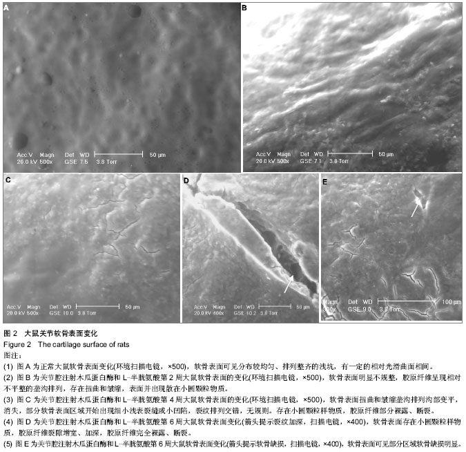
| [1] Li X,Lang W,Ye H,et al.Tougu Xiaotong capsule inhibits the tidemark replication and cartilage degradation of papain-induced osteoarthritis by the regulation of chondrocyte autophagy.Int J Mol Med.2013;31(6):1349-1356.[2] Phitak T,Pothacharoen P,Settakorn J,et al.Chondroprotective and anti-inflammatory effects of sesamin.Phytochemistry. 2012;80:77-88.[3] Kopp S,Mejersjo C,Clemenssson E.Induction of osteoarthrosis in the guinea pig knee by papain.Oral Surg Oral Med Oral Pathol.1983;55(3):259-266.[4] Pomonis JD,Boulet JM,Gottshall SL,et al.Development and pharmacological characterizati-on of a rat model of osteoarthritis pain.Pain.2005;114(3):339-346.[5] 韩冠英,凌沛学,王凤山,等.不同浓度木瓜蛋白酶建立兔膝骨关节炎模型的比较研究[J].中国骨伤,2012,5(25):424-429.[6] Stein H,Levanon D.Articular cartilage of the rabbit knee after synovectomy: a scanning electron microscopy s tudy. J Anat. 1998;192(Pt 3): 343-349.[7] 张昊, 杜宁, 任峰, 等.手法治疗实验性膝骨关节炎扫描电镜研究[J].中国中医骨伤科杂志, 2000,8(2): 1-6.[8] Ghadially FN, Ghadially JA, Oryschak AF, et al. Experimental production of ridges on rabbit articular cartilage:a scanning electron microscope s tudy. J Anat.1976;121(Pt 1): 119-132[9] 姚合梅,王政,崔瑾,等. 苗药验方皮部熏洗对骨关节炎大鼠软骨细胞凋亡与增殖的影响[J]. 浙江中医药大学学报,2013,(7): 893-896.[10] Havdrup T,Telhag H.Papain-induced changes in the knee joints of adult rabbits. Acta Orthop Scand.1977;48(2): 143-149.[11] 方斌,刘文刚,赵自明,等. 参麦注射液关节内注射治疗家兔膝骨关节炎的实验研究[J]. 风湿病与关节炎,2013,2(8): 27-30. [12] 帅明,林荔军,林昭伟,等. IL-1β、HIF-1α和VEGF在兔膝关节骨性关节炎模型滑膜中的表达[J]. 中国老年学杂志,2013,33(10): 2311-2313. [13] 高润成,王志文,袁强,等. 麝香乌龙丸对兔膝骨关节炎软骨组织形态及p38MAPK,Caspase-3表达的影响[J]. 中国实验方剂学杂志,2013,19(11):228-231. [14] 李成,赵硕,张振宇. 软骨源性形态发生蛋白缓释系统对兔膝骨关节炎的影响[J]. 山东医药,2013,53(12):18-20.[15] 王庆甫,马玉峰,殷岳衫,等. 低频超声促透中药对兔膝关节炎细胞因子的影响[J]. 北京中医药大学学报,2013,36(2): 108-112. [16] 汪福东,郭义娟,董立新,等. 抗骨增生片对兔膝骨关节炎软骨组织病理形态影响研究[J]. 实用中医药杂志,2013,29(3): 159-161. [17] 程园园,刘健,冯云霞,等. 骨关节炎大鼠心、肺功能变化与CD4~+ CD25~+ Foxp3~+ Treg的相关性分析[J]. 中国免疫学杂志,2013,29(3):227-231,235. [18] 侯小丽,赵林涛,宋延平. 金天格胶囊对骨关节炎大鼠病理形态的影响[J]. 中国实验方剂学杂志,2013,19(6): 287-290. [19] 孙鲁宁,黄桂成,赵燕华,等. 木瓜蛋白酶诱导兔膝关节骨关节炎模型滑膜中白细胞介素1、白细胞介素6、白三烯浓度变化与药物注射时间的关系[J]. 中国组织工程研究,2012,16(33): 6184-6188. [20] 孙鲁宁,黄桂成,赵燕华,等. 伤科消炎膏对木瓜蛋白酶诱导兔膝骨关节炎模型滑膜中白介素-1、白三烯、金属蛋白酶-3的影响[J]. 辽宁中医药大学学报,2012,14(9):143-145. [21] 周建中,王长峰,马勇,等. 骨关节炎模型兔膝关节腔注射丹皮酚联合针刺足三里血清白细胞介素1β、肿瘤坏死因子α的变化[J]. 中国组织工程研究,2012,16(22):4096-4099.[22] 戚晶敏,吴启富,王繁盛,等. 风湿康治疗大鼠膝骨关节炎的实验研究[J]. 热带医学杂志,2012,12(4):386-389. [23] 季卫锋,施伟峰,陈林,等. 补肾活血法防治大鼠膝骨性关节炎的实验研究[J]. 中国骨伤,2012,25(3):246-250. [24] 孙鲁宁,赵燕华,黄桂成,等. 木瓜蛋白酶诱导膝关节骨关节炎模型兔滑膜病理变化与药物注射时间的关系[J]. 中国组织工程研究与临床康复,2011,15(50): 9311-9313. [25] 李石胜,吴耀持,张峻峰,等.长针透刺膝骨关节炎模型大鼠滑膜组织中基质金属蛋白酶3的变化[J]. 中国组织工程研究与临床康复, 2011,15(50):9415-9418.[26] 谭庆远,王黎明,曲洪雪,等. 中药萃取超导透入治疗兔膝骨关节炎疗效的实验研究[J]. 中医临床研究,2011,3(3): 11-13. [27] 韩冠英,凌沛学,王凤山,等. 黄原胶注射液对兔膝骨关节炎治疗作用的实验研究[J]. 食品与药品,2011,13(11): 381-384.[28] 尹学永,王志文,赵鑫. 抗骨增生片对兔膝骨关节炎软骨组织病理形态及MMP-1、TIMP-1的影响[J]. 河北中医药学报,2011, 26(2): 3-4. [29] 许放,师咏梅,柳占彪,等. 痹祺胶囊对实验性骨关节炎大鼠NO、HYP的影响[J]. 天津中医药,2011,28(3): 237-239. [30] 钟鼎文,郭长青,嵇波,等. 膝骨关节炎大鼠丘脑及下丘脑β-EP受体含量变化及针刀松解法的影响研究[J]. 中华中医药学刊, 2011, 29(2): 276-278. [31] 郭长青,嵇波,陈幼楠,等. 针刀松解法对膝骨关节炎大鼠中枢不同部位亮氨酸-脑啡肽的影响[J]. 中国骨伤,2011,24(8):656- 658. [32] 庄超,刘瑞平,徐南伟,等. 兔骨关节炎模型血清炎症指标的动态观察[J]. 南京医科大学学报:自然科学版,2011,31(3):369-373.[33] 尹学永,王志文,赵鑫. 抗骨增生片对兔骨关节炎软骨细胞凋亡的影响[J]. 陕西中医,2011,32(8): 1083-1085. [34] 张延娇,李绍新,郭周义,等. 低强度激光联合照射口咽部和犊鼻穴对实验性膝骨关节炎兔氧自由基代谢的影响[J]. 中国组织工程研究与临床康复,2010,14(28):5208-5211. [35] 嵇波,郭长青,金燕,等. 针刀和电针对膝骨关节炎大鼠痛阈和中枢单胺类神经递质的影响[J]. 中国病理生理杂志,2010,26(6): 1091-1095. [36] 路明珠,陆文铨,伊佳,等. 不同分子量构成的玻璃酸钠对骨关节炎治疗作用的实验研究[J]. 药学实践杂志,2010,28(1):19-22. [37] 王志文,赵鑫,鲍际鹏,等. 麝香乌龙丸对兔骨关节炎软骨细胞凋亡的影响[J]. 时珍国医国药,2009,20(11):2893-2894. [38] 王翠民,尹学永,谷宁飞,等. MMP-1、TIMP-1在兔实验性膝骨关节炎中的表达[J]. 中国现代医学杂志,2009,19(18):2754-2756. [39] 邓宇,伍筱梅,任医民,等. 关节腔内注射不同蛋白酶建立兔膝骨关节炎模型的对比研究[J]. 中华关节外科杂志:电子版,2009, 3(3): 332-339.[40] 戴国钢,刘波,罗小兵等.正常兔膝关节表面结构的扫描电镜观察[J].中国组织工程研究与临床康复,2007,11(32):6391-6393.[41] Clarke IC. Articular cartilage:a review and scanning electron microscope s tudy. II. The territorial fibrillar architecture. J Ana.1974;118(2): 261-280.[42] Buckwalter JA,Mankin HJ.Articular cartilage. J Bone Joint Surg.1997;79-A(4): 600-633.[43] Weakley BS. A Beginner’s handbook in biological transmission Electron Microscopy. 2nd ed. New York: Churchill Livingstone;1981:49.[44] Soeder S, Kuhlmann A, Aigner T. Analysis of protein distribution in cartilage using immunofluorescence and laser confocal scanning microscopy. Methods Mol Med. 2004; 101:107-125.[45] Kubo T, Arai Y, Namie K, et al.Time-sequential changes in biomechanical and morphological properties of articular cartilage in cryopreserved osteochondral allografting. J Orthop Sci. 2001; 6:276-281.[46] Paulsen HU, Thomsen JS, Hougen HP, et al.A histomorphometric and scanning electron microscopy study of human condylar cartilage and bone tissue changes in relation to age. Clin Orthod Res. 1999;2:67-78.[47] Clark JM, Simonian PT. Scanning electron microscopy of ‘‘fibrillated’’ and ‘‘malacic’’ human articular cartilage: technical considerations.Microsc ResTech. 1997;37:299-313.[48] Li B, Marshall D, Roe M, et al. The electron microscope appearance of the subchondral bone plate in the human femoral head in osteoarthritis and osteoporosis. J Anat. 1999;195(1):101-210.[49] Goodwin DW, Zhu H, Dunn JF. In vitro MR imaging of hyaline cartilage: correlation with scanning electron microscopy. AJR Am J Roentgenol. 2000;174:405-409.[50] Stein H, Levanon D. Articular cartilage of the rabbit knee after synovectomy: a scanning electron microscopy study. J Anat.1998;192(3):343-349.[51] Jurvelin J, Kuusela T, Heikkila R, et al. Investigation of articular cartilage surface morphology with a semiquantitative scanning electron microscopic method.Acta Anat (Basel). 1983; 116:302-311.[52] Hong SP, Henderson CN. Articular cartilage surface changes following immobilization of the rat knee joint. A semiquantitative scanning electron-microscopic study. Acta Anat (Basel).1996;157:27-40.[53] O’Connor P, Oates K, Gardner DL, et al. Low temperature and conventional scanning electron microscopic observations of dog femoral condylar cartilage surface after anterior cruciate ligament division. Ann Rheum Dis. 1985;44:321-327.[54] Suso S, Carbonell JA, Segur JM, et al. Cartilage appearance using an environmental scanning electron microscope. Cell Preserv Technol.2004;2:51-54.[55] 史宗道, 杨峰,何志秀,等.透明质酸钠与强的松龙治疗兔实验性颞颌关节骨关节炎的研究[J].中国修复重建外科杂志,2002, 16(1):5-10.[56] 杨峰,史宗道.用木瓜蛋白酶建立兔颞颌关节骨关节炎模型的研究[J].华西口腔医学杂志,2002,20(5):330-332. [57] Guzman RE, Evans MG, Bove S, et al. Mono-iodoacetate-induced histologic changes in subchondral bone and articular cartilage of rat femorotibial joints: an animal model of osteoarthritis.Toxicol Pathol. 2003;31: 619-624.[58] Bendele AM. Animal models of osteoarthritis. J.Muscu Neur Inte. 2001;1: 363-376.[59] Fernihough JC, Gentry M, MalcangioA, et al.Pain related behaviour in two models of osteoarthritis in the rat knee.Pain, 2004;112: 83-93.[60] Pomonis JD, Boulet JM, Gottshall SL,et al. Walker. Development and pharmacological characterization of a rat model of osteoarthritis pain. Pain. 2005;114: 339-346.[61] Palmoski MJ, Bean JS. Cartilage atrophy induced by limb immobobilization.\\Greenwal RA,Diamond HS, eds. Handbook of Animal Models for the Rheumatic Diseases II. Bocha Raton, Florida:CRC Press. 1988:83-87. |