Chinese Journal of Tissue Engineering Research ›› 2013, Vol. 17 ›› Issue (37): 6541-6547.doi: 10.3969/j.issn.2095-4344.2013.37.001

    Next Articles

Gene transfection at different time affects the osteogenesis in traction area

Hu Chun-bing1, Wu Guo-ping1, Liu Xi2, Lan Yong-shu3, He Xiao-chuan1, Guo Li1   

  1. 1Department of Plastic Surgery, 3CT Room, the Affiliated Hospital of Luzhou Medical College, Luzhou  646000, Sichuan Province, China; 2Department of Radiology, Hospital of Stomatology, Luzhou Medical College, Luzhou  646000, Sichuan Province, China
  • Received:2013-04-09 Revised:2013-04-28 Online:2013-09-10 Published:2013-09-10
  • Contact: Wu Guo-ping, M.D., Professor, Department of Plastic Surgery, the Affiliated Hospital of Luzhou Medical College, Luzhou 646000, Sichuan Province, China drgpwu@yahoo.com.cn
  • About author:Hu Chun-bing★, Master, physician, Department of Plastic Surgery, the Affiliated Hospital of Luzhou Medical College, Luzhou 646000, Sichuan Province, China huchunbingdr@163.com
  • Supported by:

    National Natural Science Foundation of China, No. 30600653*; Key Project of Sichuan Provincial Education Department, No. 11ZA239*; Scientific Research Project of Sichuan Provincial Health Department, No. 100285*

Abstract:

BACKGROUND: The previous studies have shown that electroporation-mediated recombinant plasmid pIRES-hBMP2-hVEGF165 can promote the early angiogenesis and new bone formation in the traction zone.
OBJECTIVE: To investigate the effect of gene transfection at different time on the new bone formation in the traction zone during mandibular distraction in rabbits, so as to explore the optimal time for gene therapy and obtain a better treatment effect.
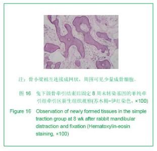
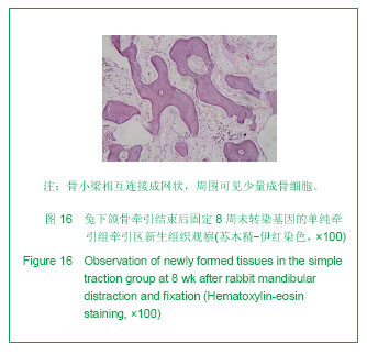
METHODS: Forty-eight New-Zealand rabbits were employed. After accomplished the bilateral mandibular osteotomy and distractor implantation under general anaesthesia, the rabbits were randomly divided into four groups. The bilateral traction zones were transfected with the 2 μg (0.1g/L) recombinant plasmids pIRES-hBMP2-hVEGF165 at latency period, traction period and consolidation period, respectively, and the rabbits in these three groups received electroporation stimulation. The simple traction group only received traction without gene transfection. The rabbits in each group received traction at 3 days after modeling with the rate of 0.8 mm/d and once per day, and continuous traction for 10 days; three rabbits were sacrificed in each group at 1, 2, 4 and 8 week. The new tissues in the mandibular traction zone were cut for histological observation and histomorphometry analysis.
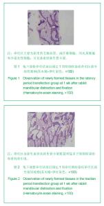
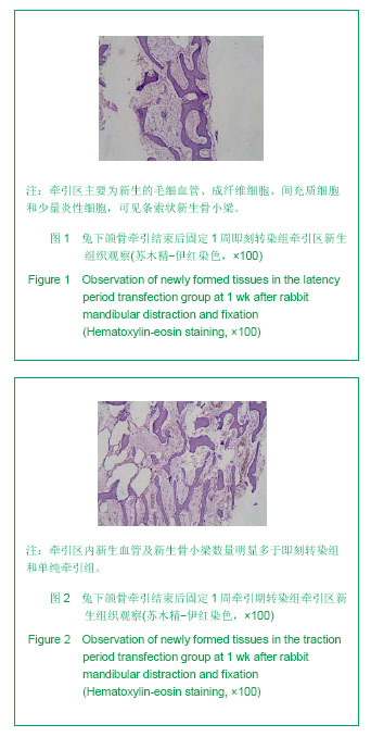
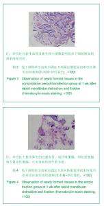
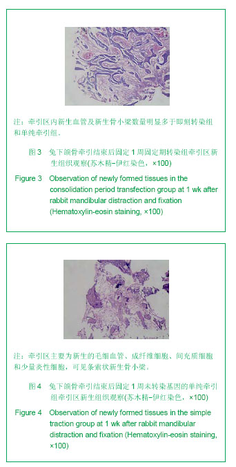
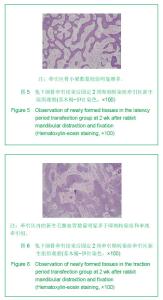
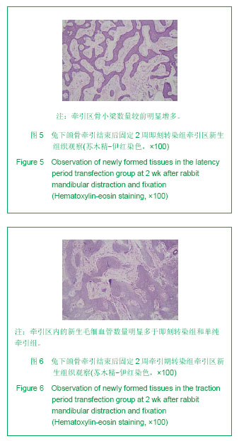
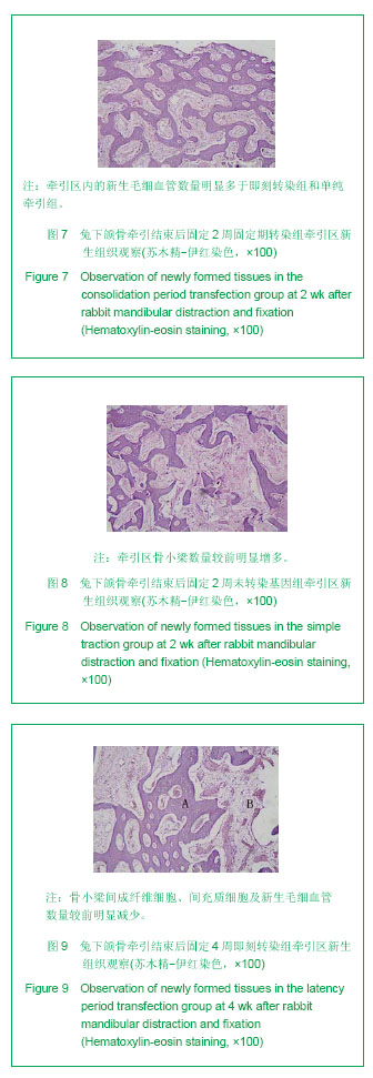
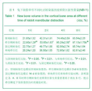
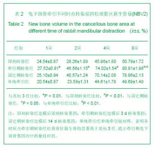
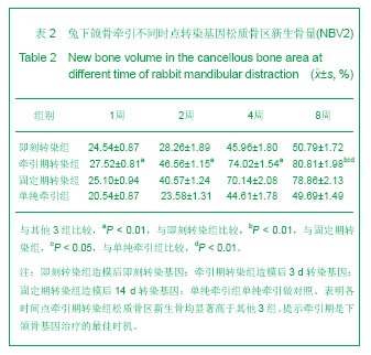
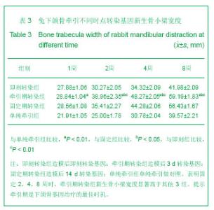
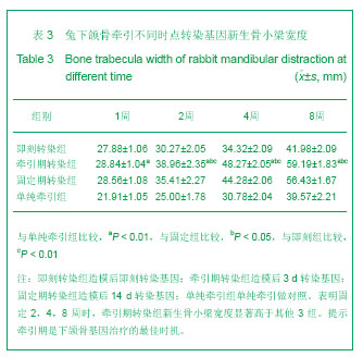
RESULTS AND CONCLUSION: Histological examination and histomorphometry analysis showed that amounts of newly formed vessels, osteoblasts and mesenchymal cells of distraction period transfection group were greater than those of the latency period transfection group, consolidation period transfection group and simple traction group, and the new bone volume and the width of bone trabecula in the distraction period transfection group were higher than those in the other three groups (P < 0.05). It is better to transfect gene at the beginning of traction (distraction period) than at other stages of traction; in this way, we can obtain more remarkable effects on new bone formation. It suggests that the distraction stage is the optimal time for gene therapy.

Key words: electroporation, gene therapy, traction, transfection

CLC Number: