Chinese Journal of Tissue Engineering Research ›› 2013, Vol. 17 ›› Issue (34): 6089-6096.doi: 10.3969/j.issn.2095-4344.2013.34.005
Previous Articles Next Articles
Tissue-engineered bone repairs sheep alveolar bone defects
Zhang Qin, Yang Chuan-bo, He Hui-yu, Cui Jie, Yang Nan, Ma Wen-yuan
- Department of Prosthodontics, First Affiliated Hospital of Xinjiang Medical University, Urumqi 830054, Xinjiang Uygur Autonomous Region, China
-
Online:2013-08-20Published:2013-08-20 -
Contact:He Hui-yu, M.D., Professor, Master’s supervisor, Chief physician, First Affiliated Hospital of Xinjiang Medical University, Urumqi 830054, Xinjiang Uygur Autonomous Region, China hehuiyu02@sina.com -
About author:Zhang Qin, Master, Attending physician, Department of Prosthodontics, First Affiliated Hospital of Xinjiang Medical University, Urumqi 830054, Xinjiang Uygur Autonomous Region, China zhangqin678ok@163.com -
Supported by:the National Natural Science Foundation of China, No. 81060088*;
the Natural Science Foundation of the Science and Technology Bureau of Xinjiang Uygur Autonomous Rgion, No. 2011211A073*;
the Youth Fund of Affiliated Hospital of Xinjiang Medical University, No. 2012QN21*
CLC Number:
Cite this article
Zhang Qin, Yang Chuan-bo, He Hui-yu, Cui Jie, Yang Nan, Ma Wen-yuan. Tissue-engineered bone repairs sheep alveolar bone defects[J]. Chinese Journal of Tissue Engineering Research, 2013, 17(34): 6089-6096.
share this article
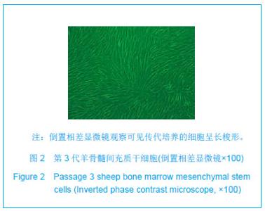
2.1 培养的骨髓间充质干细胞的形态 原代培养的骨髓间充质干细胞:接种的细胞多呈球形悬浮于培养液中,观察至第2天发现部分细胞出现贴壁现象,细胞形态多呈现椭圆形。第7天观察时可见细胞数量陡增,汇集到培养瓶底部80%左右,细胞多为长梭形,漩涡生长。对于细胞的鉴定及成骨诱导培养,由于这次实验是序列研究课题,课题组前期已对羊骨髓间充质干细胞进行成骨诱导及鉴定,证实培养的细胞为骨髓间充质干细胞[22] 。 传代培养的骨髓间充质干细胞:细胞传代培养接种24 h后可见细胞贴壁生长,逐渐生长为长梭形,胞体较原代细胞体积略大。传代培养至第3代时细胞生长旺盛,形态比较均一,无明显改变,见图2。"
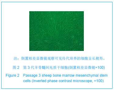
2.2 高温煅烧骨基本性能 对骨块进行脱脂、脱蛋白后,经过950 ℃煅烧的椎骨松质骨块为白垩色,表面呈现蜂窝状多孔结构,保留了骨组织间特有的多孔状空间特性。利用甘油液体置换法测定出煅烧骨材料孔隙率为(66.10±1.32)%[23]。扫描电镜观察可见煅烧骨材料的骨小梁结构完整,孔隙间相互连通,孔径范围在137.44-538.72 μm。大孔壁上可见许多微孔结构存在,微孔孔壁表面粗糙,形貌为小梁状和山嵴状,利于细胞的粘附生长,见图3。"

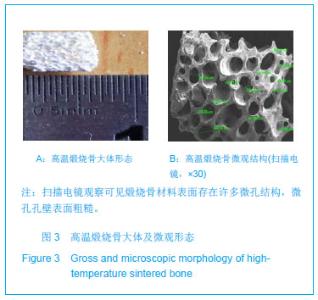
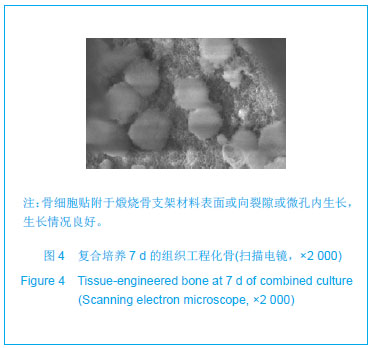
2.3 构建的组织工程化骨的形态 培养2 h后将6孔板取出,超净台内沿6孔板壁缓慢加入DMEM/F12培养液 3 mL,放入培养箱中培养。细胞-煅烧骨支架复合物培养7 d后,大体观察生物陶瓷材料上无明显变化。在高倍镜下观察可见细胞与细胞之间分界不清,细胞与煅烧骨材料结合较为紧密。扫描电镜观察所示可见细胞贴附于支架材料表面或向裂隙或微孔内生长,细胞与煅烧骨材料复合生长情况如图4。"

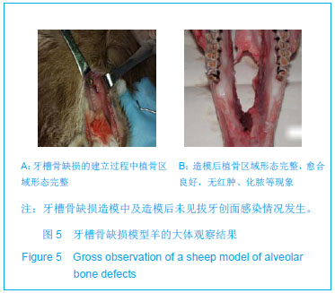
2.4 实验动物数量分析 2组实验羊均进入结果分析,无脱失。 2.5 牙槽骨缺损模型羊的大体观察结果 各组实验动物造模后均未见拔牙创面感染情况发生。造模后1周可见植骨区域的牙槽窝创面愈合良好,无红肿、化脓等现象,见图5。"
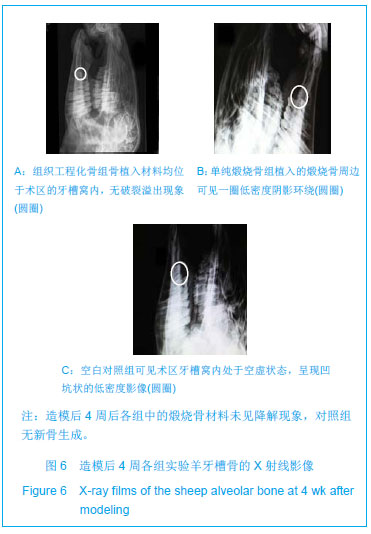
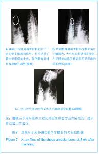
2.6 组织工程化骨修复羊牙槽骨的影像学观察 造模后4周:单纯煅烧骨组和组织工程化骨组的骨植入材料均位于术区的牙槽窝内,无破裂溢出现象。植入的煅烧骨周边可见一圈低密度阴影环绕,与周邻组织界限模糊,煅烧骨材料形态及大小无明显改变,提示植入的煅烧骨材料未见降解现象;对于未植入任何材料的空白对照组内观察可见术区牙槽窝内处于空虚状态,呈现凹坑状的低密度影像,缺牙区临近牙槽嵴高度与邻牙区牙槽嵴高度呈现出不连续状,见图6。"
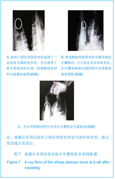
造模后8周:组织工程化骨组煅烧骨体积大小与较单纯煅烧骨组有所减小,在煅烧骨边缘可见低密度影环绕,与周围组织不连续。手术区域牙槽嵴高度与周邻组织高度相近,提示组织工程化骨组的骨材料起到了一定的骨支撑机构作用,并且诱导了新生骨组织的生成,致使煅烧骨材料有崩解的趋势,见图7A。 在单纯煅烧骨组中发现骨材料完整充填在牙槽窝内,大小形态未见明显变化,在牙槽骨缺损区域周围可见零散的高密度影,可能为煅烧骨组织的残片,见图7B。 在空白对照组中观察可见牙槽骨缺损区域牙槽嵴高度有所降低,与邻牙不连续,仍然可见术区牙槽窝呈空虚状态,见图7C。"
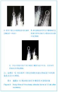
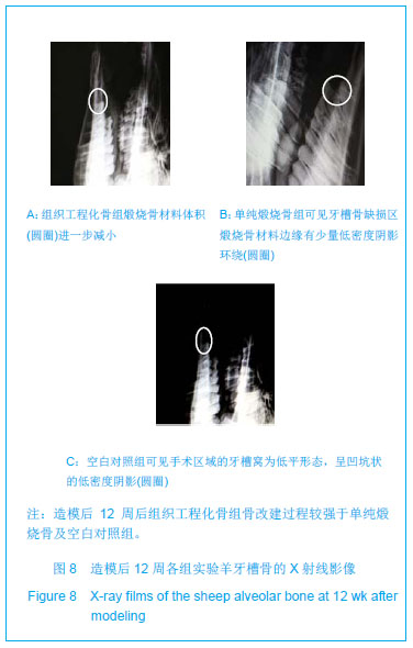
造模后12周:组织工程化骨组煅烧骨材料体积进一步减小,材料边缘可见散在低密度影,与周围骨组织较连续,牙槽嵴高度较单纯煅烧骨组及空白对照组高,牙槽窝未见凹坑状的低密度阴影,见图8A。 单纯煅烧骨组可见牙槽骨缺损区煅烧骨材料边缘有少量低密度阴影环绕,煅烧骨材料边缘模糊不清,缺损区阴影变浅与周围牙槽骨组织界限较模糊,提示有少量的骨组织形成,但效果没有组织工程化骨明显,见图8B。 空白对照组观察可见手术区域的牙槽窝为低平形态,呈凹坑状的低密度阴影,提示空白对照组骨缺损区域出现了骨组织吸收,见图8C。"
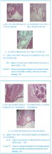
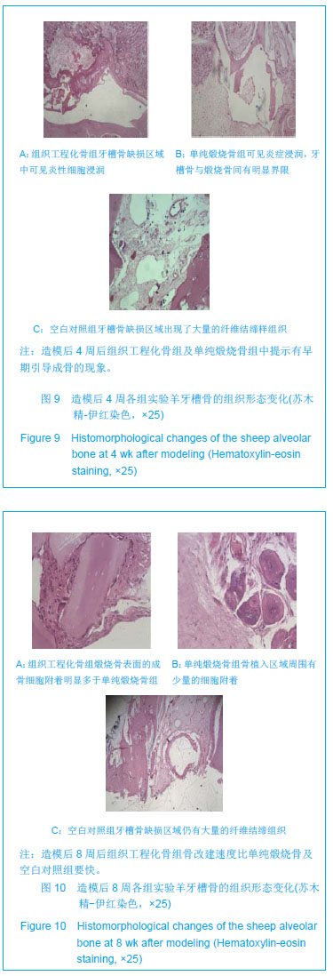
2.7 组织工程化骨修复羊牙槽骨的组织病理结果 造模后4周:单纯煅烧骨组和组织工程化骨组牙槽骨缺损区域中均可见炎性细胞浸润。在牙槽骨缺损区域周围和煅烧骨材料之间有明显的界限,视野中可发现局部有未降解的煅烧骨材料残片,可能是在制作病理切片时所致,提示新生骨组织的改建仍在进行过程中。在空白对照组牙槽骨缺损区域出现了大量的纤维结缔样组织,分布较为广泛,呈不规则状,见图9。 造模后8周:单纯煅烧骨组和组织工程化骨组中的骨植入区域周围有少量的细胞附着,经进一步观察证实为骨母细胞,为早期的骨组织细胞,提示新生牙槽骨组织正处于改建过程中。通过对比发现组织工程化骨组煅烧骨表面的成骨细胞附着明显多于单纯煅烧骨组,说明了组织工程化骨组的骨改建及诱导新生骨过程早于单纯煅烧骨组。空白对照组中观察可见牙槽骨缺损区域仍有大量的纤维结缔组织充斥,与周围的骨组织呈不连续状态,见图10。"
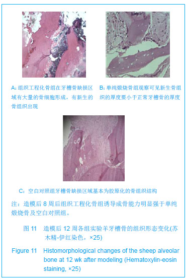
造模后12周:组织工程化骨组在牙槽骨缺损区域有大量的骨细胞形成,有新生的骨组织出现,与周围组织分界不清,呈现出融合生长状态。新生牙槽骨结构更加接近正常骨结构,骨板排列趋向规则,出现整齐的同心圆状的哈佛系统,基本具备了骨组织的正常结构。单纯煅烧骨组中观察可见新生骨组织的厚度要小于正常牙槽骨的厚度,煅烧骨组织呈片状分布于骨缺损区域,与周围组织间不连续,提示虽然有新骨组织生成,但效果不及组织工程化骨组。空白对照组中可见牙槽骨缺损区域基本为胶原化的骨组织结构,其胶原钙化程度极低,骨缺损区域的自我修复能力较差,见图11。"
| [1]张淅,姚遥,刘迎飞.牙周炎致骨缺损的植骨治疗[J].临床口腔医学杂志,2010,26(5):292-294.[2]Somerman MJ, Ouyang HJ, Berry JE, et al. Evolution of periodontal regeneration: from the roots' point of view. J Periodontal Res. 1999;34(7):420-424.[3]Kubota K, Yoshimura N, Yokota M, et al. Overview of effects of electrical stimulation on osteogenesis and alveolar bone. J Periodontol. 1995;66(1):2-6.[4]童庆春,李宁毅,张志勇,等.即刻骨移植与延期牙种植术联合应用修复牙槽骨缺损的初步研究[J].上海口腔医学,2008,17(5): 488-491.[5]朱淑倩,何家才.人工骨替代材料修复种植体周围骨缺损的研究进展[J].医学综述,2010,16(6):863-865.[6]游永刚,唐辉,徐永清.不同骨移植材料骨诱导活性的研究现状[J].现代生物医学进展,2008,8(7):1338-1339.[7]杨川博,何惠宇.牙槽骨缺损修复治疗新进展[J].中国实用口腔科杂志,2012,3(5):182-185.[8]刘世森,苏方,刘洪臣.组织工程骨修复种植部位骨量不足的临床进展[J]中华老年口腔医学杂志,2009,7(3):180-182.[9]Griffith LG, Naughton G. Tissue engineering--current challenges and expanding opportunities. Science. 2002; 295(5557):1009-1014.[10]王敏,翁雨来,胡晓洁,等.组织工程技术修复犬牙槽骨缺损的实验研究[J].中华医学杂志,2003,83(15):1339-1344.[11]许慧芬,何惠宇,唐小雪,等.不同方法制备异种骨支架材料的生物相容性评价[J].中国组织工程研究与临床康复,2011,15(47): 8749-8752.[12]Weng Y, Cao Y, Silva CA, et al. Tissue-engineered composites of bone and cartilage for mandible condylar reconstruction. J Oral Maxillofac Surg. 2001;59(2):185-190.[13]Shang Q, Wang Z, Liu W, et al. Tissue-engineered bone repair of sheep cranial defects with autologous bone marrow stromal cells. J Craniofac Surg. 2001;12(6):586-595.[14]杨川博,何惠宇,崔杰,等.骨髓基质干细胞复合煅烧骨的皮下成骨[J].中国组织工程研究,2013:17(16):2883-2890.[15]中华人民共和国科学技术部. 关于善待实验动物的指导性意见. 2006-09-30.[16]唐小雪,何惠宇,许慧芬等.两种不同分离方法对羊骨髓间充质干细胞生物活性的影响[J].中国组织工程研究与临床康复,2011, 15(49):9137-9140.[17]Erdemli O, Captug O, Bilgili H, et al. In vitro and in vivo evaluation of the effects of demineralized bone matrix or calcium sulfate addition to polycaprolactone-bioglass composites. J Mater Sci Mater Med. 2010;21(1):295-308. [18]Jähn K, Braunstein V, Furlong PI, et al. A rapid method for the generation of uniform acellular bone explants: a technical note. J Orthop Surg Res. 2010;5:32. [19]Lin FH, Liao CJ, Chen KS, et al. Preparation of a biphasic porous bioceramic by heating bovine cancellous bone with Na4P2O7.10H2O addition. Biomaterials. 1999;20(5):475-484.[20]崔杰.去抗原异种松质骨支架材料的制备及性能研究[D].乌鲁木齐:新疆医科大学.2011.[21]许慧芬,何惠宇,唐小雪,崔杰.物理联合化学或化学方法处理去抗原异种松质骨支架与骨髓间充质干细胞的细胞相容性[J].中国组织工程研究,2012,02(5):958-962.[22]唐小雪,何惠宇,许慧芬,等.两种不同分离方法对羊骨髓间充质干细胞生物活性的影响[J].中国组织工程研究与临床康复,2011, 15(49):9137-9140.[23]王文良,张华亮,初殿伟,等.自体骨髓间充质干细胞复合壳聚糖/羟基磷灰石支架修复兔膝骨软骨缺损[J].中华骨科杂志,2009, 29(1):61-64.[24]孟焕新.牙周病学[M].北京:人民卫生出版社,2008.[25]孙路,王永兰.牙周组织工程研究新进展[J].牙体牙髓牙周病学杂志,2012,3:170-174. [26]宋珂,曹颖光.多元化骨组织工程修复颌骨缺损的研究进展[J].临床口腔医学杂志,2010,26(2):125-127.[27]陈广东,杨惠林,王根林,等.骨髓间充质干细胞的成骨分化及临床应用[J].中国组织工程研究与临床康复,2010,14(49): 9272-9276.[28]Caplan AI. Mesenchymal stem cells. J Orthop Res. 1991;9(5): 641-650.[29]Derubeis AR, Cancedda R. Bone marrow stromal cells (BMSCs) in bone engineering: limitations and recent advances. Ann Biomed Eng. 2004;32(1):160-165.[30]Hasegawa N, Kawaguchi H, Hirachi A, et al. Behavior of transplanted bone marrow-derived mesenchymal stem cells in periodontal defects. J Periodontol. 2006;77(6):1003-1007.[31]Caplan AI, Dennis JE. Mesenchymal stem cells as trophic mediators. J Cell Biochem. 2006;98(5):1076-1084.[32]Maniatopoulos C, Sodek J, Melcher AH. Bone formation in vitro by stromal cells obtained from bone marrow of young adult rats. Cell Tissue Res. 1988;254(2):317-330.[33]Weng Y, Wang M, Liu W, et al. Repair of experimental alveolar bone defects by tissue-engineered bone. Tissue Eng. 2006; 12(6):1503-1513.[34]Hayashi O, Katsube Y, Hirose M, et al. Comparison of osteogenic ability of rat mesenchymal stem cells from bone marrow, periosteum, and adipose tissue. Calcif Tissue Int. 2008;82(3):238-247. [35]Prockop DJ. Marrow stromal cells as stem cells for nonhematopoietic tissues. Science. 1997;276(5309):71-74.[36]柴岗,张艳,刘伟,等.人骨髓基质细胞表面标志的研究[J].组织工程与重建外科杂志,2005,1(5):263-264.[37]Minamide A, Tamaki T, Kawakami M, et al. Experimental spinal fusion using sintered bovine bone coated with type I collagen and recombinant human bone morphogenetic protein-2. Spine (Phila Pa 1976). 1999;24(18):1863-1872.[38]Barnes B, Boden SD, Louis-Ugbo J, et al. Lower dose of rhBMP-2 achieves spine fusion when combined with an osteoconductive bulking agent in non-human primates. Spine (Phila Pa 1976). 2005;30(10):1127-1133.[39]Tamaki T, Sakurai K, Kasamatsu N. Results of basic and clinical studies of true bone ceramic. In: Urist MR, O'Connor BT, Burw ell RG, eds. Bone Grafts, Derivatives and Substitute. Oxford: Buterworth Heinemann,1994.[40]Katoh T, Sato K, Kawamura M, et al. Osteogenesis in sintered bone combined with bovine bone morphogenetic protein. Clin Orthop Relat Res. 1993;(287):266-275.[41]Lin FH, Liao CJ, Chen KS, et al. Preparation of betaTCP/HAP biphasic ceramics with natural bone structure by heating bovine cancellous bone with the addition of (NH(4))(2)HPO(4). J Biomed Mater Res. 2000;51(2):157-163.[42]Le Huec JC, Schaeverbeke T, Clement D, et al. Influence of porosity on the mechanical resistance of hydroxyapatite ceramics under compressive stress. Biomaterials. 1995;16(2): 113-118.[43]Lane JM, Bostrom MP. Bone grafting and new composite biosynthetic graft materials. Instr Course Lect. 1998;47: 525-534.[44]许永华,施新猷,胡蕴玉,等.牛煅烧骨与体外培养兔骨膜成骨细胞的相容性[J].第四军医大学学报,2000,21(4):512-514.[45]Sandberg MM, Aro HT, Vuorio EI. Gene expression during bone repair. Clin Orthop Relat Res. 1993;(289):292-312.[46]Wilson CE, Dhert WJ, Van Blitterswijk CA, et al. Evaluating 3D bone tissue engineered constructs with different seeding densities using the alamarBlue assay and the effect on in vivo bone formation. J Mater Sci Mater Med. 2002;13(12):1265- 1269.[47]Kruyt M, De Bruijn J, Rouwkema J, et al. Analysis of the dynamics of bone formation, effect of cell seeding density, and potential of allogeneic cells in cell-based bone tissue engineering in goats. Tissue Eng Part A. 2008;14(6):1081- 1088. [48]van Gaalen SM, de Bruijn JD, Wilson CE, et al. Relating cell proliferation to in vivo bone formation in porous Ca/P scaffolds. J Biomed Mater Res A. 2010;92(1):303-310. |
| [1] | Hua Haotian, Zhao Wenyu, Zhang Lei, Bai Wenbo, Wang Xinwei. Meta-analysis of clinical efficacy and safety of antibiotic artificial bone in the treatment of chronic osteomyelitis [J]. Chinese Journal of Tissue Engineering Research, 2021, 25(6): 970-976. |
| [2] | Zhang Bin, Sun Lihua, Zhang Junhua, Liu Yusan, Cui Caiyun. A modified flap immediate implant is beneficial to soft tissue reconstruction in maxillary aesthetic area [J]. Chinese Journal of Tissue Engineering Research, 2021, 25(5): 707-712. |
| [3] | Li Chenjie, Lü Linwei, Song Yang, Liu Jingna, Zhang Chunqiu. Measurement and statistical analysis of trabecular morphological parameters of titanium alloy peri-prosthesis under preload [J]. Chinese Journal of Tissue Engineering Research, 2021, 25(4): 516-520. |
| [4] | Li Li, Ma Li. Immobilization of lactase on magnetic chitosan microspheres and its effect on enzymatic properties [J]. Chinese Journal of Tissue Engineering Research, 2021, 25(4): 576-581. |
| [5] | Liu Liu, Zhou Qingzhu, Gong Zhuo, Liu Boyan, Yang Bin, Zhao Xian. Characteristics and manufacturing techniques of collagen/inorganic materials for constructing tissue-engineered bone [J]. Chinese Journal of Tissue Engineering Research, 2021, 25(4): 607-613. |
| [6] | Li Xiaozhuang, Duan Hao, Wang Weizhou, Tang Zhihong, Wang Yanghao, He Fei. Application of bone tissue engineering materials in the treatment of bone defect diseases in vivo [J]. Chinese Journal of Tissue Engineering Research, 2021, 25(4): 626-631. |
| [7] | He Jie, Chang Qi. Biological reconstruction of large bone defects after resection of malignant tumor of extremities [J]. Chinese Journal of Tissue Engineering Research, 2021, 25(3): 420-425. |
| [8] | Xing Hao, Zhang Yonghong, Wang Dong. Advantages and disadvantages of repairing large-segment bone defect [J]. Chinese Journal of Tissue Engineering Research, 2021, 25(3): 426-430. |
| [9] | Chen Siqi, Xian Debin, Xu Rongsheng, Qin Zhongjie, Zhang Lei, Xia Delin. Effects of bone marrow mesenchymal stem cells and human umbilical vein endothelial cells combined with hydroxyapatite-tricalcium phosphate scaffolds on early angiogenesis in skull defect repair in rats [J]. Chinese Journal of Tissue Engineering Research, 2021, 25(22): 3458-3465. |
| [10] | Zhou Anqi, Tang Yufei, Wu Bingfeng, Xiang Lin. Designing of periosteum tissue engineering: combination of generality and individuality [J]. Chinese Journal of Tissue Engineering Research, 2021, 25(22): 3551-3557. |
| [11] | Zhang Zhenhua, Liu Zichen, Yu Baoqing. Status and problems of polycaprolactone and its composite materials in bone tissue engineering [J]. Chinese Journal of Tissue Engineering Research, 2021, 25(22): 3571-3577. |
| [12] | Lang Limin, He Sheng, Jiang Zengyu, Hu Yiyi, Zhang Zhixing, Liang Minqian. Application progress of conductive composite materials in the field of tissue engineering treatment of myocardial infarction [J]. Chinese Journal of Tissue Engineering Research, 2021, 25(22): 3584-3590. |
| [13] | He Fan, Xiong Xiuli, Shan Xianfeng, Zhang Shutong, Hu Jian, Wang Xuejin. Guided bone regeneration in a small animal model of critical size craniofacial bone defects [J]. Chinese Journal of Tissue Engineering Research, 2021, 25(20): 3226-3231. |
| [14] | Xie Jian, Su Jiansheng. Advantages and characteristics of electrospun aligned nanofibers as scaffolds for tissue engineering [J]. Chinese Journal of Tissue Engineering Research, 2021, 25(16): 2575-2581. |
| [15] | Ji Qi, Yu Zhengwen, Zhang Jian. Problems and trends of technique and clinical application of metallic biomaterials prepared by three-dimensional printing technology [J]. Chinese Journal of Tissue Engineering Research, 2021, 25(16): 2597-2604. |
| Viewed | ||||||
|
Full text |
|
|||||
|
Abstract |
|
|||||